. 2019 Mar 19;9(1):4853.
doi: 10.1038/s41598-019-41269-z.

The acidic protein rich in leucines Anp32b is an immunomodulator of inflammation in mice

Affiliations

The acidic protein rich in leucines Anp32b is an immunomodulator of inflammation in mice

Jan Chemnitz et al. Sci Rep. .

Abstract

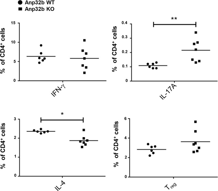
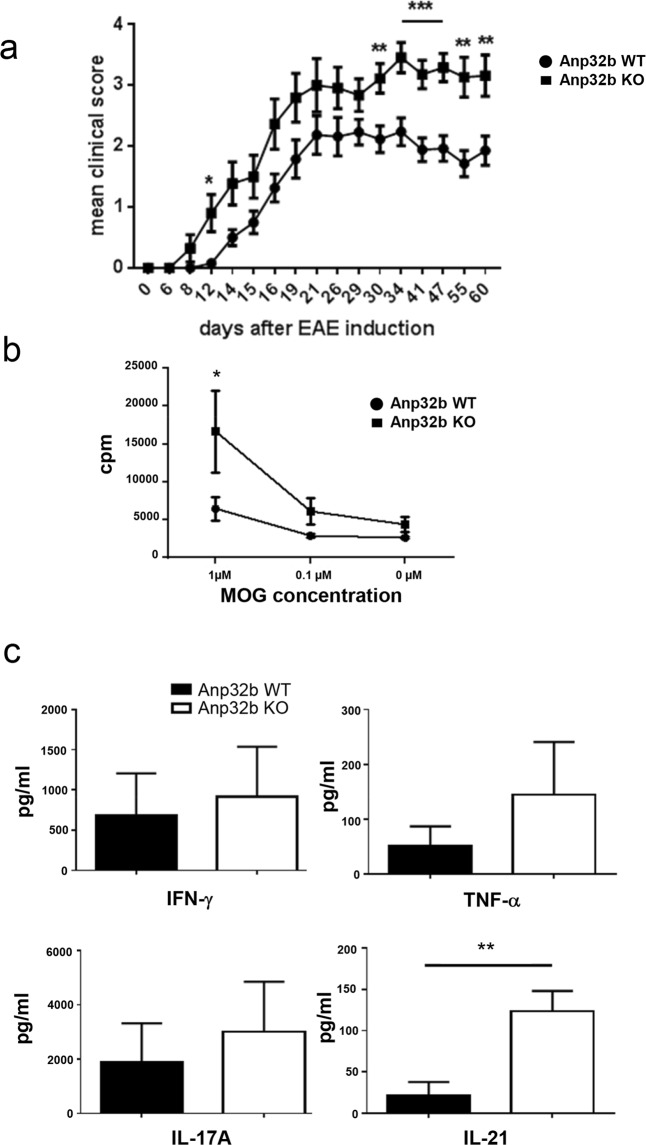
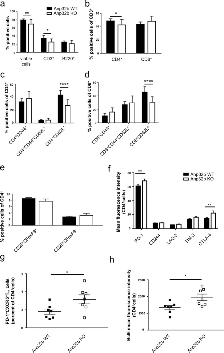
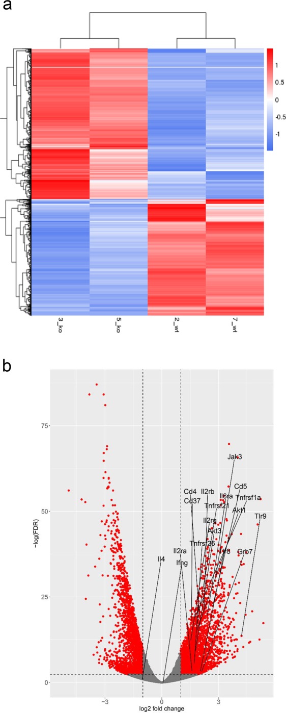
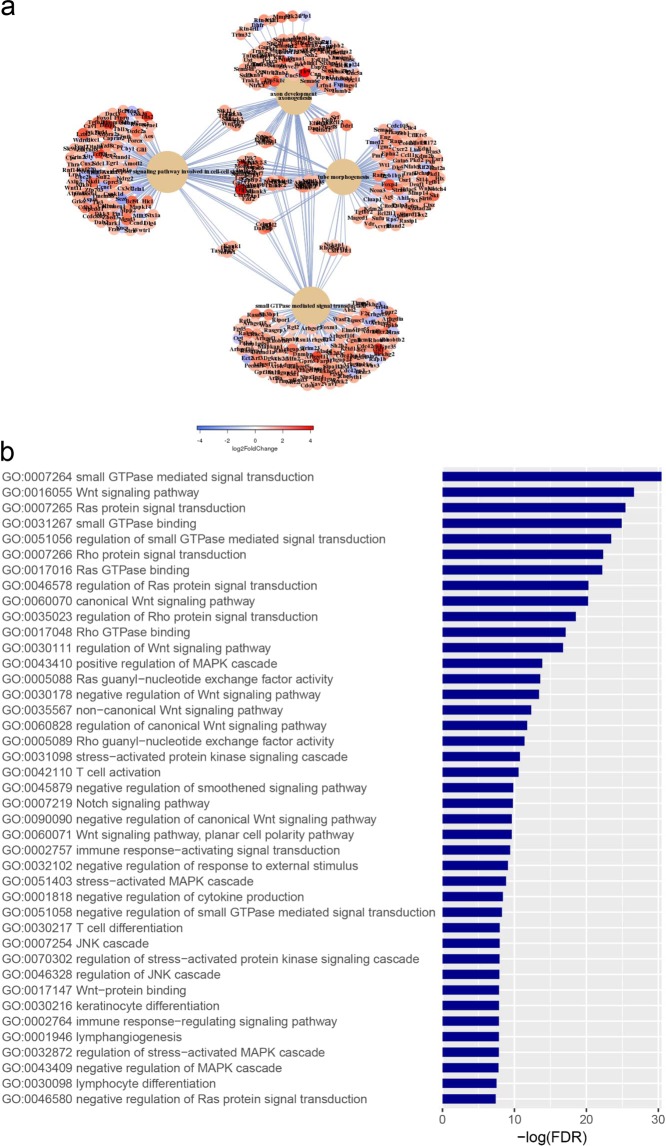
ANP32B belongs to a family of evolutionary conserved acidic nuclear phosphoproteins (ANP32A-H). Family members have been described as multifunctional regulatory proteins and proto-oncogenic factors affecting embryonic development, cell proliferation, apoptosis, and gene expression at various levels. Involvement of ANP32B in multiple processes of cellular life is reflected by the previous finding that systemic gene knockout (KO) of Anp32b leads to embryonic lethality in mice. Here, we demonstrate that a conditional KO of Anp32b is well tolerated in adult animals. However, after immune activation splenocytes isolated from Anp32b KO mice showed a strong commitment towards Th17 immune responses. Therefore, we further analyzed the respective animals in vivo using an experimental autoimmune encephalomyelitis (EAE) model. Interestingly, an exacerbated clinical score was observed in the Anp32b KO mice. This was accompanied by the finding that animal-derived T lymphocytes were in a more activated state, and RNA sequencing analyses revealed hyperactivation of several T lymphocyte-associated immune modulatory pathways, attended by significant upregulation of Tfh cell numbers that altogether might explain the observed strong autoreactive processes. Therefore, Anp32b appears to fulfill a role in regulating adequate adaptive immune responses and, hence, may be involved in dysregulation of pathways leading to autoimmune disorders and/or immune deficiencies.


Conflict of interest statement

The authors declare no competing interests.

Figures

Figure 1
Figure 1
Targeting constructs and PCR strategies. Schematic depiction of (a) the original targeting construct and (b) targeting variants in the Anp32b locus with PCR oligonucleotide primers allowing detection of the respective wild type allele, “floxed” allele and “Cre’ed” allele. (c) Targeting variants of Anp32b transcripts using PCR primers for detecting Anp32b knockout. FRT, flippase (Flp) target site; loxP, Cre target site; En2 SA, engrailed homeobox 2 splice acceptor; T2A, self-cleaving 2A peptide sequence; lacZ, β-galactosidase reporter gene; neo, neomycin resistance gene; pA, polyadenylation signal.
Figure 2
Figure 2
Detection of Anp32b KO in mice. (a) Immunohistochemical detection of Anp32b in spleens. Spleen sections from Anp32b KO and wild type (WT) animals were stained with a monoclonal antibody specific for murine Anp32b. (b) Detection of Anp32b by Western blot analysis. Single cell suspensions of murine splenocytes from Anp32b KO and WT animals were prepared and crude protein extracts were analyzed using polyclonal ANP32B antiserum.
Figure 3
Figure 3
Hematopoietic characterization of Anp32b KO mice. Splenic single cell suspensions from naïve Anp32b KO and WT animals were analyzed for cell counts, viability, T lymphocyte prevalence, activation status and differentiation. (a) Splenic T and B cell lymphocytes were analyzed based on CD3 and B220 expression. (b) CD3+ peripheral T cell subsets were analyzed with respect to their expression of CD4 and CD8 surface molecules. (c) The activation status of splenic CD4+ or (d) CD8+ peripheral T cells as well as the activation status of (e) B220+ B cells was further analyzed with respect to naive and effector memory lymphocyte subsets of the spleen. (f) Splenic cells were further analyzed for percentages of CD4+CD25+Foxp3+ Tregs and CD4+CD25+Foxp3 within the CD4+ population. Data are mean ± SD. Anp32b WT (n = 4) and Anp32b KO (n = 4). *p < 0.05; Mann-Whitney U test.
Figure 4
Figure 4
Increased Th17 CD4+ T cell population in Anp32b KO mice. Splenic CD4+ T cells of Anp32b KO mice and controls were incubated for 4–6 h with 1 mg/ml ionomycin, 20 ng/ml PMA, and 1 mg/ml Brefeldin A (BD GolgiPlug). Cells were stained to determine the percentage of Th1, Th2 and Th17 cells among the CD4+ cell population. Anp32b wild type (WT; n = 6) and Anp32b KO (n = 7). Pooled data (mean ± SEM) from two independent experiments are shown. *p < 0.05, **p < 0.01, Mann-Whitney U test was used.
Figure 5
Figure 5
Experimental autoimmune encephalomyelitis (EAE) is enhanced in Anp32b KO mice. (a) Representative disease course in Anp32b KO (n = 31) and Anp32b wild type (WT) mice (n = 26) after immunization with MOG peptide emulsified in CFA and enriched with M. tuberculosis. Clinical score measurement was performed to indicate the onset, severity and duration of the autoimmune disorder. (b) Isolated splenocytes from Anp32b KO and WT animals that had experienced 30 days of EAE were restimulated with MOG peptide at the indicated concentrations. Cell proliferation was subsequently measured after 4 days of restimulation by incorporation of [3H]methyl-thymidine. (c) Supernatants from (b) were analyzed for expression of IL-17A, TNF-α, and IFN-γ and IL-21. Data in panel (a–c) are mean ± SEM. EAE scoring significances: days 6, 8, 14, 15, 16, 19, 21, 26, 29 (NS); days 12 (p < 0.05); day 30, 55, 60 (p < 0.01) and day 34, 41, 47 (p < 0.001). Pooled data from two independent experiments are shown. *p < 0.05, **p < 0.01, ***p < 0.001, Mann–Whitney U test (a), two-way ANOVA (b), Student t test (c).
Figure 6
Figure 6
Prevalence and activation status of T cell subsets. Splenocytes of mice that had experienced 60 days of EAE were analyzed as before. (a) Splenic T and B cell lymphocytes were analyzed based on CD3 and B220 expression. (b) CD3+ peripheral T cell subset was analyzed with respect to CD4 and CD8 expression. (c) Activation status of splenic CD4+ or (d) CD8+ peripheral T cells were further analyzed with respect to naive T cells and T effector memory cell subsets. (e) Splenic cells were analyzed for the percentages of CD25+Foxp3+ Tregs and CD25+ Foxp3 within their CD4+ population. (f) MOG immunized mice were examined for the expression of T cell exhaustion hallmarks. Increased co-expression of multiple inhibitory receptors of Anp32b KO animals in comparison to WT littermates. Lymph node cells were isolated 60 days after MOG immunization. PD-1, CTLA-4, LAG-3, TIM-3 and CD244 expression was analyzed by flow cytometry. (g) PD-1+CXCR5+ among the CD4+ T cells from isolated lymph nodes as described above were defined as Tfh cells. Frequencies of Tfh are shown. (h) The transcriptional repressor, Bcl-6, that directs Tfh cell differentiation, was determined by flow cytometry within the CD4+T cells. Data are mean ± SEM. For (a–e): Anp32b WT (n = 12) and Anp32b KO (n = 8); For (f–h): Anp32b WT (n = 7) and Anp32b KO (n = 7) or indicated as individual dots in (h). *p < 0.05, **p < 0.01, ***p < 0.001 ****p < 0.0001 Mann-Whitney U test.
Figure 7
Figure 7
Transcriptome analyses from Anp32b KO splenocytes at onset of EAE. Lymphocytes from Anp32b KO mice and respective control animals were isolated at day 6 of EAE, then total mRNA was isolated and subjected to next generation sequencing. (a) Transcript levels relevant for T cell biology were included in a heat map analysis. (b) Volcano Plot of significant affected transcripts by Anp32b KO. Special Transcripts relevant for T cell responses are indicated.
Figure 8
Figure 8
Bioinformatics of gene onthologies. Transcriptome data from Fig. 7 were analyzed for significant regulated cellular pathways. (a) Schematic overview of upregulated pathways and ontologies. (b) Selection of significantly upregulated pathways (gene onthologies, GO) affecting immune signaling, lymphocyte biology, differentiation and activation.

Similar articles

Cited by

References

    1. Matilla A, Radrizzani M. The Anp32 family of proteins containing leucine-rich repeats. Cerebellum. 2005;4:7–18. doi: 10.1080/14734220410019020. - DOI - PubMed
    1. Matilla A, et al. The cerebellar leucine-rich acidic nuclear protein interacts with ataxin-1. Nature. 1997;389:974–978. doi: 10.1038/40159. - DOI - PubMed
    1. Matsuoka K, et al. A nuclear factor containing the leucine-rich repeats expressed in murine cerebellar neurons. Proc. Natl. Acad. Sci. USA. 1994;91:9670–9674. doi: 10.1073/pnas.91.21.9670. - DOI - PMC - PubMed
    1. Felsenstein J. Evolutionary trees from DNA sequences: a maximum likelihood approach. J. Mol. Evol. 1981;17:368–376. doi: 10.1007/BF01734359. - DOI - PubMed
    1. Henikoff S, Henikoff JG. Amino acid substitution matrices from protein blocks. Proc. Natl. Acad. Sci. USA. 1992;89:10915–10919. doi: 10.1073/pnas.89.22.10915. - DOI - PMC - PubMed

Publication types

MeSH terms