. 2019 Mar 19;9(1):4872.
doi: 10.1038/s41598-019-41266-2.

Glutathione-related substances maintain cardiomyocyte contractile function in hypoxic conditions

Affiliations

Glutathione-related substances maintain cardiomyocyte contractile function in hypoxic conditions

Yuri M Poluektov et al. Sci Rep. .

Abstract

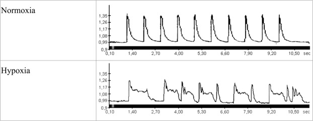
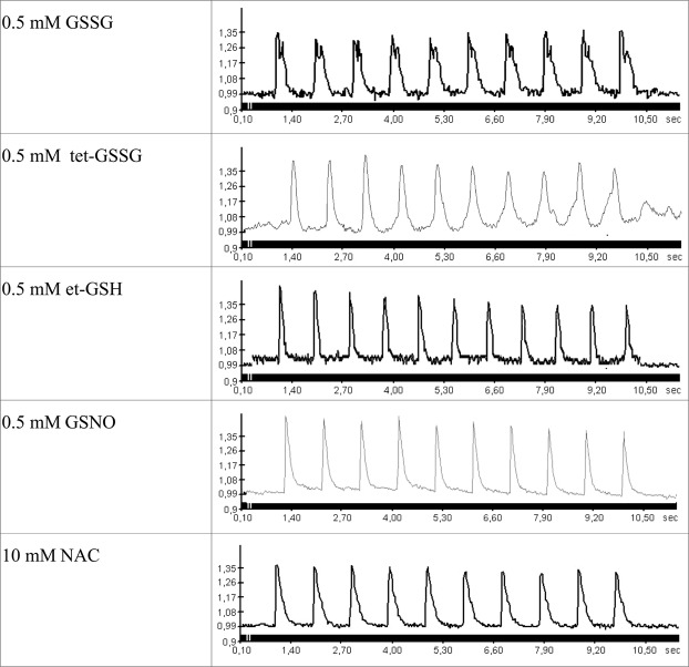
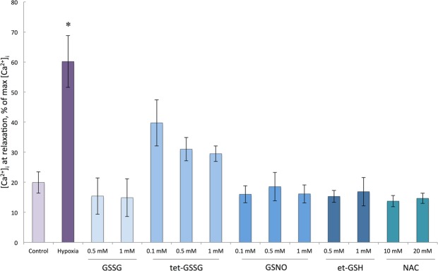
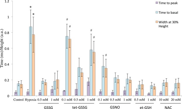
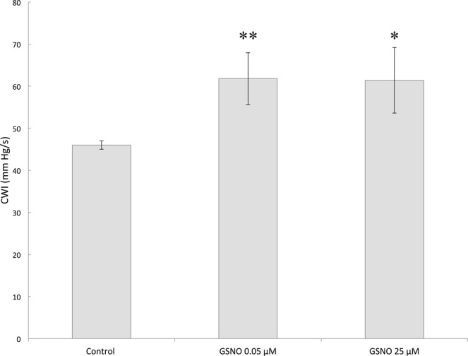
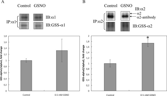
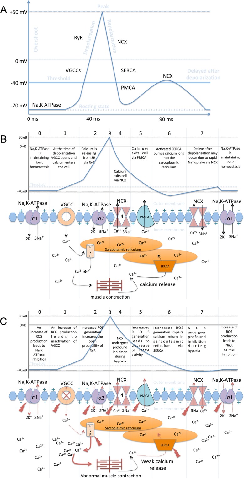
Severe hypoxia leads to decline in cardiac contractility and induces arrhythmic events in part due to oxidative damage to cardiomyocyte proteins including ion transporters. This results in compromised handling of Ca2+ ions that trigger heart contractile machinery. Here, we demonstrate that thiol-containing compounds such as N-acetylcysteine (NAC), glutathione ethyl ester (et-GSH), oxidized tetraethylglutathione (tet-GSSG), oxidized glutathione (GSSG) and S-nitrosoglutathione (GSNO) are capable of reducing negative effects of hypoxia on isolated rat cardiomyocytes. Preincubation of cardiomyocytes with 0.1 mM GSNO, 0.5 mM et-GSH, GSSG, tet-GSSG or with 10 mM NAC allows cells 5-times longer tolerate the hypoxic conditions and elicit regular Ca2+ transients in response to electric pacing. The shape of Ca2+ transients generated in the presence of GSNO, et-GSH and NAC was similar to that observed in normoxic control cardiomyocytes. The leader compound, GSNO, accelerated by 34% the recovery of normal contractile function of isolated rat heart subjected to ischemia-reperfusion. GSNO increased glutathionylation of Na,K-ATPase alpha-2 subunit, the principal ion-transporter of cardiac myocyte sarcolemma, which prevents irreversible oxidation of Na,K-ATPase and regulates its function to support normal Ca2+ ion handling in hypoxic cardiomyocytes. Altogether, GSNO appears effective cardioprotector in hypoxic conditions worth further studies toward its cardiovascular application.


Conflict of interest statement

The authors declare no competing interests.

Figures

Figure 1
Figure 1
Changes in the concentration of free intracellular Ca2+ in isolated rat cardiomyocytes during electric stimulation (1 Hz) at normal oxygen level (buffer saturated with carbogen 95% O2 and 5% CO2) and under hypoxic conditions (buffer saturated with 95% N2 and 5% CO2). 10 sec fragments of the records, at 2 min of normoxia or hypoxia, are given. The ordinate shows the fluorescence intensity of Fluo-4 in arbitrary units (a.u.).
Figure 2
Figure 2
Changes in the concentration of free intracellular Ca2+ in isolated rat cardiomyocytes during electric stimulation (1 Hz) under hypoxic conditions (buffer saturated with 95% N2 and 5% CO2) in the presence of low molecular weight thiols. 10 sec fragments of the records in the presence of 0.5 mM et-GSH, 0.5 mM GSSG, 0.5 mM tet-GSSG, GSNO and 10 mM NAC, at 3 min of hypoxia are given. Similar recordings were obtained during the next 8–12 minutes of hypoxia depending on the substance added. The ordinate shows the fluorescence intensity of Fluo-4 (a.u.).
Figure 3
Figure 3
Relative levels of basal Ca2+ in rat cardiomyocytes in control, hypoxic conditions and in hypoxic conditions in the presence of et-GSH, GSSG, tet-GSSG, GSNO and NAC. The measurements were carried out at 2 min for control normoxia and hypoxia, and at 5 min for all other exposures. Average ratio ([Ca2+]i at relaxation/[Ca2+]i at peak) × 100% is presented for each condition. For all Ca2+ transients [Ca2+]i at relaxation is determined at time point after Ca2+ peak in control normoxic cardiomyocytes when fluorescent signal reaches 20% of the peak value (on average at 250 msec). n = 11–17. Mean value ± S.D. Statistical analysis was performed using one-way ANOVA (F = 115.0, p < 0.00001) with post hoc testing (using paired samples Student’s t-test with Bonferroni correction); after a Bonferroni correction, a p-value < 0.003 was considered as statistically significant; *p < 0.001 for all conditions vs hypoxia.
Figure 4
Figure 4
Calcium peak parameters (Time to peak, Time to basal and Width at 30% height) in control, hypoxic conditions and in hypoxic conditions in the presence of et-GSH, GSSG, tet-GSSG, GSNO and NAC. n = 18–25. Mean value ± S.D. Statistical analysis was performed using one-way ANOVA (for parameter «Time to peak» F = 3.4, p < 0.00099; for parameter «Time to basal» F = 24.4; p < 0.00001; «Width at 30% height» F = 23.5, p < 0.00001) with post hoc testing using paired samples Student’s t-test with Bonferroni correction. After a Bonferroni correction, a p-value < 0.003 was considered as statistically significant; *p < 0.003 for all conditions vs hypoxia excluding conditions marked #.
Figure 5
Figure 5
Restoration of the cardiac work index (CWI) after ischemia/reperfusion in the absence and in presence of GSNO n = 5, mean ± SD. Statistical analysis was performed using one-way ANOVA (F = 12.2, p = 0.00129) with post hoc testing (using paired samples Student’s t-test with Bonferroni correction); after a Bonferroni correction, a p-value < 0.016 was considered as statistically significant; *p < 0.003; **p < 0.0005.
Figure 6
Figure 6
S-glutathionylation of α1 and α2 isoforms of Na,K-ATPase catalytic-subunit after GSNO treatment of cardiomyocytes. α1-Subunit (A) or α2-subunit (B) of Na,K-ATPase was immunoprecipitated (IP) from cell lysates by anti-α1 antibodies or anti-α2 antibodies and glutathionylation was detected with anti-glutathione (anti-GS) antibodies. The original immunoblotting readouts are presented above. Bars represent changes in the S-glutathionylated (GSS- α1/α1) or (GSS- α2/α2) form of the protein normalized to its total amount; n = 3, mean ± SD; *p < 0.05. Full-length blots are included in Supplementary materials (Supplementary Fig. 7).
Figure 7
Figure 7
Scheme depicting activation of ion-transporting enzymes in cardiomyocytes during electrochemical coupling and actomyosin contraction. (А) The projection of activation of ion-transporting enzymes on the action potential of cardiomyocytes. (B) Activation of ion-transporting enzymes during the action potential of cardiomyocytes. At rest, the membrane charge is maintained by Na,K-ATPase and Na/Ca exchanger (NCX) enzymes. During stimulation, potential-dependent membrane calcium channels (VGCC) become permeable, calcium enters the cell and activates the ryanodine receptors (RyR2) of the sarcoplasmic reticulum (SR). Calcium released from SR interacts with troponin-myosin complex, which leads to muscle contraction. Calcium released from troponin-myosin complex back to the cytosol activates the Ca-ATPase of the sarcoplasmic reticulum (SERCA2), which begins to pump Ca2+ back into the SR, in parallel Ca2 + is pumped out of the cell via membrane calcium ATPase (PMCA) and NCX. (C) Effect of acute hypoxia on ion-transporting enzymes. Increased ROS production, which occurs soon after the onset of hypoxia, leads to Na,K-ATPase inhibition. Tissue specific α2-subunit Na,K-ATPase which is important for regulating of Ca2+ levels is more redox sensitive than ubiquitous α1- subunit. So disturbance of activity of α2-containing isozyme is one of the first events during hypoxia that affect calcium transients. Inhibition of both isoforms of Na,K-ATPase results in Ca2+ overload, because the increased level of intracellular Na+ reverses NCX activity. Also, oxidative damage of VGCC leads to decline of their permeability and disruption of the Ca2+ uptake. ROS increases the open probability of RyR, which leads to excessive Ca2+ efflux into the cytosol from sarcoplasmic reticulum. PMCA and SERCA are also redox sensitive enzymes, and hypoxia leads to their inhibition, thus Ca2+ can not be driven out of the cell.

Similar articles

Cited by

References

    1. Vanek T, et al. Heart arrest and myocardial protection by four types of cardioplegic solutions. Exp. Clin. Cardiol. 2014;20:3129–3138.
    1. Giricz Z, et al. Cardioprotection by remote ischemic preconditioning of the rat heart is mediated by extracellular vesicles. J. Mol. Cell. Cardiol. 2014;68:75–78. doi: 10.1016/j.yjmcc.2014.01.004. - DOI - PubMed
    1. Eaton P, Bell RM, Cave AC, Shattock MJ. Ischemic preconditioning: a potential role for protein S-thiolation? Antioxid.Redox.Signal. 2005;7:882–888. doi: 10.1089/ars.2005.7.882. - DOI - PubMed
    1. Fuller W, Parmar V, Eaton P, Bell JR, Shattock MJ. Cardiac ischemia causes inhibition of the Na/K ATPase by a labile cytosolic compound whose production is linked to oxidant stress. Cardiovasc. Res. 2003;57:1044–1051. doi: 10.1016/S0008-6363(02)00810-6. - DOI - PubMed
    1. Bogdanova, A., Petrushanko, I. Y., Hernansanz-Agust�n, P. & Mart�nez-Ruiz, A. ‘Oxygen sensing’ by Na,K-ATPase: These miraculous thiols. Frontiers in Physiology7 (2016). - PMC - PubMed

Publication types

MeSH terms