. 2019 Mar 19;9(1):4865.
doi: 10.1038/s41598-019-41378-9.

Bacteriophage T4 capsid as a nanocarrier for Peptide-N-Glycosidase F immobilization through self-assembly

Affiliations

Bacteriophage T4 capsid as a nanocarrier for Peptide-N-Glycosidase F immobilization through self-assembly

Liang Zhang et al. Sci Rep. .

Abstract

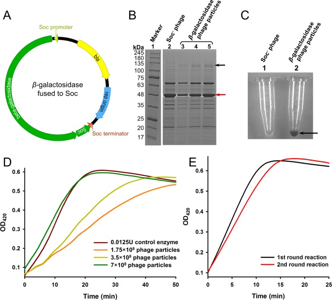
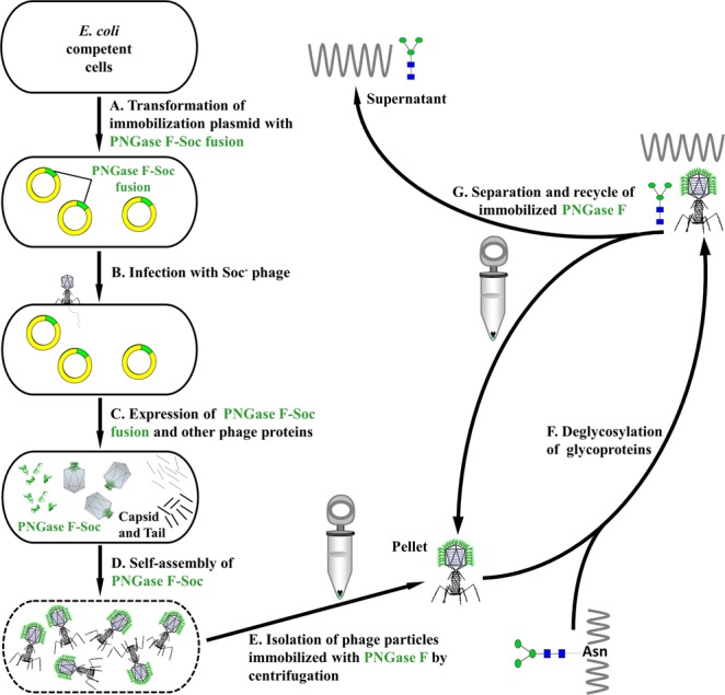
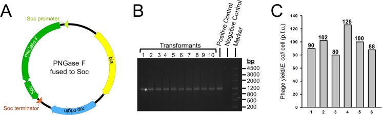
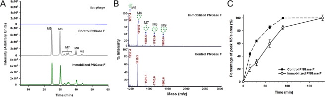
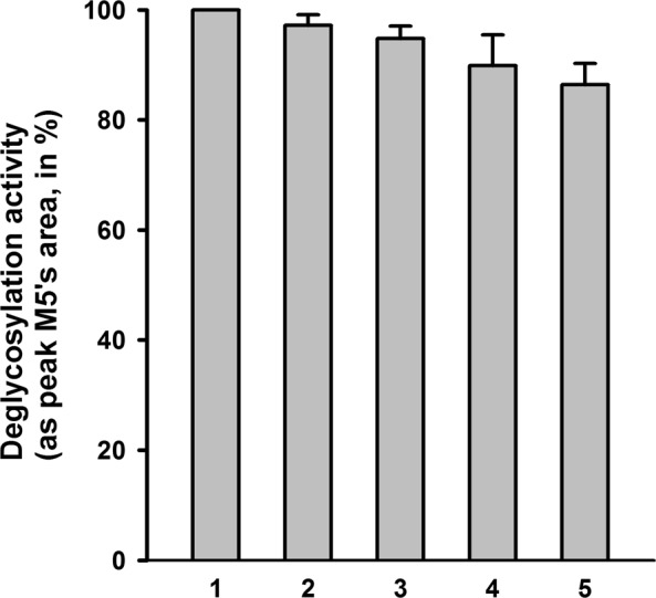
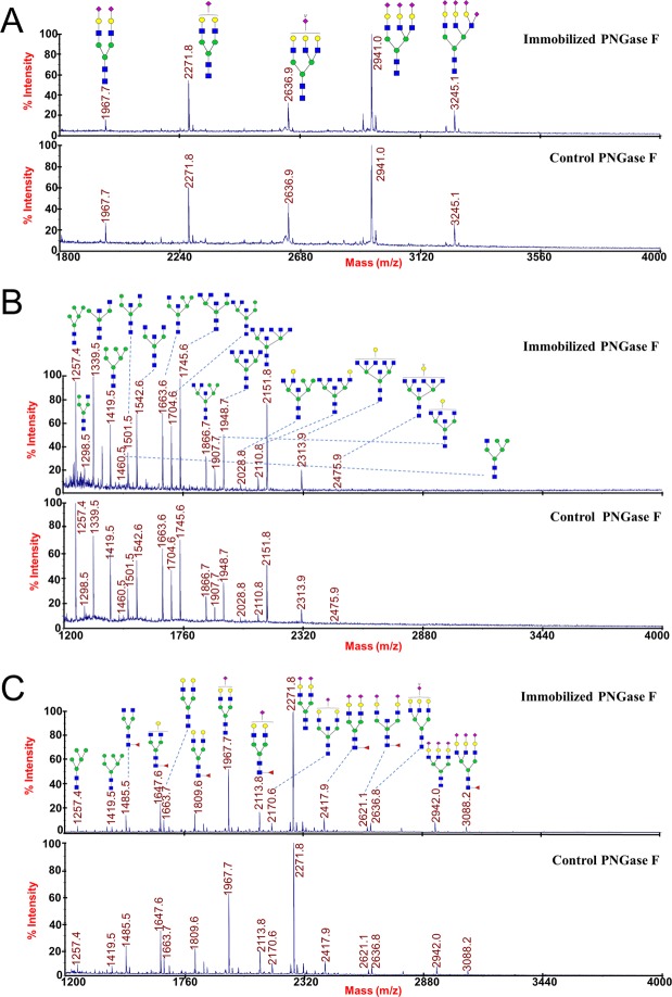
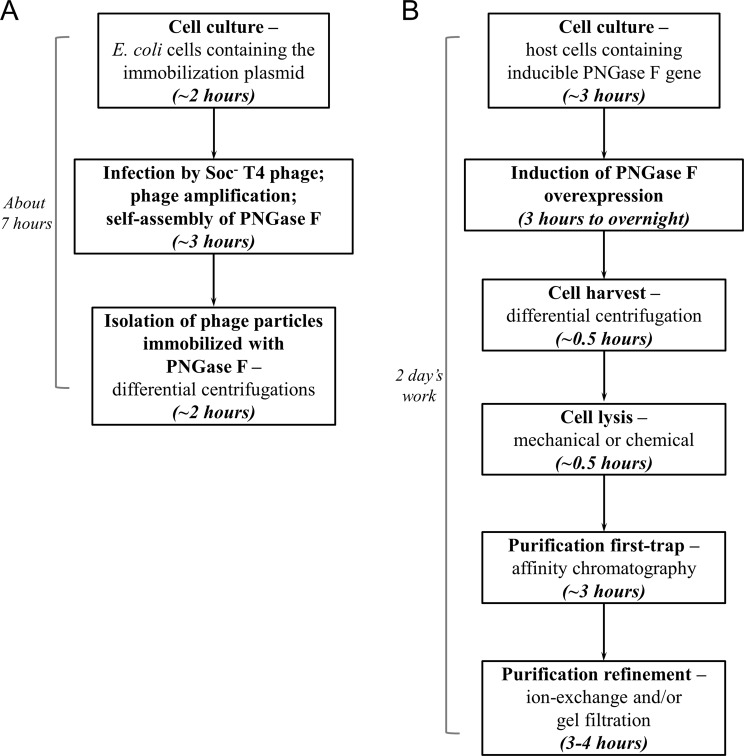
Enzyme immobilization is widely applied in biocatalysis to improve stability and facilitate recovery and reuse of enzymes. However, high cost of supporting materials and laborious immobilization procedures has limited its industrial application and commercialization. In this study, we report a novel self-assembly immobilization system using bacteriophage T4 capsid as a nanocarrier. The system utilizes the binding sites of the small outer capsid protein, Soc, on the T4 capsid. Enzymes as Soc fusions constructed with regular molecular cloning technology expressed at the appropriate time during phage assembly and self-assembled onto the capsids. The proof of principle experiment was carried out by immobilizing β-galactosidase, and the system was successfully applied to the immobilization of an important glycomics enzyme, Peptide-N-Glycosidase F. Production of Peptide-N-Glycosidase F and simultaneous immobilization was finished within seven hours. Characterizations of the immobilized Peptide-N-Glycosidase F indicated high retention of activity and well reserved deglycosylation capacity. The immobilized Peptide-N-Glycosidase F was easily recycled by centrifugation and exhibited good stability that sustained five repeated uses. This novel system uses the self-amplified T4 capsid as the nanoparticle-type of supporting material, and operates with a self-assembly procedure, making it a simple and low-cost enzyme immobilization technology with promising application potentials.


Conflict of interest statement

The authors declare no competing interests.

Figures

Figure 1
Figure 1
Immobilization of β-galactosidase on phage T4 capsid. (A) Schematic of the β-galactosidase immobilization plasmid. The circular plasmid DNA molecule is represented by a circle. The β-galactosidase and Soc genes are represented by green ribbons and the expression directions are indicated as arrow shapes. Soc promoter, Soc terminator, ampicillin antibiotic resistance gene (bla), and the replication origins are indicated. (B) SDS-PAGE analysis of the phage particles immobilized with β-galactosidase. 5 × 108, 109 and 2 × 109 phage particles assembled with β-galactosidase-Soc fusion protein (lanes 3–5) were treated with SDS-PAGE loading buffer (containing 3% SDS), boiled for 10 min, analyzed on a 4–20% gradient SDS-PAGE gel, and stained with Coomassie Brilliant Blue. Lane 1 is the protein molecular weight standard marker, with the molecular weight (in kDa) of each band indicated by a number. Line 2 is 2 × 109 Soc phage particles treated in the same way as the control. The positions of β-galactosidase-Soc fusion protein and T4 gp23 are indicated by black and red arrows, respectively. The image was cropped from different parts of the same gel, and the full-length gel is presented in Supplementary Fig. S1. (C) Enzymatic activity of the immobilized β-galactosidase. The arrow indicated color change of the reaction mixture in one of the Eppendorf tubes. (D) Quantification of immobilized β-galactosidase activity. Curves were drawn with the mean of 3 parallel reaction wells. (E) Activity of the recycled β-galactosidase. After one round of reaction with ONPG for 25 min, phage particles (7 × 108) immobilized with β-galactosidase were recycled by centrifugation for the 2nd round of reaction. Curves were drawn with the mean of 3 parallel reaction wells.
Figure 2
Figure 2
A self-assembly approach to immobilize PNGase F. (A) The PNGase F immobilization plasmid was transformed into E. coli competent cells. (B) E. coli cells containing the PNGase F immobilization plasmid were infected by Soc phage. The Soc phage had the Soc gene knocked out, so that the phage would not produce endogenous Soc protein. (C) The PNGase F-Soc fusion protein was expressed in parallel with other phage proteins that assembled the capsid and tail. The PNGase F-Soc fusion served as the alternative of Soc protein in the phage assembly process. (D) At the late stage of phage assembly, the Soc binding sites on the capsid exposed, and the PNGase F-Soc fusion self-assembled on the capsid and immobilized. At the completion of assembly, the E. coli cells lysed and phage particles immobilized with PNGase F were released. (E) Isolation of the phage particles immobilized with PNGase F at 34,500 g for 45 min by centrifugation. (F) Deglycosylation of glycoproteins with the PNGase F immobilized on the phage particles. (G) Separation and recycle of the immobilized PNGase F (pellet) from the product and/or substrate (supernatant) by centrifugation.
Figure 3
Figure 3
The PNGase F immobilization plasmid construction and maintenance in E. coli cells, and phage yield of the immobilization procedure. (A) Schematic of the PNGase F immobilization plasmid. The circular plasmid DNA molecule is represented by a circle. The PNGase F and Soc genes are represented by green ribbons and the expression directions are indicated as arrow shapes. Soc promoter, Soc terminator, ampicillin antibiotic resistance gene (bla), and the replication origins are indicated. (B) Screening of the transformants for the presence of the PNGase F-Soc ORF. Transformants 1–10 were subjects for colony PCR with primers specific to the PNGase F-Soc ORF, and the PCR products were analyzed by agarose gel electrophoresis. PCR using the PNGase F immobilization plasmid as the template served as the positive control. PCR without a template was used as the negative control. (C) Phage yield of the immobilization by self-assembly procedure. The number of phage particles produced from one E. coli cell is presented in plaque forming units (p.f.u.). Phage yields from 6 independent experiments (bars 1–6) are shown. The exact number of phage yield of each experiment is indicated on top of each bar.
Figure 4
Figure 4
Enzymatic activity of the immobilized PNGase F. (A) HPLC profiles of the N-glycans released from RNase B by the immobilized PNGase F (bottom), the control (middle), and the Soc phage (top). The peaks of the 5 N-glycans (M5 to M9) are indicated. (B) Analysis of N-glycans released from RNase B by the immobilized PNGase F (top) and the control (bottom) with MALDI-TOF MS spectra. The structures and mass-to-charge ratios of M5 to M9 are indicated. (C) Time curves of the percentages of peak M5’s areas released from RNase B by the immobilized PNGase F and the control. Error bars represent the standard deviation from 3 independent experiments.
Figure 5
Figure 5
Deglycosylation activities of the immobilized PNGase F in repeated uses. 1.25 × 1011 phage particles immobilized with PNGase F (equal to 1 U) were used to digest 10 μg of denatured RNase B at 37 °C for 1 h. After the reaction, the immobilized PNGase F was recycled in the pellet by centrifugation at 34,500 g for 45 min at 4 °C. The supernatant was used to analyze the deglycosylation activity by quantification of the released M5. The immobilized PNGase F in the pellet was washed with 1 ml buffer containing 50 mM Tris-HCl (pH 7.5) and 5 mM MgCl2 for 3 times, and then used for another round of digestion of 10 μg RNase B. The deglycosylation activities of round 1–5 (indicated under the x-axis) are presented as the peak M5’s areas normalized to the first round of digestion (set at 100%). Error bars represent the standard deviation from 3 independent experiments.
Figure 6
Figure 6
Deglycosylation of different substrates by the immobilized PNGase F. N-glycans released from fetuin (A), ovalbumin (B) and human serum (C) by the immobilized PNGase F (top) and the control (bottom) were analyzed with MALDI-TOF MS spectra. The structures and mass-to-charge ratios of the N-glycans are indicated.
Figure 7
Figure 7
Comparison of the immobilization method and traditional method to obtain PNGase F. (A) Flow chart showing the procedure of the immobilization method. The cell culture of E. coli containing the immobilization plasmid was grown for 2 h, and infected by Soc phage at a multiplicity of infection of 1. After infection, the phage amplified and PNGase F self-assembled. 3 h of incubation time ensured complete amplification and assembly of phage in the culture. The phage was isolated from the culture by differential centrifugations. The total time required was 7 h. (B) Flow chart of the procedure to produce PNGase F in the traditional way. Cell culture with the inducible PNGase F gene was grown to an appropriate density and PNGase F overexpression was induced for a time period ranging from 3 h to overnight. The cells were harvested and lysed, followed by purification steps including affinity chromatography, ion-exchange and/or gel filtration. Two days were required to produce PNGase F in the traditional way.

Similar articles

Cited by

References

    1. Kirk O, Borchert TV, Fuglsang CC. Industrial enzyme applications. Curr Opin Biotechnol. 2002;13:345–351. doi: 10.1016/S0958-1669(02)00328-2. - DOI - PubMed
    1. Zheng GW, Xu JH. New opportunities for biocatalysis: driving the synthesis of chiral chemicals. Curr Opin Biotechnol. 2011;22:784–792. doi: 10.1016/j.copbio.2011.07.002. - DOI - PubMed
    1. Choi JM, Han SS, Kim HS. Industrial applications of enzyme biocatalysis: Current status and future aspects. Biotechnol Adv. 2015;33:1443–1454. doi: 10.1016/j.biotechadv.2015.02.014. - DOI - PubMed
    1. Bornscheuer UT, et al. Engineering the third wave of biocatalysis. Nature. 2012;485:185–194. doi: 10.1038/nature11117. - DOI - PubMed
    1. Datta S, Christena LR, Rajaram YR. Enzyme immobilization: an overview on techniques and support materials. 3 Biotech. 2013;3:1–9. doi: 10.1007/s13205-012-0071-7. - DOI - PMC - PubMed

Publication types

MeSH terms

Substances