. 2019 Mar 19;9(1):4859.
doi: 10.1038/s41598-019-41227-9.

Temporal evolution of quantitative EEG within 3 days of birth in early preterm infants

Affiliations

Temporal evolution of quantitative EEG within 3 days of birth in early preterm infants

John M O'Toole et al. Sci Rep. .

Abstract

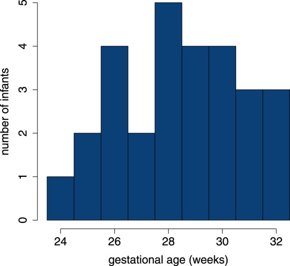
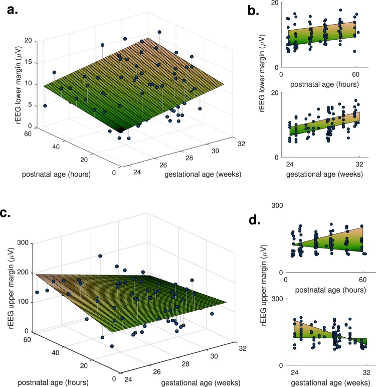
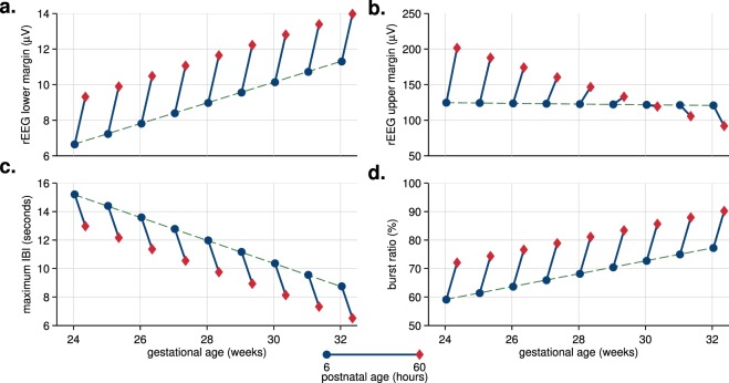
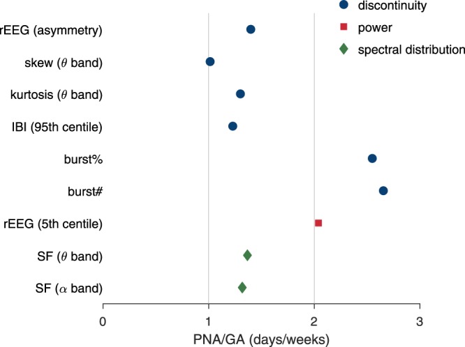
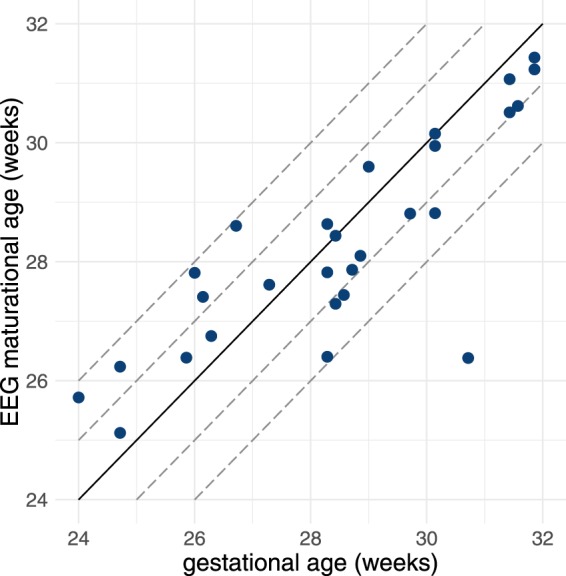
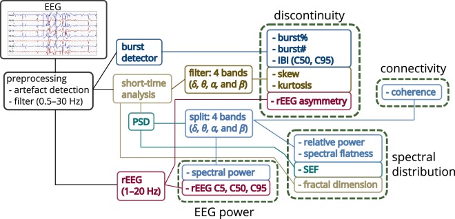
For the premature newborn, little is known about changes in brain activity during transition to extra-uterine life. We aim to quantify these changes in relation to the longer-term maturation of the developing brain. We analysed EEG for up to 72 hours after birth from 28 infants born <32 weeks of gestation. These infants had favourable neurodevelopment at 2 years of age and were without significant neurological compromise at time of EEG monitoring. Quantitative EEG was generated using features representing EEG power, discontinuity, spectral distribution, and inter-hemispheric connectivity. We found rapid changes in cortical activity over the 3 days distinct from slower changes associated with gestational age: for many features, evolution over 1 day after birth is equivalent to approximately 1 to 2.5 weeks of maturation. Considerable changes in the EEG immediately after birth implies that postnatal adaption significantly influences cerebral activity for early preterm infants. Postnatal age, in addition to gestational age, should be considered when analysing preterm EEG within the first few days after birth.


Conflict of interest statement

The authors declare no competing interests.

Figures

Figure 1
Figure 1
Distribution of gestational age.
Figure 2
Figure 2
Two features of the rEEG, lower and upper margins, as dots with estimated fixed effects (2D planes). Features are dependent on both gestational age and postnatal age as the 2D plots in (a,c) illustrate this dependency; plots (b,d) show the marginal distribution, the side-view from either the x- or y-axis of the 2D plots in (a,c). The rEEG upper margin includes the postnatal age–by–gestational age fixed effect, resulting in a nonlinear 2D plane.
Figure 3
Figure 3
Visualising the temporal trajectories in context of intra-uterine maturation: plots of postnatal-age fixed-effect (as dots-to-diamonds lines) at selected gestational ages. Four features, 2 from the range-EEG (rEEG) in (a,b) and 2 features of the burst annotation (c,d); maximum IBI (inter-burst interval) refers to the 95th centile of the IBI. All features are dependent on time and gestational age; rEEG upper margin in (b) is also dependent on time-by-gestational interaction.
Figure 4
Figure 4
Ratio of estimated fixed-effects of postnatal age (PNA) to gestational age (GA). SF: spectral flatness; rEEG: range EEG; IBI: inter-burst interval; burst%: burst ratio; burst#: number of bursts. Frequency bands defined as 3–8 Hz for θ and 8–15 Hz for α bands.
Figure 5
Figure 5
Algorithm’s estimation of EEG maturational age from testing results comparative to gestational age for n = 28 infants. Correlation coefficient is r = 0.83. Dashed lines represent 1 and 2 weeks deviation from the ideal 1:1 representation (solid line).
Figure 6
Figure 6
Outline of process to generate quantitative EEG. Features grouped as measures EEG power, spectral distribution, connectivity, and discontinuity. Frequency bands (FB): 0.5–3 Hz (δ), 3–8 Hz (θ), 8–15 Hz (α), and 15–30 Hz (β); C5, C50, and C95: 5th, 50th, and 95th centiles; SEF: spectral edge frequency; rEEG: range EEG; IBI: inter-burst interval; burst%: burst ratio; burst#: number of bursts.

Similar articles

Cited by

References

    1. Kidokoro H, et al. Brain Injury and Altered Brain Growth in Preterm Infants: Predictors and Prognosis. Pediatrics. 2014;134:e444–e453. doi: 10.1542/peds.2013-2336. - DOI - PubMed
    1. Pichler G, Schmölzer GM, Urlesberger B. Cerebral Tissue Oxygenation during Immediate Neonatal Transition and Resuscitation. Front. Pediatr. 2017;5:1–5. doi: 10.3389/fped.2017.00029. - DOI - PMC - PubMed
    1. Plomgaard AM, et al. Early biomarkers of brain injury and cerebral hypo- and hyperoxia in the SafeBoosC II trial. PLoS One. 2017;12:e0173440. doi: 10.1371/journal.pone.0173440. - DOI - PMC - PubMed
    1. Kenosi M, et al. Monitoring cerebral oxygenation of preterm infants using a neonatal specific sensor. J. Perinatol. 2017 doi: 10.1038/s41372-017-0007-5. - DOI - PubMed
    1. Pavlidis E, Lloyd RO, Boylan GB. EEG–A Valuable Biomarker of Brain Injury in Preterm Infants. Dev. Neurosci. 2017;39:23–35. doi: 10.1159/000456659. - DOI - PubMed

Publication types