Review
. 2012 Dec 4:2:175-187.
doi: 10.2147/DNND.S38353. eCollection 2012.

Pathogenic mechanisms of neurodegeneration based on the phenotypic expression of progressive forms of immune-mediated neurologic disease

Affiliations
Review

Pathogenic mechanisms of neurodegeneration based on the phenotypic expression of progressive forms of immune-mediated neurologic disease

Michael C Levin et al. Degener Neurol Neuromuscul Dis. .

Abstract

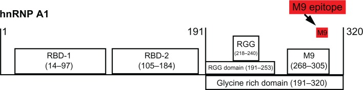
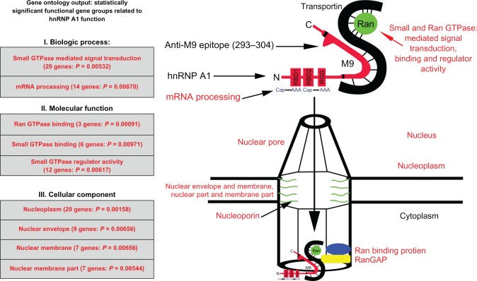
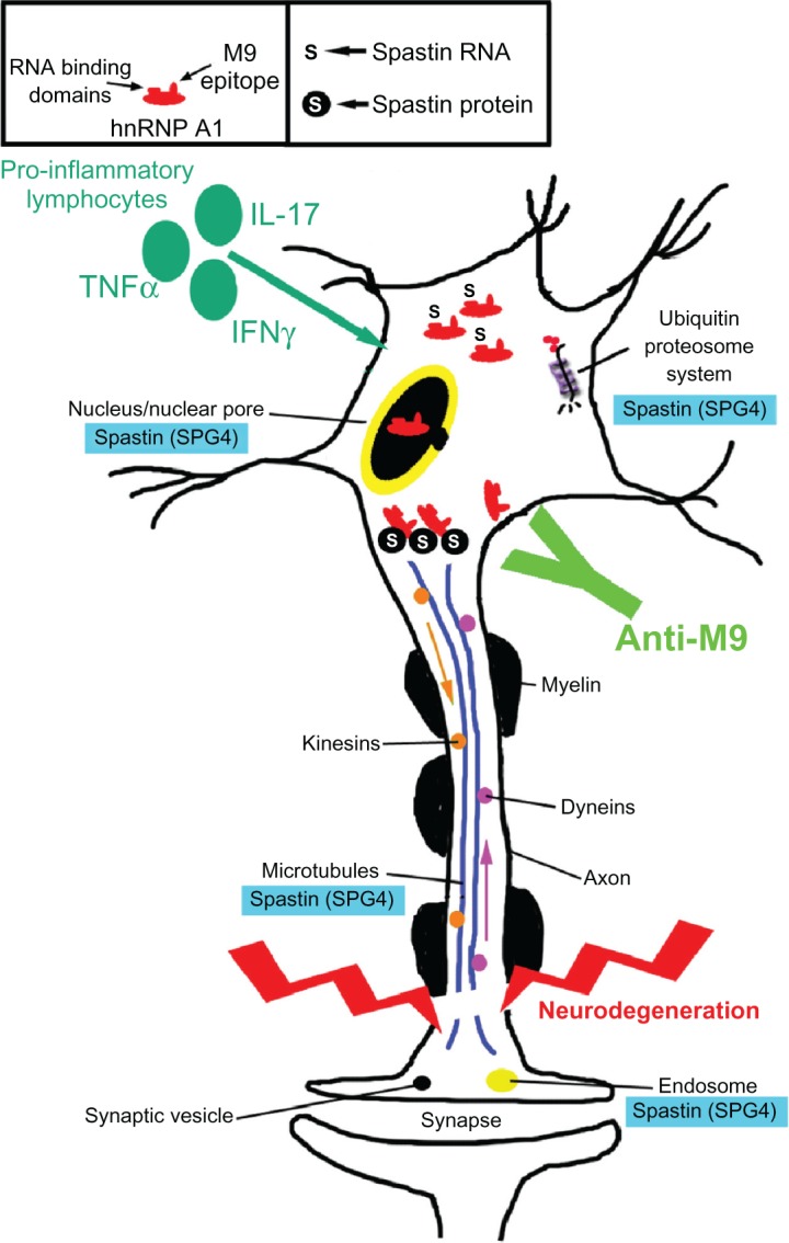
Considering there are no treatments for progressive forms of multiple sclerosis (MS), a comprehensive understanding of the role of neurodegeneration in the pathogenesis of MS should lead to novel therapeutic strategies to treat it. Many studies have implicated viral triggers as a cause of MS, yet no single virus has been exclusively shown to cause MS. Given this, human and animal viral models of MS are used to study its pathogenesis. One example is human T-lymphotropic virus type 1-associated myelopathy/tropical spastic paraparesis (HAM/TSP). Importantly, HAM/TSP is similar clinically, pathologically, and immunologically to progressive MS. Interestingly, both MS and HAM/TSP patients were found to make antibodies to heterogeneous nuclear ribonucleoprotein (hnRNP) A1, an RNA-binding protein overexpressed in neurons. Anti-hnRNP A1 antibodies reduced neuronal firing and caused neurodegeneration in neuronal cell lines, suggesting the autoantibodies are pathogenic. Further, microarray analyses of neurons exposed to anti-hnRNP A1 antibodies revealed novel pathways of neurodegeneration related to alterations of RNA levels of the spinal paraplegia genes (SPGs). Mutations in SPGs cause hereditary spastic paraparesis, genetic disorders clinically indistinguishable from progressive MS and HAM/TSP. Thus, there is a strong association between involvement of SPGs in neurodegeneration and the clinical phenotype of progressive MS and HAM/TSP patients, who commonly develop spastic paraparesis. Taken together, these data begin to clarify mechanisms of neurodegeneration related to the clinical presentation of patients with chronic immune-mediated neurological disease of the central nervous system, which will give insights into the design of novel therapies to treat these neurological diseases.

Keywords: RNA-binding protein; autoimmunity; heterogeneous nuclear ribonucleoprotein A1 (hnRNP A1); human T-lymphotropic virus type 1 (HTLV-1); multiple sclerosis; neurodegeneration; spastic paraparesis.


Conflict of interest statement

Disclosure Drs Michael Levin and Sangmin Lee have a patent pending titled “Biomarker for neurodegeneration in neurological disease.” All other authors report no conflicts of interest in this paper.

Figures

Figure 1
Figure 1
Human T-lymphotropic virus type 1-associated myelopathy/tropical spastic paraparesis (HAM/TSP) and multiple sclerosis (MS) immunoglobulin G (IgG) react with the same epitope in M9. Schematic of heterogeneous nuclear ribonucleoprotein (hnRNP) A1 shows the RNA-binding domains (RBDs), the RGG domain, and M9. Overlapping protein-fragment experiments showed HAM/TSP and MS IgG reacted with an epitope (amino acids [AA] 293-GQYFAKPRNQGG-304, red box), which is contained within the M9 sequence (AA 268–305).,
Figure 2
Figure 2
Genes identified by Gene Ontology are directly related to heterogeneous nuclear ribonucleoprotein (hnRNP) A1 function. The gene categories and some individual genes affected by the anti-M9 antibodies are shown in red type. The complete M9 sequence (amino acids [AA] 268–305) is elongated for emphasis and bordered by the black lines within hnRNP A1. Transportin binds the M9 region at AA 263–289. AA 293–304, which are recognized by the multiple sclerosis and human T-lymphotropic virus type 1-associated myelopathy/tropical spastic paraparesis immunoglobulin G anti-M9 immune response, are not bound to transportin, and thus are available for antibody binding. Abbreviation: GTP, guanosine triphosphate.
Figure 3
Figure 3
Spastin mRNA is bound to heterogeneous nuclear ribonucleoprotein (hnRNP) A1 in neuronal cells. Upper panel: agarose gel. Compared to the neuronal lysate without immunoprecipitation or input (lane 3), there is an enriched spastin mRNA signal following immunoprecipitation with anti-hnRNP A1 mouse monoclonal antibodies (lane 1). In contrast, spastin mRNA was not isolated following immunoprecipitation with a nonspecific control antibody – mouse IgG (lane 2). Lane 4 used spastin primers without lysate (control for DNA contamination). Lower panel: the image was analyzed using ImageQuant software, which provided relative fluorescent intensity of the bands.
Figure 4
Figure 4
Heterogeneous nuclear ribonucleoprotein (hnRNP) A1 localization following TNF-α exposure in neurons. Without TNF-α exposure, hnRNP A1 is localized to nuclei (*) in dNT-2 neurons (left panels) (blue nuclear stain is diamidino-2-phenylindole; green stain is immunohistochemistry using an anti-hnRNP A1 antibody). Following exposure to TNF-α (400 ng/mL, 50 mM glutamate, 30 minutes), hnRNP A1 is also found in the cytoplasm (arrowheads) and neuronal processes (arrow) of neurons (lower right panel). Abbreviation: TNF, tumor necrosis factor.
Figure 5
Figure 5
Potential contribution of the anti-heterogeneous nuclear ribonucleoprotein (hnRNP) A1 M9 immune response to neurodegeneration in immune-mediated neurological disease. Multiple sclerosis and human T-lymphotropic virus type 1-associated myelopathy/tropical spastic paraparesis patients develop antibodies to an epitope contained within the M9 region of hnRNP A1 (box). hnRNP A1 has been shown to interact molecularly with spastin RNA and protein (box and figure). The anti-M9 immune response altered spastin RNA levels, which may alter spastin function at multiple sites within neurons (blue boxes). The combination of proinflammatory cytokines and the anti-M9 immune response might contribute to neurodegeneration in immune-mediated neurological disease. Abbreviations: TNF, tumor necrosis factor; IFN, interferon; IL, interleukin; SPG, spinal paraplegia gene.

References

    1. Dutta R, Trapp BD. Pathogenesis of axonal and neuronal damage in multiple sclerosis. Neurology. 2007;68(22 Suppl 3):S22–S31. discussion S43–S54. - PubMed
    1. Peterson JW, Trapp BD. Neuropathobiology of multiple sclerosis. Neurol Clin. 2005;23(1):107–129. vi–vii. - PubMed
    1. Noseworthy J, Lucchinetti C, Rodgriguez M, Weinshenker B. Multiple sclerosis. N Engl J Med. 2000;343(13):938–952. - PubMed
    1. Lassmann H, Bruck W, Lucchinetti CF. The immunopathology of multiple sclerosis: an overview. Brain Pathol. 2007;17(2):210–218. - PMC - PubMed
    1. Filippi M, Rovaris M, Rocca MA. Imaging primary progressive multiple sclerosis: the contribution of structural, metabolic, and functional MRI techniques. Mult Scler. Jun. 2004;10(Suppl 1):S36–S44. discussion S44–S45. - PubMed