. 2019 Mar 5:11:34.
doi: 10.3389/fnagi.2019.00034. eCollection 2019.

Pathological Tau From Alzheimer's Brain Induces Site-Specific Hyperphosphorylation and SDS- and Reducing Agent-Resistant Aggregation of Tau in vivo

Affiliations

Pathological Tau From Alzheimer's Brain Induces Site-Specific Hyperphosphorylation and SDS- and Reducing Agent-Resistant Aggregation of Tau in vivo

Jin Miao et al. Front Aging Neurosci. .

Abstract

Neurofibrillary tangles (NFTs) made up of hyperphosphorylated tau are a histopathological hallmark of Alzheimer's disease (AD) and related tauopathies. Hyperphosphorylation of tau is responsible for its loss of normal physiological function, gain of toxicity and its aggregation to form NFTs. Injection of misfolded tau seeds into mouse brain induces tau aggregation, but the nature of tau phosphorylation in pathologic tau seeded pathology is unclear. In the present study, we injected hyperphosphorylated and oligomeric tau isolated from AD brain (AD P-tau) into hippocampus of human tau transgenic mice and found that in addition to tau aggregation/pathology, tau was hyperphosphorylated at Ser202/Thr205, Thr212, Ser214, Thr217, Ser262, and Ser422 in AD P-tau injected hippocampus and at Ser422 in the contralateral hippocampus and in the ipsilateral cortex. AD P-tau-induced AD-like high molecular weight aggregation of tau that was SDS- and reducing agent-resistant and site-specifically hyperphosphorylated in the ipsilateral hippocampus. There were no detectable alterations in levels of tau phosphatases or tau kinases in AD P-tau-injected brains. Furthermore, we found that hyperphosphorylated tau was easier to be captured by AD P-tau and that aggregated tau was more difficult to be dephosphorylated than the non-aggregated tau by protein phosphatase 2A (PP2A). Based on these findings, we speculate that AD P-tau seeds hyperphosphorylated tau to form aggregates, which resist to the dephosphorylation by PP2A, resulting in hyperphosphorylation and pathology of tau.

Keywords: AD P-tau; Alzheimer’s disease; hyperphosphorylation of tau; propagation of tau pathology; tau pathology.


Figures

Figure 1
Figure 1
Alzheimer’s hyperphosphorylated and oligomeric tau (AD P-tau)-seeded tau aggregates/pathology in the hippocampus in Tg/hTau mice. (A–D) AD P-tau (0.55 μg) or as a control saline was injected unilaterally into the hippocampus of 9–11-month-old Tg/hTau or Tau−/− mice. Coronal brain sections were immunostained with tau antibodies, AT8 (pSer202/Thr205-tau) (A), anti-pSer262-tau (C), and PHF-1 (pSer396/404) (D) 10 weeks after injection and immunostaining of ipsilateral (Ips) and contralateral (Cont) hippocampi of Tg/hTau or Tau−/− mice were captured. AT8 staining in 3–4 sections of both ipsilateral and contralateral hippocampi was quantified with ImageJ software from each of three Tg/hTau mice injected with AD P-tau and statistically analyzed with paired student t-test (B), *p < 0.05. (E) CA1 region of Tg/hTau mouse ipsilateral hippocampus double immunostained with AT8 (Red) and anti-pSer422-tau (Green) and counterstained with TO-PRO 3 iodide for nucleus. Scale bar 100 μm and insert scale bar 20 μm.
Figure 2
Figure 2
AD P-tau-induced site-specific hyperphosphorylation of tau in the hippocampus in Tg/hTau mice. AD P-tau (0.55 μg) was injected unilaterally into the hippocampus in 9–11-month-old Tg/hTau and Tau−/− mice. Ten weeks post injection, phosphorylation of tau in the hippocampus was analyzed by Western blots developed with the indicated phosphorylation-dependent and site-specific tau antibodies (A,C), normalized with total tau and analyzed with unpaired student t-test, and are presented as scattered dots with mean ± SD (B,D), and analyzed with unpaired student t-test; *p < 0.05; **p < 0.01; ***p < 0.001; ****p < 0.0001. Arrow head of AT8 blots indicates the heavy chain of IgG (50 kDa) and *indicates the non-specific band.
Figure 3
Figure 3
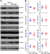
Tau phosphorylation in the ipsilateral cortex in Tg/hTau mice injected with AD P-tau. Phosphorylation of tau at Ser199, Thr205, Ser214, Thr217, Ser262, Ser396, Ser404 and Ser422 in the ipsilateral cortex was analyzed by Western blots (A) and analyzed with unpaired student t-test. Data are presented as scattered dots with mean ± SD (B); *p < 0.05.
Figure 4
Figure 4
SDS- and β-mercaptoethanol-resistant high molecular weight tau (HMW-tau) in AD P-tau injected hippocampus. Phosphorylation of tau and SDS- and β-mercaptoethanol-resistant HMW-tau in saline or AD P-tau injected hippocampus was analyzed by Western blots developed with anti-pThr217, anti-pSer262, PHF-1, and anti-pSer422 antibodies with shorter (left) and longer (right) exposure to X-ray film (A). Shorter exposure blots were used for quantification of phosphorylation of middle molecular weight tau (MMW-tau), whereas longer exposure blots were used for quantification of SDS- and β-mercaptoethanol-resistant HMW-tau. *Indicates a non-specific band. The data from ipsilateral and contralateral hippocampi in Tg/hTau mice injected with saline were pooled as control since there was no significant difference in tau phosphorylation between them. Levels of phosphorylated MMW-tau (B) and HMW-tau (C) werestatistically analyzed with one-way ANOVA post Tukey’s multiple comparisons test andare presented as mean ± SD. *p < 0.05; **p < 0.01; ****p < 0.0001.
Figure 5
Figure 5
SDS- and β-mercaptoethanol-resistant HMW-tau in AD P-tau injected hippocampus of Tg/hTau mice. Ipsilateral hippocampi from Tg/hTau and Tau−/− mice injected with AD P-tau were analyzed by Western blots developed with AT8 (anti-pS202/T205-tau), anti-pT212-tau,anti-pT217-tau, anti-pS262-tau, and anti-pS422-tau to study site-specific SDS- and β-mercaptoethanol-resistant HMW-tau. *Points the non-specific bands and arrow head indicates the heavy chain of IgG.
Figure 6
Figure 6
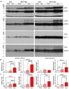
Expression of tau phosphatases in AD P-tau-injected hippocampus. PP1, protein phosphatase 2A (PP2A), demethylated PP2A, PP2B and PP5 in the ipsilateral hippocampus in Tg/hTau and Tau−/− mice injected with AD P-tau or saline were analyzed by Western blots developed with antibodies indicated (A). The levels of demethylated PP2A and PP2A (B) and PP1, PP2B and PP5 (C) were statistically analyzed with two-way ANOVA post Sidak’s multiple comparisons test after normalized with PP2A (for DM-PP2A) or GAPDH (for PPs) and are presented as mean ± SD. ****p < 0.0001.
Figure 7
Figure 7
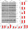
Expression of tau kinases in AD P-tau-injected hippocampi in Tg/hTau and Tau−/− mice. Cdk5, inactive form of GSK-3β (phospho-GSK-3β), Dyrk1A, active form of Erk (phospho-Erk), active form of Jnk/SAPK (phospho-Jnk/SAPK), active form of AMPK (phospho-AMPK), PKA catalytic subunit, active form of AKT (phosphorylated at Ser473), (phospho-AKT), active form of P70S6k (phospho-P70S6K), and CK1ε in ipsilateral hippocampus of Tg/htau and Tau−/− mice injected with AD P-tau or saline were analyzed by Western blots (A). The levels of proline-directed protein kinases (PDPKs; B) and non-PDPKs (C) were statistically analyzed with two-way ANOVA post Sidak’s multiple comparisons test and are presented as mean ± SD. *p < 0.05; ****p < 0.0001.
Figure 8
Figure 8
Effects of hyperphosphorylation of tau on its capture by AD P-tau and aggregation of tau on its dephosphorylation by PP2A. (A,B) Effect of hyperphosphorylation of tau on its capture by AD P-tau. Tau441 tagged with HA was overexpressed in HEK-293FT cells for 48 h. The cells were treated with 100 nM okadaic acid (OA) for 2 h to induce hyperphosphorylation of tau and lysed in PBS containing a cocktail of proteinase and phosphatase inhibitors by probe sonication. AD P-tau was dotted on nitrocellulose membrane and incubated with the 15,000× g extract from OA treated cells (OA-tau) or control treated cells (Con-tau) overnight. AD P-tau captured HA-tau441 was analyzed by anti-HA, followed by HRP-anti-mouse IgG and ECL (A) and plotted against various amount of AD P-tau (B). (C,D) Effect of aggregation of tau on its dephosphorylation by PP2A. Sarkosyl insoluble tau (SI-tau), AD P-tau, and heat stable monomeric tau (HS-tau) were isolated from AD cerebral cortex and analyzed by Western blots developed with R134d (pan-tau), anti-pS199-tau and PHF-1 (C). SI-tau and HS-tau were incubated with PP2A (20 mU/ml) for various time points. The phosphorylation of tau at Ser199 was analyzed by dot-blots (D) and plotted against time points of dephosphorylation reaction (E).

Similar articles

Cited by

References

    1. Ahmed Z., Cooper J., Murray T. K., Garn K., McNaughton E., Clarke H., et al. . (2014). A novel in vivo model of tau propagation with rapid and progressive neurofibrillary tangle pathology: the pattern of spread is determined by connectivity, not proximity. Acta Neuropathol. 127, 667–683. 10.1007/s00401-014-1254-6 - DOI - PMC - PubMed
    1. Alafuzoff I., Iqbal K., Friden H., Adolfsson R., Winblad B. (1987). Histopathological criteria for progressive dementia disorders: clinical-pathological correlation and classification by multivariate data analysis. Acta Neuropathol. 74, 209–225. 10.1007/BF00688184 - DOI - PubMed
    1. Alonso A. D., Grundke-Iqbal I., Iqbal K. (1996). Alzheimer’s disease hyperphosphorylated tau sequesters normal tau into tangles of filaments and disassembles microtubules. Nat. Med. 2, 783–787. - PubMed
    1. Alonso A. C., Zaidi T., Grundke-Iqbal I., Iqbal K. (1994). Role of abnormally phosphorylated tau in the breakdown of microtubules in Alzheimer disease. Proc. Natl. Acad. Sci. U S A 91, 5562–5566. - PMC - PubMed
    1. Arriagada P. V., Growdon J. H., Hedley-Whyte E. T., Hyman B. T. (1992). Neurofibrillary tangles but not senile plaques parallel duration and severity of Alzheimer’s disease. Neurology 42, 631–639. - PubMed

LinkOut - more resources