. 2019 Mar 5:10:166.
doi: 10.3389/fphys.2019.00166. eCollection 2019.

Impaired Contracture of 3D Collagen Constructs by Fibronectin-Deficient Murine Fibroblasts

Affiliations

Impaired Contracture of 3D Collagen Constructs by Fibronectin-Deficient Murine Fibroblasts

Joël Beyeler et al. Front Physiol. .

Abstract

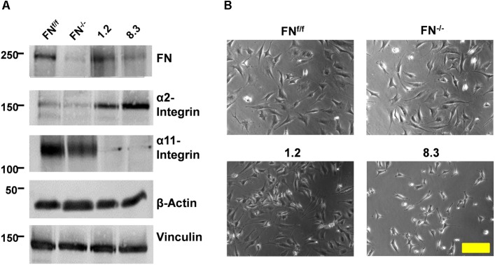
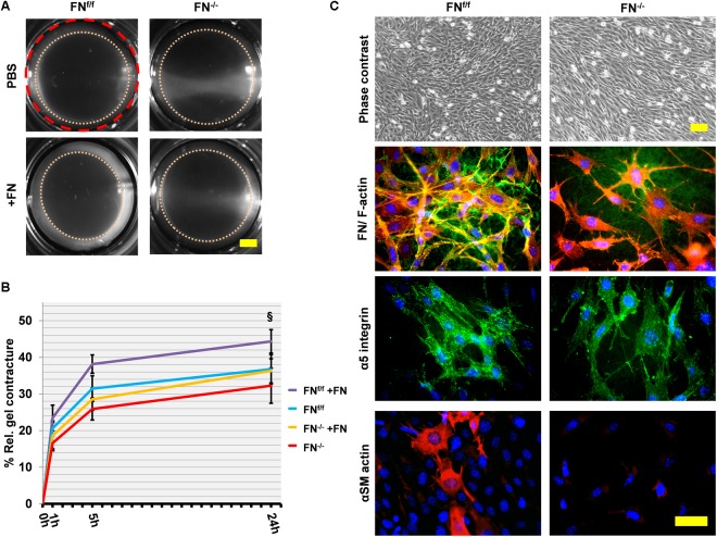
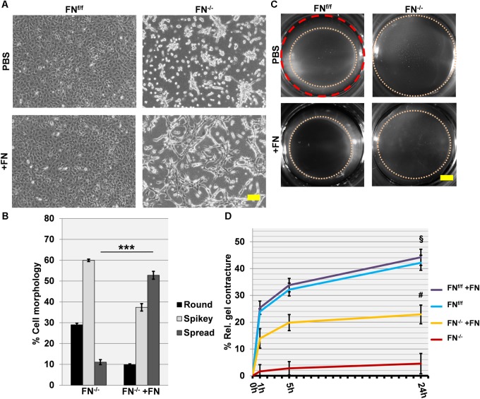
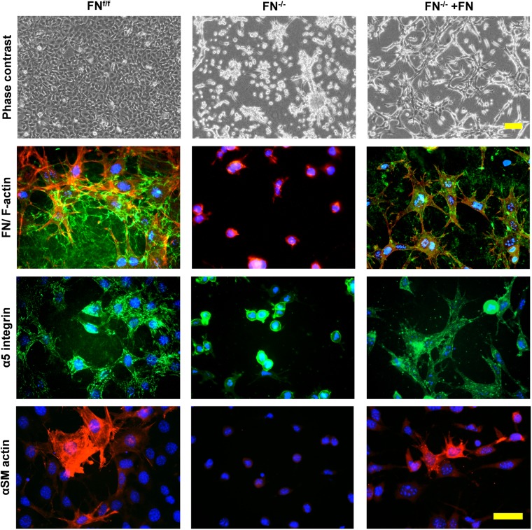
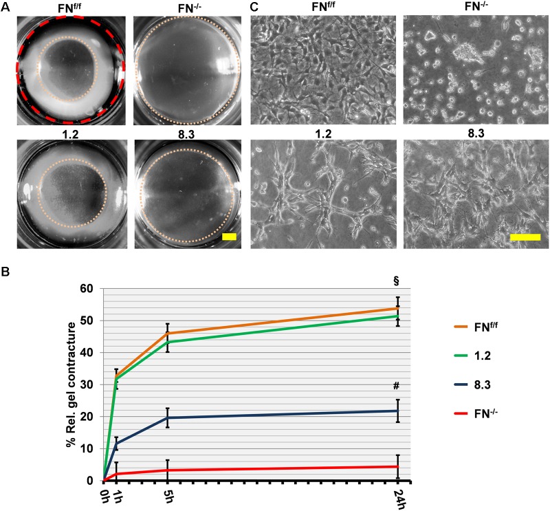
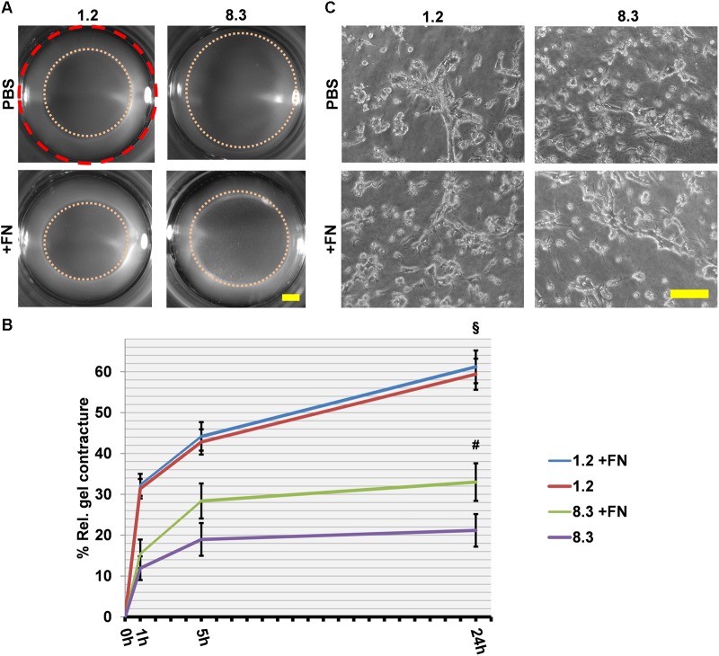
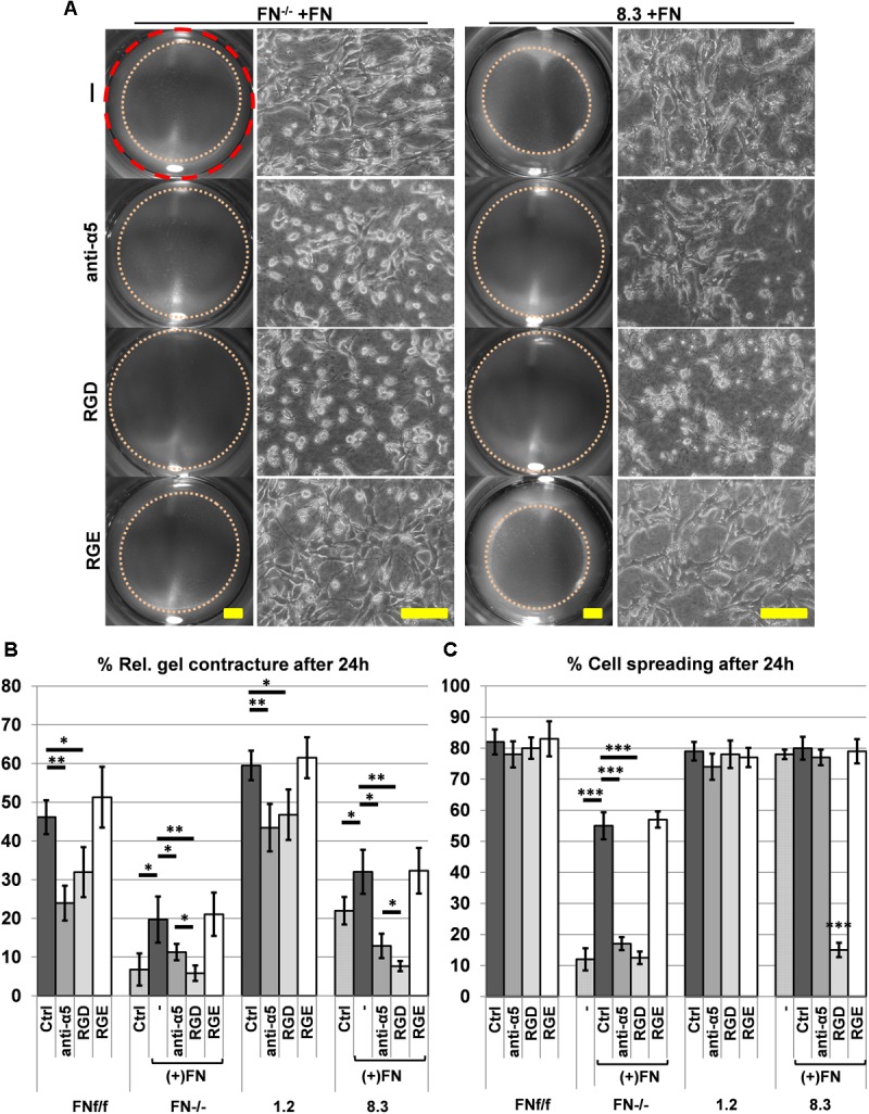
Fibronectin (FN) is an extracellular matrix glycoprotein that is abundantly expressed by fibroblasts in contracting wounds, where it mediates cell adhesion, migration and proliferation. FN also efficiently binds to collagen. Therefore, we and others hypothesized that FN and its cellular receptor integrin α5β1 might be involved in collagen matrix contracture by acting as linkers. However, there are conflicting reports on this issue. Moreover, several publications suggest an important role of collagen-binding integrin receptors α2β1 and α11β1 in collagen matrix contracture. Therefore, the aim of the present study was to determine the contributions of FN-integrin α5β1 interactions relative to those of collagen receptors α2β1 and α11β1 in this process. To assess the role of cellular FN directly, we employed FN-deficient mouse fibroblasts, subjected them to a collagen gel contracture assay in vitro, and compared them to their wildtype counterparts. Exogenous FN was removed from serum-containing medium. For dissecting the role of major collagen receptors, we used two FN-deficient mouse fibroblast lines that both possess integrin α5β1 but differ in their collagen-binding integrins. Embryo-derived FN-null fibroblasts, which express α11- but no α2-integrin, barely spread and tended to cluster on collagen gels. Moreover, FN-null fibroblasts required exogenously added FN to assemble α5β1-integrin in fibrillar adhesion contacts, and to contract collagen matrices. In contrast, postnatal kidney fibroblasts were found to express α2- but barely α11-integrin. When FN expression was suppressed in these cells by shRNA transfection, they were able to spread on and partially contract collagen gels in the absence of exogenous FN. Also in this case, however, collagen contracture was stimulated by adding FN to the medium. Antibody to integrin α5β1 or RGD peptide completely abolished collagen contracture by FN-deficient fibroblasts stimulated by FN addition. We conclude that although collagen-binding integrins (especially α2β1) can mediate contracture of fibrillar collagen gels by murine fibroblasts to some extent, full activity is causally linked to the presence of pericellular FN and its receptor integrin α5β1.

Keywords: alpha5-integrin; collagen; collagen contraction; collagen contracture; fibroblasts; fibronectin.


Figures

FIGURE 1
FIGURE 1
Expression of FN and collagen-binding integrins by FN-deficient vs. control fibroblasts. (A) Immunoblots of cell extracts obtained from FNf/f, FN−/−, clone 1.2, and clone 8.3 fibroblasts. Blots were probed with antibodies to fibronectin (FN), α2-integrin, α11-integrin, and β-actin and vinculin for loading control. The immunoblot experiments were performed three times; yielding identical results (see Supplementary Figure S3). (B) Phase contrast images of FNf/f, FN−/−, clone 1.2, and clone 8.3 fibroblasts on culture dishes in 10% FCS medium (Scale bar: 100 μm).
FIGURE 2
FIGURE 2
Fibrin contracture and immunofluorescence staining of FNf/f and FN−/− fibroblasts on fibrin 24 h after gel detachment. (A) Representative images of fibrin gels 24 h after release. The initial area is given by the dashed red circle. After gel release, a “contracture ring” (pink dotted circle) defined the borders of the measured area with time (Scale bar 2 mm). (B) The graph indicates the relative mean fibrin gel contracture of control (FNf/f) and fibronectin null (FN−/−) fibroblasts on standard (FN-containing) fibrin gels. Data are from at least three independent experiments done in quintuplicates and expressed relative to the initial area (red dashed line) in percent (±SD; significance after 24 h was tested by two-way ANOVA, followed by Tukey’s multiple comparisons test) 0, 1, 5, and 24 h after gel detachment. Overall there was no significant difference between FN−/− and FNf/f fibroblasts on fibrin gels. Adding exogenous FN (20 μg/ml, +FN) slightly but significantly increased the contracture ability of FNf/f cells (§p < 0.05). (C) Immunofluorescence staining of cell cultures on fibrin 24 h after gel detachment: Cells spread and grew easily on fibrin gels as seen in the phase contrast images (top panels; scale bar: 100 μm). FNf/f but not FN−/− fibroblasts assembled a dense FN-network (green) and formed longer fibrillar adhesions containing α5 integrin receptors (green). F-Actin (red). Certain FNf/f fibroblasts were positive for αSM-actin (bottom row, Texas Red staining) indicating the existence of myofibroblasts, which were not observed in FN−/− fibroblast cultures (bottom panels; scale bar: 50 μm).
FIGURE 3
FIGURE 3
Cell spreading on collagen gels of FNf/f and FN−/− fibroblasts, and their ability to contract a collagen matrix. (A) In contrast to wildtype fibroblasts (FNf/f, top left panel), fibronectin-null (FN−/−) cells did not spread and tended to cluster on collagen gels after 24 h in FN-depleted conditions (top right). Spreading of FNf/f fibroblasts was not influenced by adding 20 μg/ml purified FN to the medium (bottom left). A representative image shows partial rescue of cell spreading by FN−/− cells in the presence of exogenous FN (bottom right) (Scale bar: 100 μm). (B) Mean percentage (±SD; significance was tested by a paired t-test) of “round” (black bar), “spikey” (light gray bar) and “spread” (dark gray bar) FN−/− fibroblasts on collagen gels 24 h after seeding with or without exogenous FN (∗∗∗p < 0.001). (C) Representative images of collagen gels 24 h after release; the initial area is given by the dashed red circle: After gel release, a “contracture ring” (pink dotted circle) defined the borders of the measured area with time (Scale bar: 2 mm). (D) The graph indicates the relative mean gel contracture of FNf/f and FN−/− fibroblasts in the absence or presence of 20 ug/ml FN added to the collagen gel (+FN), 0, 1, 5, and 24 h after gel detachment. Data are from at least three independent experiments done in quintuplicates and are expressed as contracture relative to the initial area in percent (±SD; significance after 24 h was tested by two-way ANOVA, followed by Tukey’s multiple comparisons test). FNf/f fibroblasts exerted roughly a nine-fold higher collagen contracture ability after 24 h compared to FN−/− cells (§ p < 0.001). Addition of exogenous FN did not affect control fibroblasts. Collagen contracture exerted by FN−/− cells was partially rescued by adding exogenous FN to the collagen gels, which caused a five-fold increase (#p < 0.001).
FIGURE 4
FIGURE 4
Immunofluorescence staining of FNf/f and FN−/− fibroblasts on contracted collagen gels. Control (FNf/f) fibroblasts spread extensively on native collagen, whereas FN-null (FN−/−) cells appeared roundish and tended to form clusters. In the presence of exogenous FN, FN−/− fibroblasts increasingly spread as seen on the phase contrast images (top panels; scale bar 100 μm). The assembly of a FN network (green) and α5 integrin in focal adhesions (green) by FN−/− cells in the presence of exogenous FN could also be observed (middle panels; F-actin in red). Mature myofibroblasts were present amongst FNf/f fibroblasts; these were characterized by a well-organized alpha-smooth muscle actin (αSM-actin) positive cytoskeleton (bottom panels; Texas Red staining). A fraction of FN−/− fibroblasts showed higher αSM-actin expression in the presence of exogenous FN (bottom panels; scale bar 50 μm).
FIGURE 5
FIGURE 5
Collagen gel contracture by FN-knockdown fibroblast clones 1.2 and 8.3. (A) Representative images of collagen gel contracture are shown as described above (Scale bar: 2 mm). (B) The graph indicates the mean gel contracture relative to the initial area (red dashed circle) in percent by fibroblast clones 1.2 (normal FN expression) and 8.3 (70% reduced FN expression), compared to control (FNf/f) and fibronectin-null (FN−/−) fibroblasts. Data are from at least three independent experiments (±SD; significance after 24 h was tested by two-way ANOVA, followed by Tukey’s multiple comparisons test), 0, 1, 5, and 24 h after gel detachment. Clone 1.2 showed similar collagen contracture ability as FNf/f fibroblasts, whereas clone 8.3 exerted a contracture that was substantially reduced compared to the control cells (§ p < 0.001), but higher than that of FN−/− cells (#p < 0.001). (C) Phase contrast images of fibroblasts on collagen gels 24 h after release: Cells from clone 1.2 did not spread to the same extent as FNf/f fibroblasts on native collagen (Scale bar: 100 μm).
FIGURE 6
FIGURE 6
Collagen gel contracture by FN knockdown fibroblast clones 1.2 and 8.3 with or without exogenous FN. (A) Representative images of gel cultures are shown as described above (Scale bar: 2 mm). (B) The graph indicates the relative mean gel contracture in relation to the initial area (red dashed circle) in percent from at least three independent experiments (±SD significance after 24 h was tested by two-way ANOVA, followed by Tukey’s multiple comparisons test) 0, 1, 5, and 24 h after gel detachment. The collagen gel contracture exerted by FN-deficient clone 8.3 supplemented with exogenous FN is statistically greater than that by clone 8.3 in FN-depleted conditions (#p < 0.001). Clone 1.2 contracted collagen gels more extensively than did clone 8.3 with or without exogenous FN (§ p < 0.001). (C) Phase contrast images of fibroblast clones 1.2 and 8.3 on collagen gels 24 h after detachment: No differences can be seen in cell spreading with or without exogenous FN (Scale bar 100 μm).
FIGURE 7
FIGURE 7
Effect of blocking integrin α5β1 function on collagen gel contracture and cell spreading by normal and FN-deficient fibroblasts. Cells were cultured on collagen gels in 3% FN-depleted FCS/DMEM without (Ctrl: control) or with 20 μg/ml exogenous FN (+FN). Cultures were either left untreated (–), or alternatively a function blocking antibody against integrin α5 (anti-α5, 10 μg/ml), adhesion-blocking peptide GRGDSP (RGD, 1 mM), or control peptide GRGESP (RGE, 1 mM) were added to the medium. (A) Representative images of contracted collagen gels (Scale bar: 2 mm) and the respective spreading behavior of FN-deficient (FN−/−, clone 8.3) cell lines after 24 h in the presence of exogenous FN without (-; RGE) or with (anti-α5, RGD) inhibitors added (Scale bar: 100 μm). Note that both anti-α5 integrin antibody and GRGDSP peptide reduce FN-mediated cell spreading and matrix contracture on collagen gel. (B) Mean gel contracture relative to the initial area in percent from three independent experiments (±SD significance after 24 h was tested by two-way ANOVA, followed by Tukey’s multiple comparisons test) done in triplicates 24 h after gel detachment. Both, anti-α5 integrin antibody (anti-α5) and peptide GRGDSP (RGD) significantly reduced collagen gel contracture by control fibroblasts (FNf/f, clone 1.2) in FN-depleted medium, and completely inhibited contracture ability of FN-deficient cells (FN−/−, clone 8.3) stimulated by exogenous FN (p < 0.05; ∗∗p < 0.01). (C) Mean rate of spread cells relative to the total amount of cells in percent from three independent experiments (±SD; significance after 24 h was tested by two-way ANOVA, followed by Tukey’s multiple comparisons test) after 24 h of incubation on collagen gels. Cell spreading of FN−/− fibroblasts was significantly reduced when anti-α5 integrin antibody (anti-α5) and GRGDSP (RGD) where added to the culture media. 8.3 showed reduced spreading when incubated with the RDG peptide but not with the anti-α5 integrin antibody (∗∗∗p < 0.001).

Similar articles

Cited by

References

    1. Abe M., Sogabe Y., Syuto T., Yokoyama Y., Ishikawa O. (2007). Evidence that PI3K, Rac, Rho, and Rho kinase are involved in basic fibroblast growth factor-stimulated fibroblast-Collagen matrix contraction. J. Cell Biochem. 102 1290–1299. 10.1002/jcb.21359 - DOI - PubMed
    1. Asaga H., Kikuchi S., Yoshizato K. (1991). Collagen gel contraction by fibroblasts requires cellular fibronectin but not plasma fibronectin. Exp. Cell Res. 193 167–174. 10.1016/0014-4827(91)90552-6 - DOI - PubMed
    1. Barczyk M. M., Lu N., Popova S. N., Bolstad A. I., Gullberg D. (2013). alpha11beta1 integrin-mediated MMP-13-dependent collagen lattice contraction by fibroblasts: evidence for integrin-coordinated collagen proteolysis. J. Cell Physiol. 228 1108–1119. 10.1002/jcp.24261 - DOI - PubMed
    1. Broberg A., Heino J. (1996). Integrin alpha2beta1-dependent contraction of floating collagen gels and induction of collagenase are inhibited by tyrosine kinase inhibitors. Exp. Cell Res. 228 29–35. 10.1006/excr.1996.0295 - DOI - PubMed
    1. Calderwood D. A., Tuckwell D. S., Eble J., Kuhn K., Humphries M. J. (1997). The integrin alpha1 A-domain is a ligand binding site for collagens and laminin. J. Biol. Chem. 272 12311–12317. 10.1074/jbc.272.19.12311 - DOI - PubMed