Review
. 2019 Mar 5:10:194.
doi: 10.3389/fphys.2019.00194. eCollection 2019.

Primary Vitamin D Target Genes of Human Monocytes

Affiliations
Review

Primary Vitamin D Target Genes of Human Monocytes

Veijo Nurminen et al. Front Physiol. .

Abstract

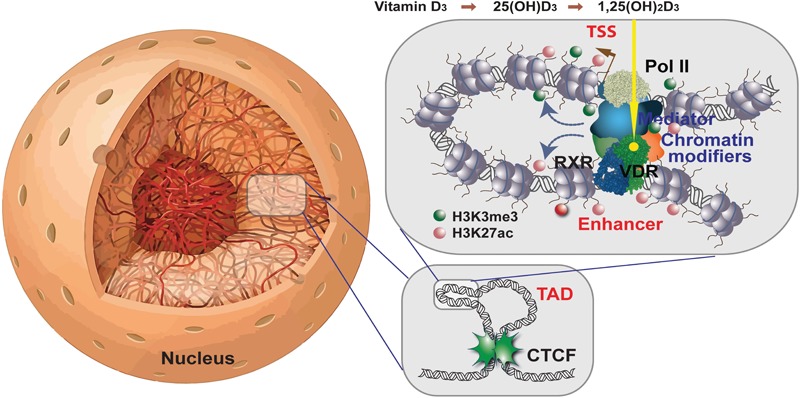
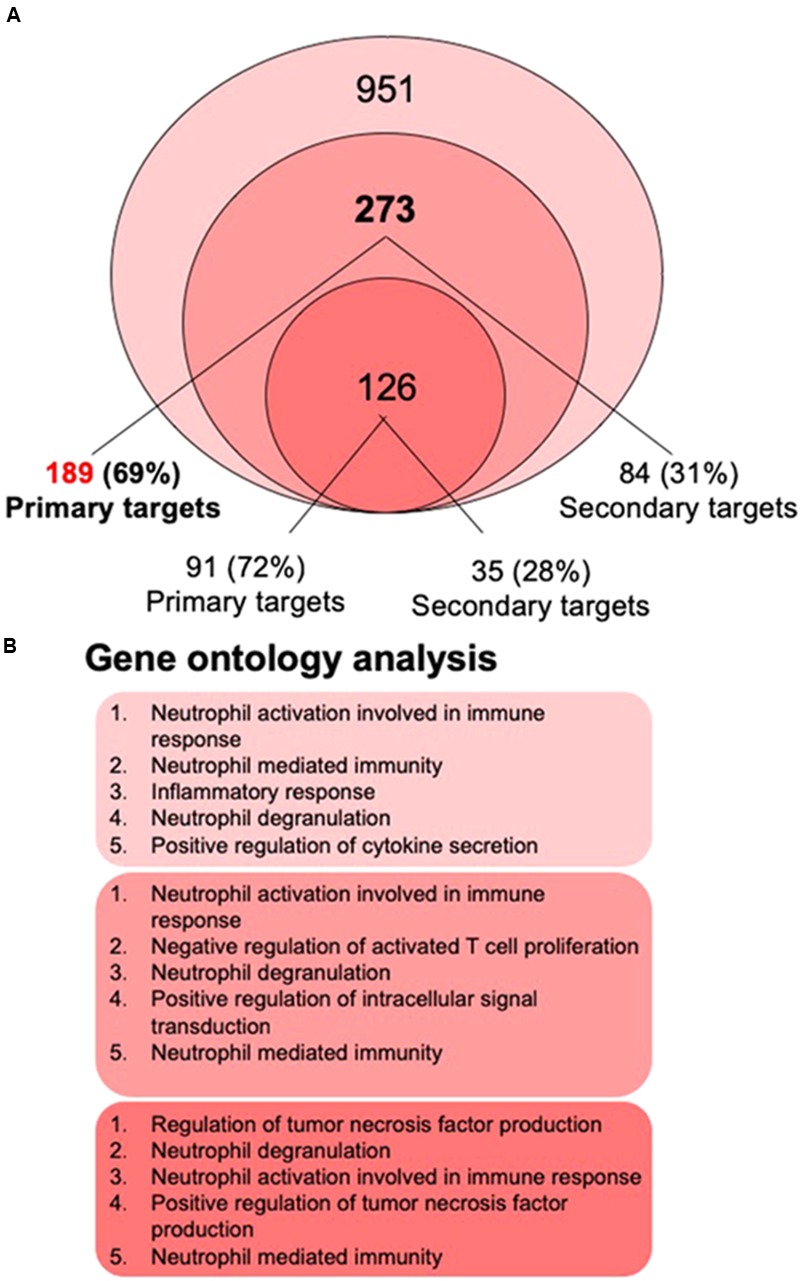
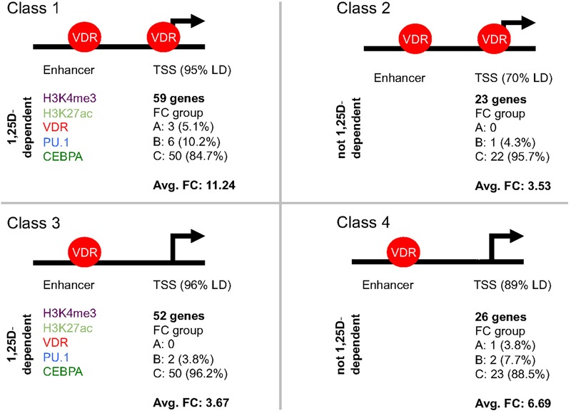
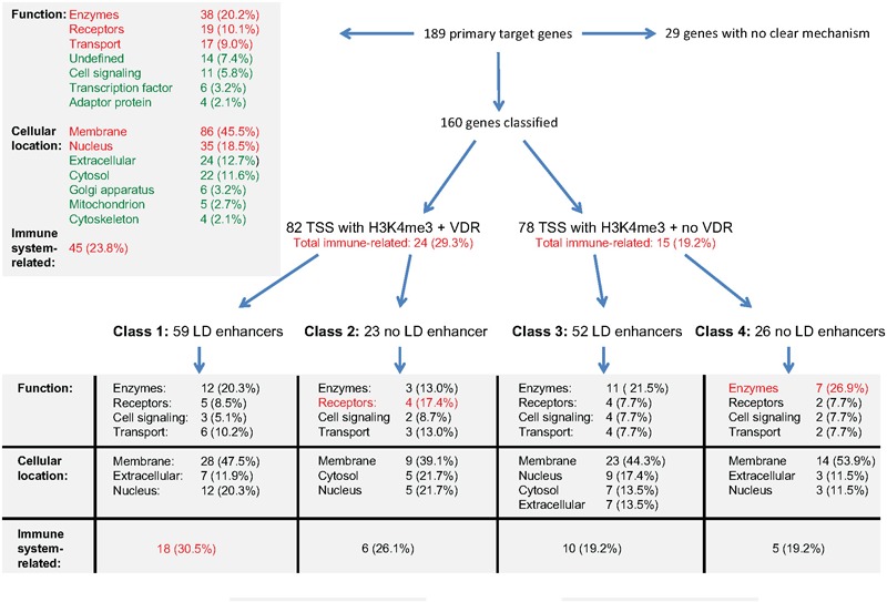
The molecular basis of vitamin D signaling implies that the metabolite 1α,25-dihydroxyvitamin D3 (1,25(OH)2D3) of the secosteroid vitamin D3 activates the transcription factor vitamin D receptor (VDR), which in turn modulates the expression of hundreds of primary vitamin D target genes. Since the evolutionary role of nuclear receptors, such as VDR, was the regulation of cellular metabolism, the control of calcium metabolism became the primary function of vitamin D and its receptor. Moreover, the nearly ubiquitous expression of VDR enabled vitamin D to acquire additional physiological functions, such as the support of the innate immune system in its defense against microbes. Monocytes and their differentiated phenotypes, macrophages and dendritic cells, are key cell types of the innate immune system. Vitamin D signaling was most comprehensively investigated in THP-1 cells, which are an established model of human monocytes. This includes the 1,25(OH)2D3-modulated cistromes of VDR, the pioneer transcription factors PU.1 and CEBPA and the chromatin modifier CTCF as well as of the histone markers of promoter and enhancer regions, H3K4me3 and H3K27ac, respectively. These epigenome-wide datasets led to the development of our chromatin model of vitamin D signaling. This review discusses the mechanistic basis of 189 primary vitamin D target genes identified by transcriptome-wide analysis of 1,25(OH)2D3-stimulated THP-1 cells and relates the epigenomic basis of four different regulatory scenarios to the physiological functions of the respective genes.

Keywords: VDR; epigenome; gene regulation; monocytes; transcriptome; vitamin D; vitamin D target genes.


Figures

FIGURE 1
FIGURE 1
Vitamin D signaling in the context of chromatin. Chromatin within the nucleus forms a 3D architecture (left). Two CTCF proteins bound at adjacent chromatin boundaries form a complex defining a TAD (right bottom). Enhancers and TSS regions that are located within the same TAD can get into physical contact within DNA looping (right top). 1,25(OH)2D3-activated VDR forms a heterodimeric complex with RXR on enhancer regions carrying appropriate binding sites. In this way, chromatin modifiers are activated that change histone marks (shown here are H3K4me3 modifications marking active TSS regions and H3K27ac indicating active chromatin) and the mediator complex forms a bridge to the basal transcriptional machinery with RNA polymerase II as its core. This finally leads to mRNA transcription of respective vitamin D target genes.
FIGURE 2
FIGURE 2
Gene ontology analysis. The most recent 1,25(OH)2D3-dependent transcriptome dataset of THP-1 cells (Nurminen et al., 2019) identified 951 genes, 273 of which overlap with the re-analyzed first RNA-seq dataset (Seuter et al., 2016; Neme et al., 2017) (A). 69% of the 273 genes are primary vitamin D targets (189 genes listed in Supplementary Table S1), while from the 126 genes that were also found by microarrays 72% are primary vitamin D targets. Gene ontology analysis using the webtool Enrichr (Chen et al., 2013) was performed for the lists of 951, 273, and 126 members and indicated that the top five biological pathways for each of the three gene sets relate to innate immunity (B).
FIGURE 3
FIGURE 3
Classification of primary vitamin D target genes. The 160 primary vitamin D target genes that show H3K4me3 marks at their TSS regions can be segregated into four classes. The structure of the genes into TSS and enhancer regions is schematically depicted. Genes of classes 1 and 2 display VDR binding to their TSS regions, while for those of classes 3 and 4 no VDR binding can be detected. The 59 genes of class 1 and the 52 genes of class 3 have significant (p < 0.05) effects of 1,25(OH)2D3 on H3K4me3, H3K27ac, VDR, PU.1 and/or CEBPA binding strength on their enhancer region, while this is not observed for the 23 genes of class 2 and the 26 genes of class 4. In addition, for the respective classes the distribution of the genes of into the fold change (FC) groups A, B and C (Supplementary Figure S2) as well as the average FC is indicated.
FIGURE 4
FIGURE 4
Integration of gene regulatory scenarios with biological function. The main relations between the epigenomic profiles of primary vitamin D target genes and the function, cellular location and relation to immunity of their encoded proteins is outlined. More details are provided in the text.

Similar articles

Cited by

References

    1. Ali T., Renkawitz R., Bartkuhn M. (2016). Insulators and domains of gene expression. Curr. Opin. Genet. Dev. 37 17–26. 10.1016/j.gde.2015.11.009 - DOI - PubMed
    1. Beisel C., Paro R. (2011). Silencing chromatin: comparing modes and mechanisms. Nat. Rev. Genet. 12 123–135. 10.1038/nrg2932 - DOI - PubMed
    1. Bendik I., Friedel A., Roos F. F., Weber P., Eggersdorfer M. (2014). Vitamin D: a critical and essential micronutrient for human health. Front. Physiol. 5:248 10.3389/fphys.2014.00248 - DOI - PMC - PubMed
    1. Bouillon R., Suda T. (2014). Vitamin D: calcium and bone homeostasis during evolution. Bonekey Rep. 3:480. 10.1038/bonekey.2013.214 - DOI - PMC - PubMed
    1. Carlberg C. (2014). Genome-wide (over)view on the actions of vitamin D. Front. Physiol. 5:167. 10.3389/fphys.2014.00167 - DOI - PMC - PubMed

LinkOut - more resources