

The Selection of a Hepatocyte Cell Line Susceptible to Plasmodium falciparum Sporozoite Invasion That Is Associated With Expression of Glypican-3
- PMID: 30891005
- PMCID: PMC6413710
- DOI: 10.3389/fmicb.2019.00127
The Selection of a Hepatocyte Cell Line Susceptible to Plasmodium falciparum Sporozoite Invasion That Is Associated With Expression of Glypican-3
Abstract
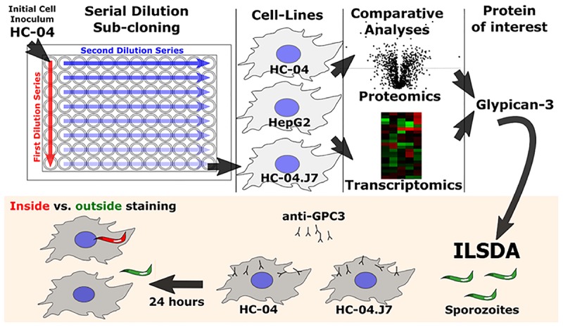
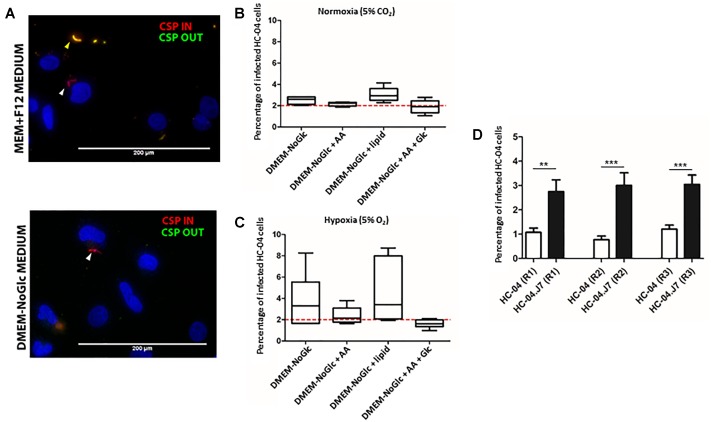
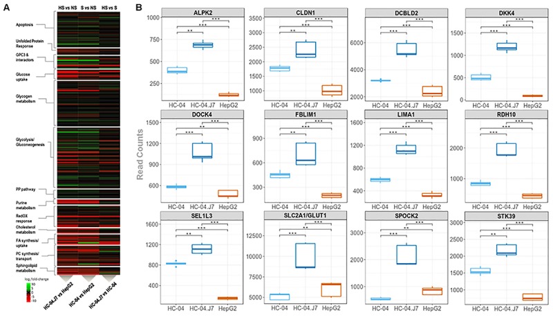
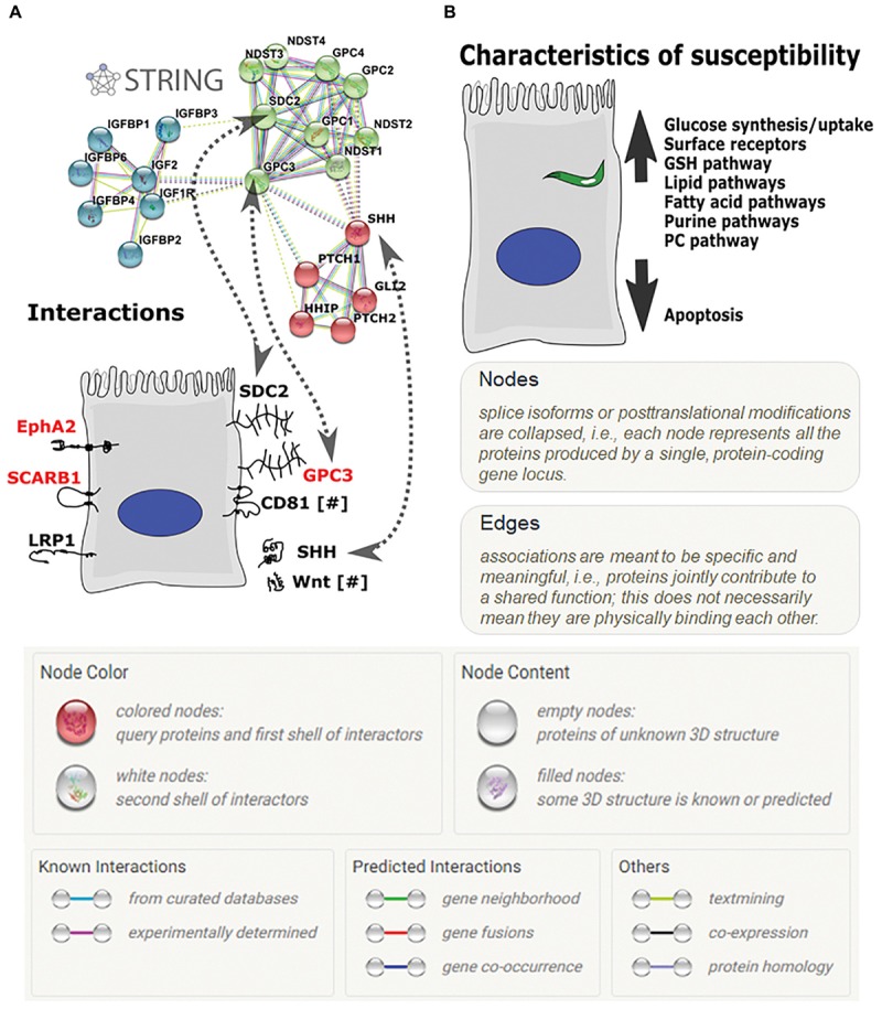
In vitro studies of liver stage (LS) development of the human malaria parasite Plasmodium falciparum are technically challenging; therefore, fundamental questions about hepatocyte receptors for invasion that can be targeted to prevent infection remain unanswered. To identify novel receptors and to further understand human hepatocyte susceptibility to P. falciparum sporozoite invasion, we created an optimized in vitro system by mimicking in vivo liver conditions and using the subcloned HC-04.J7 cell line that supports mean infection rates of 3-5% and early development of P. falciparum exoerythrocytic forms-a 3- to 5-fold improvement on current in vitro hepatocarcinoma models for P. falciparum invasion. We juxtaposed this invasion-susceptible cell line with an invasion-resistant cell line (HepG2) and performed comparative proteomics and RNA-seq analyses to identify host cell surface molecules and pathways important for sporozoite invasion of host cells. We identified and investigated a hepatocyte cell surface heparan sulfate proteoglycan, glypican-3, as a putative mediator of sporozoite invasion. We also noted the involvement of pathways that implicate the importance of the metabolic state of the hepatocyte in supporting LS development. Our study highlights important features of hepatocyte biology, and specifically the potential role of glypican-3, in mediating P. falciparum sporozoite invasion. Additionally, it establishes a simple in vitro system to study the LS with improved invasion efficiency. This work paves the way for the greater malaria and liver biology communities to explore fundamental questions of hepatocyte-pathogen interactions and extend the system to other human malaria parasite species, like P. vivax.
Keywords: Plasmodium falciparum; glypican-3; hepatocyte; in vitro model; liver stage; malaria; omics.
Figures

Similar articles
-
AMA1 and MAEBL are important for Plasmodium falciparum sporozoite infection of the liver.Cell Microbiol. 2017 Sep;19(9). doi: 10.1111/cmi.12745. Epub 2017 May 18. Cell Microbiol. 2017. PMID: 28371168
-
The acute transcriptomic and proteomic response of HC-04 hepatoma cells to hepatocyte growth factor and its implications for Plasmodium falciparum sporozoite invasion.Mol Cell Proteomics. 2014 May;13(5):1153-64. doi: 10.1074/mcp.M113.035584. Epub 2014 Feb 16. Mol Cell Proteomics. 2014. PMID: 24532842 Free PMC article.
-
A novel immortalized hepatocyte-like cell line (imHC) supports in vitro liver stage development of the human malarial parasite Plasmodium vivax.Malar J. 2018 Jan 25;17(1):50. doi: 10.1186/s12936-018-2198-4. Malar J. 2018. PMID: 29370800 Free PMC article.
-
Functional, immunological and three-dimensional analysis of chemically synthesised sporozoite peptides as components of a fully-effective antimalarial vaccine.Curr Med Chem. 2011;18(29):4470-502. doi: 10.2174/092986711797287575. Curr Med Chem. 2011. PMID: 22029724 Review.
-
Developmental biology of sporozoite-host interactions in Plasmodium falciparum malaria: implications for vaccine design.Clin Microbiol Rev. 2006 Oct;19(4):686-707. doi: 10.1128/CMR.00063-05. Clin Microbiol Rev. 2006. PMID: 17041140 Free PMC article. Review.
Cited by
-
Sporozoite motility as a quantitative readout for anti-CSP antibody inhibition.Sci Rep. 2022 Oct 13;12(1):17194. doi: 10.1038/s41598-022-22154-8. Sci Rep. 2022. PMID: 36229488 Free PMC article.
-
Evidence for infectious merozoites of Plasmodium falciparum from natural isolates of cultured hepatoma cells infected with sporozoites.PLoS One. 2025 Mar 19;20(3):e0319901. doi: 10.1371/journal.pone.0319901. eCollection 2025. PLoS One. 2025. PMID: 40106488 Free PMC article.
-
Dual RNA Sequencing Meta-analysis in Plasmodium Infection Identifies Host-Parasite Interactions.mSystems. 2021 Apr 20;6(2):e00182-21. doi: 10.1128/mSystems.00182-21. mSystems. 2021. PMID: 33879496 Free PMC article.
-
Bioengineered Liver Cell Models of Hepatotropic Infections.Viruses. 2021 Apr 27;13(5):773. doi: 10.3390/v13050773. Viruses. 2021. PMID: 33925701 Free PMC article. Review.
-
Host cell CRISPR genomics and modelling reveal shared metabolic vulnerabilities in the intracellular development of Plasmodium falciparum and related hemoparasites.Nat Commun. 2024 Jul 21;15(1):6145. doi: 10.1038/s41467-024-50405-x. Nat Commun. 2024. PMID: 39034325 Free PMC article.
References
Grants and funding
LinkOut - more resources
Full Text Sources
Molecular Biology Databases
Research Materials