

MR imaging of paratesticular bilateral leiomyoma: A case report
- PMID: 30891109
- PMCID: PMC6407094
- DOI: 10.1016/j.radcr.2019.02.019
MR imaging of paratesticular bilateral leiomyoma: A case report
Abstract
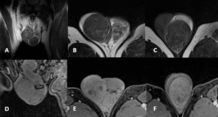
Paratesticular leiomyoma is a rare benign neoplasm that may arise from smooth muscle cells contained in either the epididymis, the spermatic cord, or the tunica albuginea. Usually patients present a palpable, asymptomatic mass, with a higher prevalence among the fourth and fifth decade of life. In this case report we describe a 57-year-old man with bilateral scrotal palpable masses evaluated with ultrasound and magnetic resonance imaging that were suggestive for leiomyoma. The lesions were surgically removed and pathology revealed no signs of malignancy confirming the diagnostic hypothesis of leiomyoma. Ultrasound is considered the imaging modality of choice for the initial evaluation of testicular masses since it allows an accurate localization (ie testicular vs paratesticular) and can identify signs of malignancy. Magnetic resonance imaging is less frequently performed but can considerably improve lesion characterization.
Keywords: Diagnostics; Leiomyoma; MRI; Paratesticular; US.
Figures


References
-
- Siristatidis C., Vaidakis D., Rigos I., Chrelias G., Papantoniou N. Leiomyoma and infertility. Minerva Ginecol. 2016;68(3):283–296. - PubMed
-
- Akbar S.A., Sayyed T.A., Jafri S.Z., Hasteh F., Neill J.S. Multimodality imaging of paratesticular neoplasms and their rare mimics. Radiographics. 2003;23(6):1461–1476. - PubMed
-
- Woodward P.J., Schwab C.M., Sesterhenn I.A. From the archives of the AFIP: extratesticular scrotal masses: radiologic-pathologic correlation. Radiographics. 2003;23(1):215–240. - PubMed
-
- Newman P.L., Fletcher C.D. Smooth muscle tumours of the external genitalia: clinicopathological analysis of a series. Histopathology. 1991;18(6):523–529. - PubMed
-
- Aganovic L., Cassidy F. Imaging of the scrotum. Radiol Clin North Am. 2012;50(6):1145–1165. - PubMed
Publication types
LinkOut - more resources
Full Text Sources
