

Case Reports
doi: 10.1093/jscr/rjz068.
eCollection 2019 Mar.
Pseudocyst of the pancreas masquerading as spontaneous pneumomediastinum
Affiliations
- PMID: 30891176
- PMCID: PMC6415621
- DOI: 10.1093/jscr/rjz068
Item in Clipboard
Case Reports
Pseudocyst of the pancreas masquerading as spontaneous pneumomediastinum
J Surg Case Rep.
.
Abstract
Pseudocyst of the pancreas extending into the thorax represents a rare but potentially catastrophic diagnosis. It can be difficult to both diagnose and manage, with only limited management suggestions within the literature. While pleural effusion is a common complication of pancreatitis, transthoracic extension of a pseudocyst is a rare phenomenon. Herein we discuss a patient with a difficult to recognize extension of pancreatic pseudocyst into the left hemithorax, with unique imaging findings. He had good response to trans-gastric and percutaneous drainage and ultimately proceeded to thoracotomy and decortication. Around this case, the options for investigation and management are discussed.
Figures

Chest X-ray prior to transgastric drainage. Suggestive of Left pleural effusion/ lower lobe collapse.
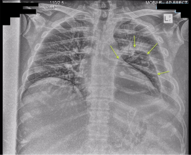
Chest X-ray post-transgastric drainage. Area of residual space highlighted with arrows.
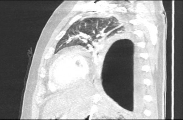
Sagittal view CT Chest revealing large residual space with loss of left lung volume due to the pseudocyst. Image post-transgastric drainage.

Area of communication between the drained Pseudocyst and the Left Pleura. Space noted with arrow.

Sagittal view CT Chest post-intercostal catheter drainage of the Pancreatico-pleural fistula. Smaller residual space can be noted compared to Image 3, with some ongoing loss of lung volume and small fluid collection.

CT one month post-decortication with complete re-expansion of the left lung and minor post-operative changes noted posteriorly.
References
-
- Loy JJ, Brooks MJ, Mahon D. Pseudoanuerysm of the thoracic aorta as a complication of pancreatic pseudocyst. EJVES Extra 2011;22:e31–e33.
-
- Ajmera AV, Judge TA. Mediastinal extension of pancreatic pseudocyst—a case with review of topic and management guidelines. Am J Ther 2012;19:152–156. - PubMed
-
- Marshall GT, Howell DA, Hansen BL, Amberson SM, Abourjaily GS, Bredenberg CE. Multidisciplinary approach to pseudoaneurysms complicating pancreatic pseudocysts. Arch Surg 1996;131:278–272. - PubMed
-
- Ali T, Srinivasan N, Le V, Chimpiri AR, Tierney WM. Pancreaticopleural fistula. Pancreas 2009;38:e26–31. - PubMed
Publication types
LinkOut - more resources
Full Text Sources
