

Layer boundary evolution method for macular OCT layer segmentation
- PMID: 30891330
- PMCID: PMC6420297
- DOI: 10.1364/BOE.10.001064
Layer boundary evolution method for macular OCT layer segmentation
Abstract
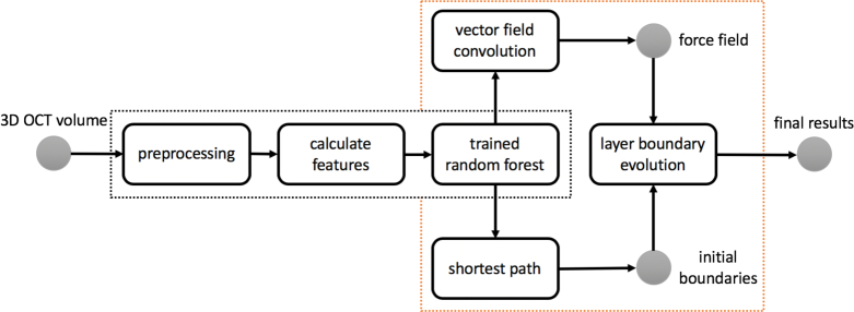
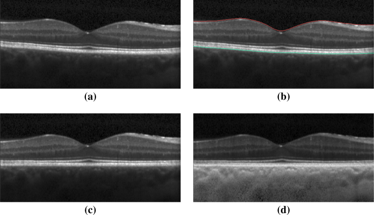
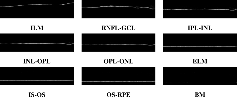
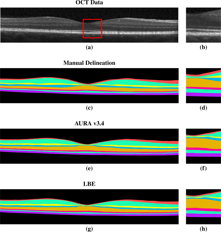
Optical coherence tomography (OCT) is used to produce high resolution depth images of the retina and is now the standard of care for in-vivo ophthalmological assessment. It is also increasingly being used for evaluation of neurological disorders such as multiple sclerosis (MS). Automatic segmentation methods identify the retinal layers of the macular cube providing consistent results without intra- and inter-rater variation and is faster than manual segmentation. In this paper, we propose a fast multi-layer macular OCT segmentation method based on a fast level set method. Our framework uses contours in an optimized approach specifically for OCT layer segmentation over the whole macular cube. Our algorithm takes boundary probability maps from a trained random forest and iteratively refines the prediction to subvoxel precision. Evaluation on both healthy and multiple sclerosis subjects shows that our method is statistically better than a state-of-the-art graph-based method.
Conflict of interest statement
The authors declare that there are no conflicts of interest related to this article.
Figures
References
-
- Saidha S., Eckstein C., Ratchford J. N., “Optical coherence tomography as a marker of axonal damage in multiple sclerosis,” Int. J. Clin. Rev. (2010).10.5275/ijcr.2010.10.01 - DOI
-
- Saidha S., Sotirchos E. S., Ibrahim M. A., Crainiceanu C. M., Gelfand J. M., Sepah Y. J., Ratchford J. N., Oh J., Seigo M. A., Newsome S. D., Balcer L. J., Frohman E. M., Green A. J., Nguyen Q. D., Calabresi P. A., “Microcystic macular oedema, thickness of the inner nuclear layer of the retina, and disease characteristics in multiple sclerosis: a retrospective study,” The Lancet Neurol. 11, 963–972 (2012).10.1016/S1474-4422(12)70213-2 - DOI - PMC - PubMed
-
- Saidha S., Syc S. B., Durbin M. K., Eckstein C., Oakley J. D., Meyer S. A., Conger A., Frohman T. C., Newsome S., Ratchford J. N., Frohman E. M., Calabresi P. A., “Visual dysfunction in multiple sclerosis correlates better with optical coherence tomography derived estimates of macular ganglion cell layer thickness than peripapillary retinal nerve fiber layer thickness,” Multiple Scler. J. 17, 1449–1463 (2011).10.1177/1352458511418630 - DOI - PubMed
-
- DeBuc D. C., Somfai G. M., “Early detection of retinal thickness changes in diabetes using optical coherence tomography,” Med. Sci. Monit. 16, MT15–MT21 (2010). - PubMed
Grants and funding
LinkOut - more resources
Full Text Sources
Other Literature Sources