. 2019 Feb 23:6:215-221.
doi: 10.1016/j.toxrep.2019.02.003. eCollection 2019.

Inhibition of lipid accumulation by the ethyl acetate fraction of Distylium racemosum in vitro and in vivo

Affiliations

Inhibition of lipid accumulation by the ethyl acetate fraction of Distylium racemosum in vitro and in vivo

Hye-Ran Kim et al. Toxicol Rep. .

Abstract

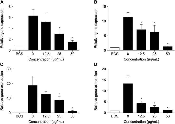
This study confirms the anti-obesity effect of the ethyl acetate fraction of Distylium racemosum (DRE), a member of Hamamelidaceae, that naturally grows on Jeju Island, on adipocyte differentiation in 3T3-L1 cells. This study further demonstrated that DRE exhibits anti-obesity effects in C57BL/6 obese mice. The degree of adipocyte differentiation was determined using Oil red O stain; results indicated a decrease in fat globules, which was dependent on DRE concentration, when pre-adipocytes were treated with differentiation-inducing agents. In addition, this significantly reduced the expression of the adipogenic transcription factor and related genes. C57BL/6 obese mice treated with DRE showed a lower rate of body weight gain than the high-fat diet (HFD) group mice. Further, the level of serum triglyceride in the DRE treatment group was lower than that in the HFD group. The findings show that DRE are capable of suppressing adipocyte accumulation; therefore, DRE may represent a promising source of functional materials for the anti-obesity.

Keywords: Distylium racemosum; Ethyl acetate fraction; Obesity.


Figures

Fig. 1
Fig. 1
Effects of fraction from Distylium racemosum on 3T3-L1 cell viability. (A) Hexane fraction, (B) Ethyl acetate fraction, (C) Butanol fraction, (D) Water fraction. 3T3-L1 cells were treated with the indicated concentrations (0, 12.5, 25, 50, and 100 μg/mL) of D. racemosum fraction for 48 h. Cell viability was measured by MTT assay. Data are expressed as mean ± standard deviation (n = 3). Significant differences from control are indicated (p < 0.05).
Fig. 2
Fig. 2
Effects of ethyl acetate fraction from Distylium racemosum on lipid accumulation in 3T3-L1 cells. Lipid accumulation was measured by Oil red O staining. All photographs were taken using a microscope. Lipid accumulation was inhibited by DRE (magnification ×400).
Fig. 3
Fig. 3
Effects of fraction from Distylium racemosum on lipid accumulation in 3T3-L1 cells. (A) Hexane fraction, (B) Ethyl acetate fraction, (C) Butanol fraction, (D) Water fraction. 3T3-L1 cells were treated with the indicated concentrations (0, 12.5, 25, and 50 μg/mL) of D. racemosum fraction. Lipid accumulation was measured by Oil red O stain. The cells were eluted into 100% isopropanol and OD was measured at 500 nm. Data are expressed as mean ± standard deviation (n = 3). Significant differences from control are indicated (p < 0.05).
Fig. 4
Fig. 4
Effects of ethyl acetate fraction from Distylium racemosum on the gene expression. (A) SREBP1c, (B) PPARγ, (C) C/EBPα, (D) FAS. Expression of adipogenic transcription factors was measured by qRT-PCR. SREBP1c, PPARγ, C/EBPα, and FAS genes were inhibited by DRE in a dose-dependent manner. Data are expressed as mean ± standard deviation (n = 3). Significant differences from control are indicated (p < 0.05).
Fig. 5
Fig. 5
Effects of ethyl acetate fraction from Distylium racemosum on triglyceride level. Levels of triglycerides were compared after a 6-week DRE treatment. Data are expressed as mean ± standard deviation (n = 8). Significant differences from the HFD group are indicated (p < 0.05).
Fig. 6
Fig. 6
Morphologic characteristics of liver tissue. (A) ND group, (B) HFD group, (C) HFD with DRE 50 mg/kg, and (D) HFD with orlistat 50 mg/kg. Morphologic characteristics of liver tissue were observed through hematoxylin and eosin staining. Difference in liver tissue after the oral administration of DRE was confirmed compared with the HFD group (magnification ×100).
Fig. 7
Fig. 7
Morphologic characteristics of adipose tissue. (A) ND group, (B) HFD group, (C) HFD with DRE 50 mg/kg, and (D) HFD with orlistat 50 mg/kg. Morphologic characteristics of adipose tissue were observed through hematoxylin and eosin staining. Difference in adipose tissue after the oral administration of DRE was confirmed compared with the HFD group (magnification ×100).

Similar articles

Cited by

References

    1. Kang J.W., Nam D., Kim K.H., Huh J.E., Lee J.D. Effect of gambisan on the inhibition of adipogenesis in 3T3-L1 adipocytes. Evid. Complement. Alternat. Med. 2013;2013:789067. - PMC - PubMed
    1. Dhurandhar N.V. A framework for identification of infections that contribute to human obesity. Lancet Infect. Dis. 2011;11:963–969. - PubMed
    1. WHO . 2015. Obesity and Overweight.http://www.who.int/mediacentre/factsheets/fs311/en/
    1. WHO Expert Consultation Appropriate body-mass index for Asian populations and its implications for policy and intervention strategies. Lancet. 2004;363:157–163. - PubMed
    1. Greenberg A.S., Obin M.S. Obesity and the role of adipose tissue in inflammation and metabolism. Am. J. Clin. Nutr. 2006;83:461S–465S. - PubMed

LinkOut - more resources