Review
. 2019 Mar 5:9:46.
doi: 10.3389/fcimb.2019.00046. eCollection 2019.

From Entry to Early Dissemination- Toxoplasma gondii's Initial Encounter With Its Host

Affiliations
Review

From Entry to Early Dissemination- Toxoplasma gondii's Initial Encounter With Its Host

Estefania Delgado Betancourt et al. Front Cell Infect Microbiol. .

Abstract

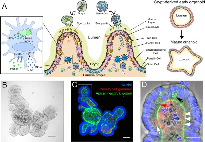
Toxoplasma gondii is a zoonotic intracellular parasite, able to infect any warm-blooded animal via ingestion of infective stages, either contained in tissue cysts or oocysts released into the environment. While immune responses during infection are well-studied, there is still limited knowledge about the very early infection events in the gut tissue after infection via the oral route. Here we briefly discuss differences in host-specific responses following infection with oocyst-derived sporozoites vs. tissue cyst-derived bradyzoites. A focus is given to innate intestinal defense mechanisms and early immune cell events that precede T. gondii's dissemination in the host. We propose stem cell-derived intestinal organoids as a model to study early events of natural host-pathogen interaction. These offer several advantages such as live cell imaging and transcriptomic profiling of the earliest invasion processes. We additionally highlight the necessity of an appropriate large animal model reflecting human infection more closely than conventional infection models, to study the roles of dendritic cells and macrophages during early infection.

Keywords: Apicomplexa; Paneth cells; Toxoplasma gondii; innate response; intestinal epithelial barrier; intestinal organoids.


Figures

Figure 1
Figure 1
(A) Simplified scheme of the intestinal mucosa with its different cell populations and its derived organoids, and its infection with T. gondii bradyzoites or sporozoites. Center: After oral uptake of oocysts or tissue cysts into the small intestine sporozoites or bradyzoites leave the cyst and enter the intestinal epithelium to reach the lamina propria. The principle strategies used by the parasite to cross this barrier are either via invasion of the intestinal epithelial cells (a) or by transepithelial migration (b). Left: Infected murine DC. T. gondii ROP5 and ROP18 phosphorylate host IRGs, thus protecting the PV from degradation. Extracellular profilin is recognized by the cell via TLRs 11 and 12, inducing the release of IL-12 and TNF-α. Right: Extracellular matrix-embedded crypt (generated via physical disruption of the intestine) leads under appropriate culture conditions to the initial formation of a crypt-derived organoid which then can differentiate into a more complex “mature” intestinal organoid. (B–D) Microscopic images of a mouse small intestine-derived organoid infected with T. gondii RH strain tachyzoites. (B) Representative bright-field confocal image of a mouse IO after 7 days in culture. Scale bar 50 μm. (C) Projection of a confocal z-stack of the IO shown in C, stained with FITC-phalloidin for apical F-actin (green), TRITC-labeled UEA-1 lectin for Paneth cell granules (red) and DAPI for nuclei (blue). Note that due to the projection of the stack fluorescent signals might appear mis-localized within the organoid compared to the single plane shown in (D). Scale bar 50 μm. (D) Enlarged view of an organoid villus-like structure (white square in C) of a single plane. Parasites (identifiable by their GFP-tagged green tubular mitochondrion (Thomsen-Zieger et al., ; black arrow) had replicated in IECs for 48 h. Paneth cells, identifiable by their multiple granules (white arrow) can be detected in the villus-like structure. Due to its granular appearance the red arrow indicates a possible Paneth cell containing replicating parasites. The IO's lumen is filled with cell debris from apoptotic cells, constantly shed as part of the high turnover rate of IECs. The red structures in the lumen marked with white arrow heads might indicate Paneth cell degranulation, as previously described by Farin et al. (2014). Scale bar 20 μm.

References

    1. Ahmed N., French T., Rausch S., Kühl A., Hemminger K., Dunay I. R., et al. . (2017). Toxoplasma co-infection prevents Th2 differentiation and leads to a Helminth-specific Th1 response. Front. Cell. Infect. Microbiol. 7:341. 10.3389/fcimb.2017.00341 - DOI - PMC - PubMed
    1. Allaire J. M., Crowley S. M., Law H. T., Chang S. Y., Ko H. J., Vallance B. A. (2018). The intestinal epithelium: central coordinator of mucosal immunity. Trends Immunol. 39, 677–696. 10.1016/j.it.2018.04.002 - DOI - PubMed
    1. Andrade W. A., Souza Mdo C., Ramos-Martinez E., Nagpal K., Dutra M. S., Melo M. B., et al. . (2013). Combined action of nucleic acid-sensing Toll-like receptors and TLR11/TLR12 heterodimers imparts resistance to Toxoplasma gondii in mice. Cell Host Microbe 13, 42–53. 10.1016/j.chom.2012.12.003 - DOI - PMC - PubMed
    1. Barragan A., Brossier F., Sibley L. D. (2005). Transepithelial migration of Toxoplasma gondii involves an interaction of intercellular adhesion molecule 1 (ICAM-1) with the parasite adhesin MIC2. Cell. Microbiol. 7, 561–568. 10.1111/j.1462-5822.2005.00486.x - DOI - PubMed
    1. Barragan A., Sibley L. D. (2002). Transepithelial migration of Toxoplasma gondii is linked to parasite motility and virulence. J. Exp. Med. 195, 1625–1633. 10.1084/jem.20020258 - DOI - PMC - PubMed

Publication types