. 2019 Feb 12:2019:4250940.
doi: 10.1155/2019/4250940. eCollection 2019.

Histomorphometric Analysis of Callus Formation Stimulated by Axial Dynamisation in a Standardised Ovine Osteotomy Model

Affiliations

Histomorphometric Analysis of Callus Formation Stimulated by Axial Dynamisation in a Standardised Ovine Osteotomy Model

K M Reich et al. Biomed Res Int. .

Abstract

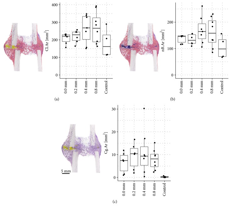
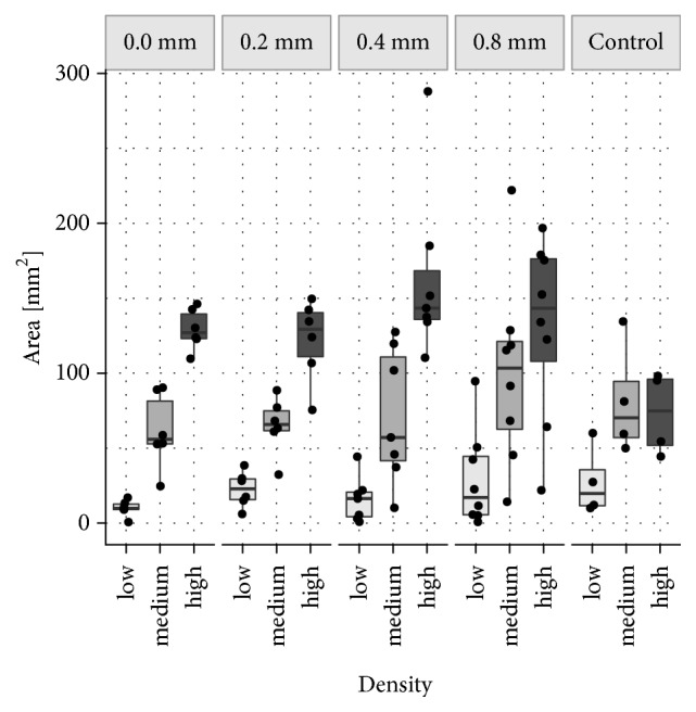
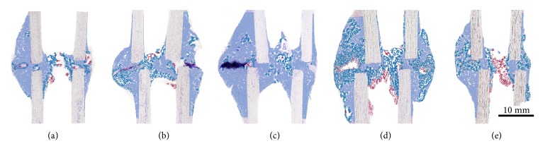
The cyclic axial dynamisation of a stabilised fracture is intended to promote callus formation and bone healing. Most studies focused on biomechanical properties or the quantity of new bone formation. Far less is known about the quality of newly formed callus tissues, such as tissue distribution and arrangement within the callus. The aim of this current study was to investigate the effect of cyclic, axial dynamisation on the quantity and quality of callus in an established delayed fracture healing model. In 41 sheep transverse osteotomies with a gap size of 3 mm were stabilised with a unilateral external fixator. In 32 of these, fracture ends were axially stimulated with displacement amplitudes of 0.8 mm, 0.4 mm, 0.2 mm, or 0.0 mm, respectively, for six weeks. In the remaining 9 sheep of the control group, an additional external fixator was mounted to achieve almost total rigidity. Animal material originating from a past animal experiment was reanalysed in this study. Histological thin-ground sections were histomorphometrically analysed regarding the histological structure and composition of the defect region. A slight tendency towards an increase in size of total callus area, area of new bone (nB.Ar), and cartilage (Cg.Ar) was detected with increasing displacement amplitudes compared to the control group. At the anterior callus side nB.Ar and Cg.Ar were significantly larger than at the posterior side in all groups independent of treatment. Regarding the quality of callus, areas of very compact bone were predominant in the treatment groups whereas in the control group a slight shift to more porous bone was observed. No difference of callus compactness was observed between the anterior and the posterior side. The established method to assess the local compactness of callus areas is a useful tool to quantitatively determine the spatial distribution of new bone tissue within the callus. The application of this method in combination with biomechanical testing might reveal interesting relations between tissue distribution and bone strength that, with traditional histomorphometry, cannot be identified.


Figures

Figure 1
Figure 1
(a) Total Callus Area (Cl.Ar). Total callus area (comprising newly formed bone, cartilage, and fibrous tissue within the ROI) increased with the increase of displacement amplitudes in the treatment groups. Largest Cl.Ar was found in the 0.4 mm group and the 0.8 mm group, smallest callus area in the control group. (b) New Bone Area (nB.Ar) within the total callus. Formation of new bone within the total callus area was lowest in the control group. Highest nB.Ar was found in the 0.4 mm group and the 0.8 mm group. (c) Cartilage Area (Cg.Ar) within the total callus. Presence of cartilage in the total callus area was very low in all groups. Variation within the groups was very high since cartilage was not present in all specimens. In the treatment groups Cg.Ar was slightly increased compared to the control group.
Figure 2
Figure 2
Size of Different Bone Density Areas (nB.Dn). Mineralised callus areas were classified as low nB.Dn (areas less than 33% new bone), medium nB.Dn (areas with 33 to 66% new bone), and high nB.Dn (areas exceeding 66% new bone). In this box plot diagram, the average size of low, medium, and high bone density areas is depicted for the 0.0 mm, 0.2 mm, 0.4 mm, and 0.8 mm group as well as for the control group.
Figure 3
Figure 3
Areas of Different Bone Densities (nB.Dn) within the Callus. Histological images were semiautomatically segmented and areas of different bone densities were colour encoded. Mineralised callus areas with less than 33% new bone were defined as regions with low nB.Dn (red), areas with 33 to 66% new bone as regions of medium nB.Dn (turquoise), and areas exceeding 66% were classified as high nB.Dn (light blue). For better visualisation, histological images are superimposed by the respective false colour images. The distribution of areas with different bone densities over the total callus area was very similar between the groups and showed a consistent inherent pattern. Besides, treatment groups (a-d) show a very similar proportional distribution: a relatively small area of low nB.Dn (5-10%), approximately 30% medium nB.Dn, and 50-65% high nB.Dn. In the control group (e), a shift to less dense bone was observed: the upwards trend observed in the treatment groups levelled off at the medium density level in the control group (approximately 45% medium nB.Dn). Areas with high nB.Dn were less common (approximately 42%) as compared to the treatment groups. (a) 0.0 mm group, (b) 0.2 mm group, (c) 0.4 mm group, (d) 0.8 mm group, and (e) control group. Anterior side is on the left and posterior side is on the right.

References

    1. Claes L., Recknagel S., Ignatius A. Fracture healing under healthy and inflammatory conditions. Nature Reviews Rheumatology. 2012;8(3):133–143. doi: 10.1038/nrrheum.2012.1. - DOI - PubMed
    1. Perren S. M. Physical and biological aspects of fracture healing with special reference to internal fixation. Clinical Orthopaedics and Related Research. 1979:175–196. - PubMed
    1. Goodship A. E. Mechanical stimulus to bone. Annals of the Rheumatic Diseases. 1992;51(1):4–1. doi: 10.1136/ard.51.1.4. - DOI - PMC - PubMed
    1. Wolff J. In: Das Gesetz der Transformation der Knochen. Hirschwald A., editor. Berlin: 1892.
    1. Santolini E., West R., Giannoudis P. V. Risk factors for long bone fracture non-union: a stratification approach based on the level of the existing scientific evidence. Injury. 2015;46(Supplement 8):S8–S19. doi: 10.1016/S0020-1383(15)30049-8. - DOI - PubMed