. 2019 Apr 1;30(4):597-603.
doi: 10.1093/annonc/mdz046.

Ultra-deep next-generation sequencing of plasma cell-free DNA in patients with advanced lung cancers: results from the Actionable Genome Consortium

Affiliations

Ultra-deep next-generation sequencing of plasma cell-free DNA in patients with advanced lung cancers: results from the Actionable Genome Consortium

B T Li et al. Ann Oncol. .

Abstract

Background: Noninvasive genotyping using plasma cell-free DNA (cfDNA) has the potential to obviate the need for some invasive biopsies in cancer patients while also elucidating disease heterogeneity. We sought to develop an ultra-deep plasma next-generation sequencing (NGS) assay for patients with non-small-cell lung cancers (NSCLC) that could detect targetable oncogenic drivers and resistance mutations in patients where tissue biopsy failed to identify an actionable alteration.

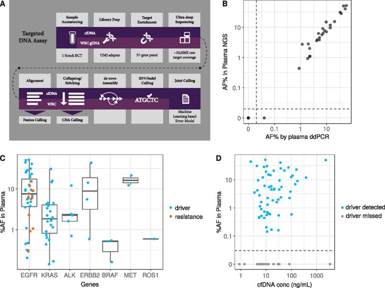
Patients and methods: Plasma was prospectively collected from patients with advanced, progressive NSCLC. We carried out ultra-deep NGS using cfDNA extracted from plasma and matched white blood cells using a hybrid capture panel covering 37 lung cancer-related genes sequenced to 50 000× raw target coverage filtering somatic mutations attributable to clonal hematopoiesis. Clinical sensitivity and specificity for plasma detection of known oncogenic drivers were calculated and compared with tissue genotyping results. Orthogonal ddPCR validation was carried out in a subset of cases.

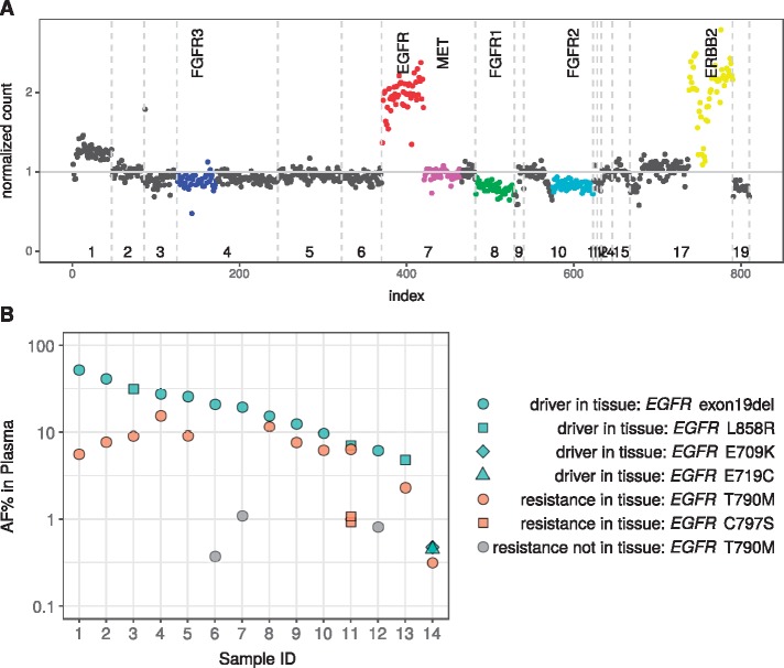
Results: In 127 assessable patients, plasma NGS detected driver mutations with variant allele fractions ranging from 0.14% to 52%. Plasma ddPCR for EGFR or KRAS mutations revealed findings nearly identical to those of plasma NGS in 21 of 22 patients, with high concordance of variant allele fraction (r = 0.98). Blinded to tissue genotype, plasma NGS sensitivity for de novo plasma detection of known oncogenic drivers was 75% (68/91). Specificity of plasma NGS in those who were driver-negative by tissue NGS was 100% (19/19). In 17 patients with tumor tissue deemed insufficient for genotyping, plasma NGS identified four KRAS mutations. In 23 EGFR mutant cases with acquired resistance to targeted therapy, plasma NGS detected potential resistance mechanisms, including EGFR T790M and C797S mutations and ERBB2 amplification.

Conclusions: Ultra-deep plasma NGS with clonal hematopoiesis filtering resulted in de novo detection of targetable oncogenic drivers and resistance mechanisms in patients with NSCLC, including when tissue biopsy was inadequate for genotyping.

Keywords: lung cancer; next-generation sequencing; oncogenic drivers; plasma cell-free DNA.


Figures

Figure 1.
Figure 1.
Plasma next-generation sequencing (NGS) assay workflow, comparison of variant allelic fraction with orthogonal plasma ddPCR, and variant detection in cfDNA. (A) Workflow describing the targeted DNA plasma cell-free DNA (cfDNA) assay. Additional details are provided in the ‘Patients and Methods’ section. (B) Allele frequency (AF) in plasma as determined by NGS were compared those calculated by ddPCR in a subset of group 1 and 3 samples with NGS-identified EGFR or KRAS mutations. Plasma AF as measured by NGS (y-axis, log scale) was plotted against plasma AF as measured by ddPCR (x-axis, log scale). Correlation was calculated using Pearson’s correlation coefficient (0.98, P < 0.001). Dotted line indicates a break in the axis to allow representation of variants which were not detected by either assay. (C) Box plot of plasma AF (y-axis) by gene (x-axis) according to mutation type (driver, blue; resistance, orange) depicting median as well as first and third quartiles. Data falling outside the Q1–Q3 range were plotted as outliers (outside of the vertical lines). (D) Correlation of plasma variant AF (y-axis, log scale) and total cfDNA concentration (x-axis, log scale) (Spearman’s correlation = 0.3 P-value = 0.006) for samples in which the driver was detected (blue dots) or missed (gray dots). Dotted line indicates a break in the vertical axis to allow representation of samples in which the driver identified by tissue-based testing was not detected by cfDNA analysis.
Figure 2.
Figure 2.
Capturing drug resistance mutations in plasma cfDNA. (A) Plot depicting one patient with EGFR and ERBB2 amplifications in plasma after treatment with erlotinib. Normalized sequencing read counts in comparison to healthy samples (y-axis) were plotted against chromosome number (x-axis); each dot represents an exon or intron target region covered by the 37-gene panel. Vertical dotted lines indicate chromosome boundaries. Specific genes are indicated by color: FGFR3, blue; EGFR, red; MET, pink; FGFR1, green; FGFR2, aqua; ERBB2, yellow. (B) Relationship between allelic fraction of driver and resistance mutations detected in plasma. The allele frequency (AF) (percent) of plasma cell-free DNA (cfDNA) variants detected in plasma (y-axis) was plotted for individual samples (x-axis). Driver and resistance mutations detected in tissue and plasma cfDNA are indicated by green and orange dots, respectively; resistance mutations identified in plasma cfDNA but not detected in tissue are indicated in gray. Shapes indicate specific mutations.

References

    1. Sholl LM, Aisner DL, Varella-Garcia M. et al. Multi-institutional oncogenic driver mutation analysis in lung adenocarcinoma: the Lung Cancer Mutation Consortium Experience. J Thorac Oncol 2015; 10(5): 768–777. - PMC - PubMed
    1. Bettegowda C, Sausen M, Leary RJ. et al. Detection of circulating tumor DNA in early- and late-stage human malignancies. Sci Transl Med 2014; 6: 224ra224. - PMC - PubMed
    1. Zehir A, Benayed R, Shah RH. et al. Mutational landscape of metastatic cancer revealed from prospective clinical sequencing of 10,000 patients. Nat Med 2017; 23(6): 703–713. - PMC - PubMed
    1. Oxnard GR, Paweletz CP, Kuang Y. et al. Noninvasive detection of response and resistance in EGFR-mutant lung cancer using quantitative next-generation genotyping of cell-free plasma DNA. Clin Cancer Res 2014; 20(6): 1698–1705. - PMC - PubMed
    1. Oxnard GR, Thress KS, Alden RS. et al. Association between plasma genotyping and outcomes of treatment with osimertinib (AZD9291) in advanced non-small-cell lung cancer. J Clin Oncol 2016; 34(28): 3375–3382. - PMC - PubMed

Publication types

MeSH terms