Review
. 2019 Jun;76(12):2369-2382.
doi: 10.1007/s00018-019-03071-y. Epub 2019 Mar 19.

Extracellular vesicle isolation methods: rising impact of size-exclusion chromatography

Affiliations
Review

Extracellular vesicle isolation methods: rising impact of size-exclusion chromatography

Marta Monguió-Tortajada et al. Cell Mol Life Sci. 2019 Jun.

Abstract

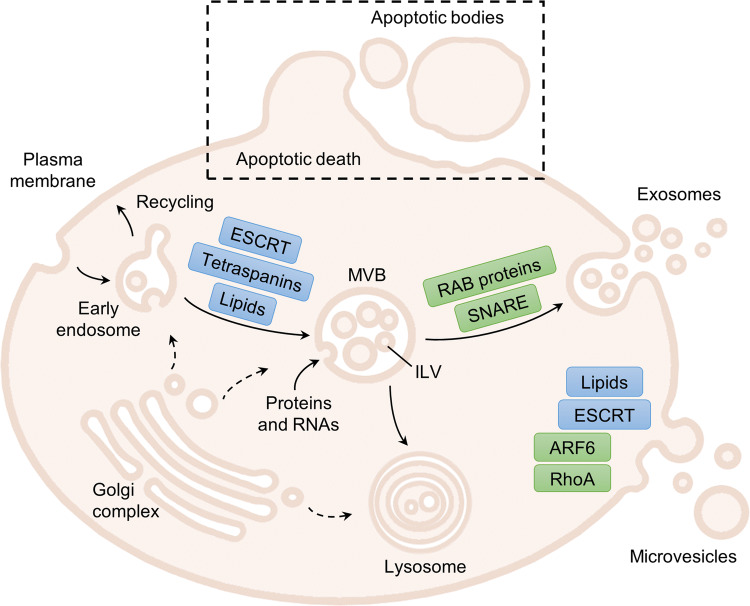
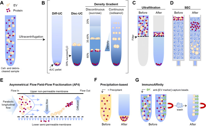
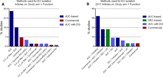
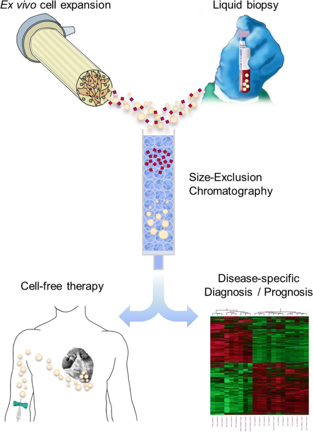
Extracellular vesicles (EVs) include a variety of nanosized vesicles released to the extracellular microenvironment by the vast majority of cells transferring bioactive lipids, proteins, mRNA, miRNA or non-coding RNA, as means of intercellular communication. Remarkably, among other fields of research, their use has become promising for immunomodulation, tissue repair and as source for novel disease-specific molecular signatures or biomarkers. However, a major challenge is to define accurate, reliable and easily implemented techniques for EV isolation due to their nanoscale size and high heterogeneity. In this context, differential ultracentrifugation (dUC) has been the most widely used laboratory methodology, but alternative procedures have emerged to allow purer EV preparations with easy implementation. Here, we present and discuss the most used of the different EV isolation methods, focusing on the increasing impact of size exclusion chromatography (SEC) on the resulting EV preparations from in vitro cultured cells-conditioned medium and biological fluids. Comparatively, low protein content and cryo-electron microscopy analysis show that SEC removes most of the overabundant soluble plasma proteins, which are not discarded using dUC or precipitating agents, while being more user friendly and less time-consuming than gradient-based EV isolation. Also, SEC highly maintains the major EVs' characteristics, including vesicular structure and content, which guarantee forthcoming applications. In sum, together with scaling-up possibilities to increase EV recovery and manufacturing following high-quality standards, SEC could be easily adapted to most laboratories to assist EV-associated biomarker discovery and to deliver innovative cell-free immunomodulatory and pro-regenerative therapies.

Keywords: Exosomes; Isolation methods; Nanomedicine; Purification; Theranostics.


Conflict of interest statement

The authors declare no competing financial interest.

Figures

Fig. 1
Fig. 1
Schematic representation of EV biogenesis and secretion. The proteins or family of proteins implicated in the formation (in blue) and release (in green) of EVs are shown. Exosomes originate within the endocytic pathway, by invagination of the endosomal membrane, forming intraluminal vesicles (ILVs) in a multivesicular body (MVB). ILV generation relies on the ESCRT machinery, tetraspanins and/or lipid-mediated membrane curvature, and are loaded with proteins and RNAs (miRNA, mRNA…) from the originating cell and/or the endocytic pathway. Then, the RAB proteins mediate the trafficking through microtubules, docking to sub-membrane actin, and the SNARE proteins cause the fusion of MVBs with the plasma membrane, to release exosomes. Alternatively, early endosomes can recycle back to the plasma membrane, and MVBs can end up in the lysosome or autophagosome to degrade and recycle its cargo. Microvesicles are instead shed directly from the plasma membrane. Although a clear mechanism has not been fully defined, the loss of lipid asymmetry is important for the curvature of plasma membrane, while components of the ESCRT and SNARE machineries have been also related to the outward budding of the plasma membrane. Then, cytoskeletal remodelling through cleavage or depolymerization of cytoskeletal proteins (ARF6, RhoA) is needed for microvesicle release. The third type of EVs that can be found, apoptotic bodies, are generated upon apoptotic cell death. They are generally bigger than exosomes and microvesicles and carry “eat-me” and DAMP signals like damaged DNA. ARF6 ADP-ribosylation factor 6, DAMP damage-associated molecular pattern, ESCRT endosomal sorting complex required for transport, ILV intraluminal vesicle, MVB multivesicular body, RAB Ras-related proteins in brain (member of the superfamily of GTPases), SNARE soluble N-ethylmaleimide-sensitive fusion attachment protein (SNAP) receptors, RhoA Ras-homologue family member A GTPase. Original graphical artwork
Fig. 2
Fig. 2
Graphical summary of mainly used EV isolation methods. a The starting sample is a cell- and debris-cleared biofluid containing EVs and proteins in suspension. b Ultracentrifugation renders an EV pellet that also contains proteins (dUC pellet), which can be further purified by discontinuous ultracentrifugation (disc-UC), like floatation in a sucrose cushion, or by density gradient (DG) ultracentrifugation: in a discontinuous gradient using different sucrose solutions or in a continuous, self-making gradient using solutions of iodixanol (optiprep). This way, proteins and the different EV populations are separated by their density. Ultrafiltration (c), size-exclusion chromatography (SEC; d) and asymmetrical flow filed-flow fractionation (AF4; e) separate molecules by their hydrodynamic radius (size). c Ultrafiltration is a dead-end filtration system that allows the separation of molecules according to the molecular weight cutoff (size) of the filter pore used. It renders a mixed sample of EVs and proteins, but allows great sample volume reduction. d In SEC, the first to elute are the molecules bigger than the matrix pores (EVs), while smaller particles within the fractionation range (proteins) get slowed down by entering the matrix bead pores and so elute later on. e In AF4, a cross-flow (field) perpendicular to the longitudinal laminar flow forces particles towards the semipermeable membrane. Particles smaller than the membrane pore are removed through the membrane. Retained ones migrate away due to diffusion and flow in the equilibrium position of the two forces (field and diffusion) according to their size. The velocity of the longitudinal flow increases parabolically, thus smaller particles, in the centre of the flow, are carried faster and elute before bigger ones. This way, proteins and differently sized EV populations are separated. f Precipitation-based isolation relies on the addition of water-excluding precipitants like PEG to concentrate all particles in one pellet. g Immunoaffinity isolation is based on EV capture using a specific antibody that recognizes an EV-specific marker, coupled to beads that can be separated by centrifugation or magnetically (like depicted). Given the lack of pan-EV markers, not all EV populations are isolated. Original graphical artwork
Fig. 3
Fig. 3
The methodology used for EV isolation is shifting towards a broader use of SEC. EV isolation protocols used in articles published and registered in the EV-TRACK database with the study aim on the function of EVs (a, n = 937) or only accounting articles published in 2017 (b, n = 34). dUC is by far the most used EV isolation method and used also in combination with other methods as starting steps to get rid of cell debris and large vesicles. Commercial methods mainly refer to proprietary methods based on precipitating agents and appear overall as the second most used after dUC. DG is mostly used as a validation technique (valid.) or in combination with dUC—to get rid of the gradient-forming solution—for downstream functional assays. While SEC is absent as one of the methods of choice when looking at all the literature (a), it appears as the second method of choice for EV isolation when only articles published in 2017 are considered (b), always as a combination method as the one described in Sect. 2. DG density gradient, dUC differential ultrafiltration, SEC size exclusion chromatography, UF ultrafiltration. Data source: EV-TRACK database, August 2018 [89]
Fig. 4
Fig. 4
Potential EV-based clinical applications. Highly purified EV preparations extracted from both ex vivo cultured cells-conditioned medium and body fluids by using SEC are greatly expected to be part of emerging cell-free therapies and source of novel biomarkers for disease-specific diagnosis and/or prognosis, respectively. In brief, donor cells seeded in specialized bioreactors designed to highly increase cell growth area and better reproduce three-dimensional conditions may produce safe and multifunctional EVs for potential therapeutic administration in standardized, large-scale and high-quality context. In addition, SEC obtains very purified EV preparations derived from almost all body fluids, including peripheral blood samples, for further multiple screening with disease-specific markers by using e.g. omics technologies

References

    1. Yáñez-Mó M, Siljander PR-M, Andreu Z, et al. Biological properties of extracellular vesicles and their physiological functions. J Extracell Vesicles. 2015;4:27066. doi: 10.3402/jev.v4.27066. - DOI - PMC - PubMed
    1. Shah R, Patel T, Freedman JE. Circulating extracellular vesicles in human disease. N Engl J Med. 2018;379:958–966. doi: 10.1056/NEJMra1704286. - DOI - PubMed
    1. Rani S, Ryan AE, Griffin MD, Ritter T. Mesenchymal stem cell-derived extracellular vesicles: toward cell-free therapeutic applications. Mol Ther. 2015;23:812–823. doi: 10.1038/mt.2015.44. - DOI - PMC - PubMed
    1. Fais S, O’Driscoll L, Borras FE, et al. Evidence-based clinical use of nanoscale extracellular vesicles in nanomedicine. ACS Nano. 2016 doi: 10.1021/acsnano.5b08015. - DOI - PubMed
    1. Kowal J, Tkach M, Théry C. Biogenesis and secretion of exosomes. Curr Opin Cell Biol. 2014;29:116–125. doi: 10.1016/j.ceb.2014.05.004. - DOI - PubMed

LinkOut - more resources