. 2019 Mar;24(3):1-9.
doi: 10.1117/1.JBO.24.3.036007.

Optical biopsy of head and neck cancer using hyperspectral imaging and convolutional neural networks

Affiliations

Optical biopsy of head and neck cancer using hyperspectral imaging and convolutional neural networks

Martin Halicek et al. J Biomed Opt. 2019 Mar.

Abstract

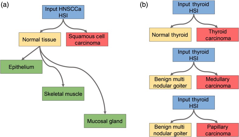
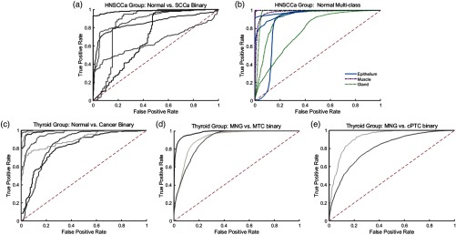
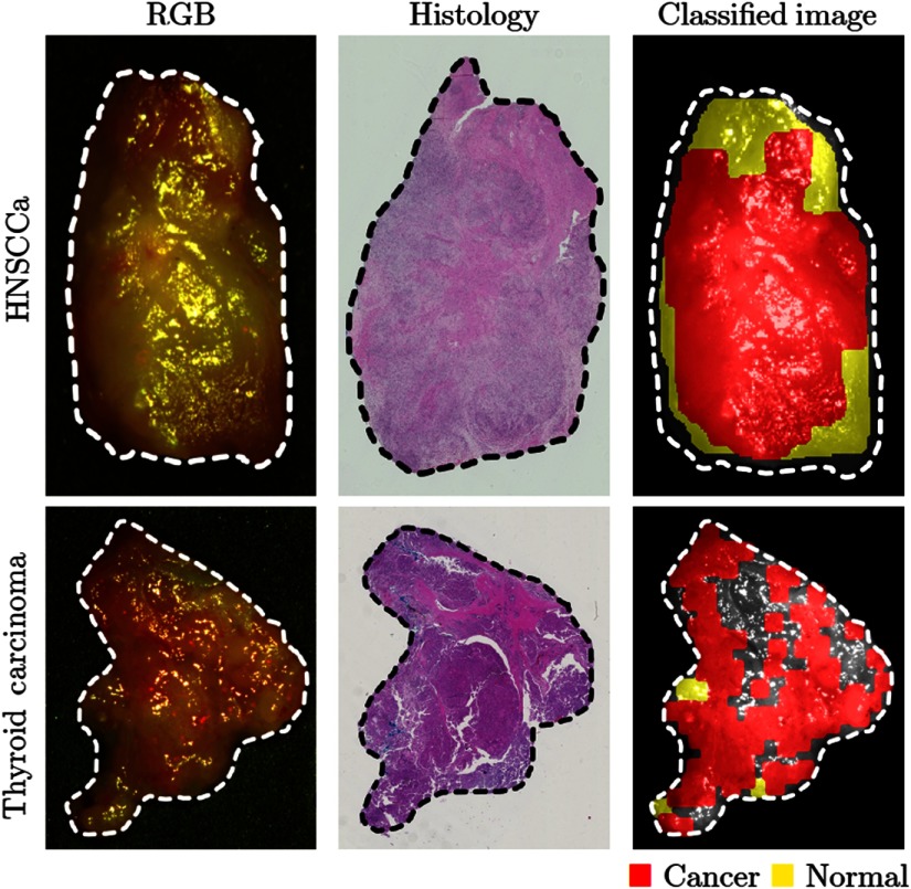
For patients undergoing surgical cancer resection of squamous cell carcinoma (SCCa), cancer-free surgical margins are essential for good prognosis. We developed a method to use hyperspectral imaging (HSI), a noncontact optical imaging modality, and convolutional neural networks (CNNs) to perform an optical biopsy of ex-vivo, surgical gross-tissue specimens, collected from 21 patients undergoing surgical cancer resection. Using a cross-validation paradigm with data from different patients, the CNN can distinguish SCCa from normal aerodigestive tract tissues with an area under the receiver operator curve (AUC) of 0.82. Additionally, normal tissue from the upper aerodigestive tract can be subclassified into squamous epithelium, muscle, and gland with an average AUC of 0.94. After separately training on thyroid tissue, the CNN can differentiate between thyroid carcinoma and normal thyroid with an AUC of 0.95, 92% accuracy, 92% sensitivity, and 92% specificity. Moreover, the CNN can discriminate medullary thyroid carcinoma from benign multinodular goiter (MNG) with an AUC of 0.93. Classical-type papillary thyroid carcinoma is differentiated from MNG with an AUC of 0.91. Our preliminary results demonstrate that an HSI-based optical biopsy method using CNNs can provide multicategory diagnostic information for normal and cancerous head-and-neck tissue, and more patient data are needed to fully investigate the potential and reliability of the proposed technique.

Keywords: classification; convolutional neural network; deep learning; head and neck cancer; hyperspectral imaging; optical biopsy; squamous cell carcinoma.


Figures

Fig. 1
Fig. 1
Tissue classification scheme. (a) For classification of the HNSCCa group, first a binary classification is considered to test the ability of the classifier to distinguish normal samples from SCCa samples. Next, histologically confirmed normal samples are subclassified as squamous epithelium, skeletal muscle, and mucosal salivary glands. (b) For classification of the thyroid group, first a binary classification is considered to test the ability of the classifier to distinguish normal thyroid samples from thyroid carcinoma of multiple types. In addition, thyroid HSI classification is tested to discriminate MNG from MTC and to discriminate MNG from classical-type PTC.
Fig. 2
Fig. 2
Normalized spectral signatures that were averaged between all patients of the classes of tissues that were included in this study. Presented by anatomical location: (a) normal tissue and SCCa of the upper aerodigestive tract, and (b) normal, benign, and carcinoma of the thyroid.
Fig. 3
Fig. 3
Modified inception module for use in the 2-D CNN architecture for classifying HSI of tissues from the upper aerodigestive tract.
Fig. 4
Fig. 4
CNN architectures implemented for classification of (a) HSI of thyroid tissue and (b) tissue from the upper aerodigestive tract.
Fig. 5
Fig. 5
Classification results of HSI as ROC curves for HNSCCa and thyroid experiments generated using leave-one-out cross validation. (a) Binary classification of SCCa and normal head-and-neck tissue; (b) multiclass subclassification of normal aerodigestive tract tissues; (c) binary classification of normal thyroid and thyroid carcinomas; (d) binary classification of MNG and MTC; (e) binary classification of MNG and classical PTC.
Fig. 6
Fig. 6
Representative results of binary cancer classification. (a) HSI-RGB composite images with cancer ROI outlined. (b) Respective histological gold standard with corresponding ROI outlined. (c) Artificially colored CNN classification results. True positive results representing correct cancer identification are visualized in red, and false negatives representing incorrect normal identification are shown in yellow. Tissue shown in grayscale represents tissue that is not classified due to the tissue surface containing glare pixels causing insufficient area to produce the necessary patch size for classification.
Fig. 7
Fig. 7
Representative results of subclassification of normal oral tissues. (a) HSI-RGB composites are shown with ROI of the tissue type outlined. (b) Respective histological gold standard with corresponding ROI outlined. (c) Artificially colored CNN classification results of the ROI only. True positive results representing correct tissue subtype are visualized in blue, and false negatives are shown in red. Tissue within the ROI that is shown in grayscale represents tissue that is not classified due to glare pixels or insufficient area to produce the necessary patch size.

References

    1. Vigneswaran N., Williams M. D., “Epidemiologic trends in head and neck cancer and aids in diagnosis,” Oral Maxillofac. Surg. Clin. N. Am. 26(2), 123–141 (2014).10.1016/j.coms.2014.01.001 - DOI - PMC - PubMed
    1. Ringash J., “Survivorship and quality of life in head and neck cancer,” J. Clin. Oncol. 33(29), 3322–3327 (2015).JCONDN10.1200/JCO.2015.61.4115 - DOI - PubMed
    1. Ajila V., et al. , “Human papilloma virus associated squamous cell carcinoma of the head and neck,” J. Sex Transm. Dis. 2015, 791024 (2015).10.1155/2015/791024 - DOI - PMC - PubMed
    1. Joseph L. J., et al. , “Racial disparities in squamous cell carcinoma of the oral tongue among women: a SEER data analysis,” Oral Oncol. 51(6), 586–592 (2015).EJCCER10.1016/j.oraloncology.2015.03.010 - DOI - PubMed
    1. Yao M., et al. , “Current surgical treatment of squamous cell carcinoma of the head and neck,” Oral Oncol. 43(3), 213–223 (2007).EJCCER10.1016/j.oraloncology.2006.04.013 - DOI - PubMed

Publication types

MeSH terms