. 2019 Jun 1;316(6):L1150-L1164.
doi: 10.1152/ajplung.00302.2018. Epub 2019 Mar 20.

Vascular TSP1-CD47 signaling promotes sickle cell-associated arterial vasculopathy and pulmonary hypertension in mice

Affiliations

Vascular TSP1-CD47 signaling promotes sickle cell-associated arterial vasculopathy and pulmonary hypertension in mice

Enrico M Novelli et al. Am J Physiol Lung Cell Mol Physiol. .

Abstract

Pulmonary hypertension (PH) is a leading cause of death in sickle cell disease (SCD) patients. Hemolysis and oxidative stress contribute to SCD-associated PH. We have reported that the protein thrombospondin-1 (TSP1) is elevated in the plasma of patients with SCD and, by interacting with its receptor CD47, limits vasodilation of distal pulmonary arteries ex vivo. We hypothesized that the TSP1-CD47 interaction may promote PH in SCD. We found that TSP1 and CD47 are upregulated in the lungs of Berkeley (BERK) sickling (Sickle) mice and patients with SCD-associated PH. We then generated chimeric animals by transplanting BERK bone marrow into C57BL/6J (n = 24) and CD47 knockout (CD47KO, n = 27) mice. Right ventricular (RV) pressure was lower in fully engrafted Sickle-to-CD47KO than Sickle-to-C57BL/6J chimeras, as shown by the reduced maximum RV pressure (P = 0.013) and mean pulmonary artery pressure (P = 0.020). The afterload of the sickle-to-CD47KO chimeras was also lower, as shown by the diminished pulmonary vascular resistance (P = 0.024) and RV effective arterial elastance (P = 0.052). On myography, aortic segments from Sickle-to-CD47KO chimeras showed improved relaxation to acetylcholine. We hypothesized that, in SCD, TSP1-CD47 signaling promotes PH, in part, by increasing reactive oxygen species (ROS) generation. In human pulmonary artery endothelial cells, treatment with TSP1 stimulated ROS generation, which was abrogated by CD47 blockade. Explanted lungs of CD47KO chimeras had less vascular congestion and a smaller oxidative footprint. Our results show that genetic absence of CD47 ameliorates SCD-associated PH, which may be due to decreased ROS levels. Modulation of TSP1-CD47 may provide a new molecular approach to the treatment of SCD-associated PH.

Keywords: oxidant stress; pulmonary hypertension; sickle cell disease; thrombospondin-1; transgenic mouse models.


Conflict of interest statement

J. S. Isenberg serves as Chair of the Scientific Advisory Board of Radiation Control Technologies, Inc. (RCTI, Garden City, NJ) and has equity interest in RCTI and Tioma Therapeutics (St. Louis, MO) that have licensed CD47 technology for development. M. T. Gladwin is a co-inventor on pending patent applications and planned patents directed to the use of recombinant neuroglobin and heme-based molecules as antidotes for CO poisoning, which have recently been licensed by Globin Solutions, Inc. M. T. Gladwin is a shareholder, advisor, and director in Globin Solutions, Inc. Additionally, and unrelated to CO poisoning, M. T. Gladwin is a co-inventor on patents directed to the use of nitrite salts in cardiovascular diseases, which have been licensed by United Therapeutics and Hope Pharmaceuticals, and is a co-investigator in a research collaboration with Bayer Pharmaceuticals to evaluate riociguate as a treatment for patients with SCD. None of the other authors has any conflicts of interest, financial or otherwise, to disclose.

Figures

Fig. 1.
Fig. 1.
Expression of thrombospondin 1 (TSP1) and CD47 in lungs of 4- and 9- to 11-mo-old Berkeley (BERK) sickling (Sickle) mice. A and B: Western blotting of lung lysates from 4-mo-old female C57BL/6J control (C57BL) and female Sickle mice and 9- to 11-mo-old female Sickle, BERK nonsickling hemizygous control (Hemi), C57BL, CD47 knockout (CD47KO), and TSP1 knockout (TSP1KO) mice (n = 20) for TSP1 and CD47. CD47KO and TSP1KO mice were used as positive and negative controls. Each lane represents an individual animal, and all data are shown. Densitometry results were normalized to β-actin. Values are means ± SE. Unpaired t-test was applied for comparison between each group.
Fig. 2.
Fig. 2.
Expression of thrombospondin 1 (TSP1) and its receptor CD47 in lungs of 13- to 14-mo-old Berkeley (BERK) sickling (Sickle) mice. Left: Western blots of lung lysates from 13- to 14-mo-old female Sickle, BERK nonsickling hemizygous control (Hemi), C57BL/6J control (C57BL), CD47 knockout (CD47KO), and TSP1 knockout (TSP1KO) mice (n = 26) for TSP1 and CD47. CD47KO and TSP1KO mice were used as positive and negative controls. Each lane represents an individual animal, and all data are shown. Pulmonary TSP1 and CD47 were significantly increased in Sickle compared with Hemi and C57BL mice. Right: densitometry results normalized to β-actin. Values are means ± SE. *P < 0.05, **P < 0.01, ***P < 0.001, ****P < 0.0001 (by one-way ANOVA with multiple comparisons).
Fig. 3.
Fig. 3.
Thrombospondin 1 (TSP1) and CD47 are upregulated in lungs of patients with sickle cell disease (SCD)-associated pulmonary hypertension (PH). A and B: TSP1 and CD47 expression was measured by immunofluorescence in lung sections from 6 patients with SCD-associated PH (SCD-PH) and 6 control patients without PH or overt lung disease. A: expression of TSP1 (red; von Willebrand factor stained green, and DAPI stained blue) was increased in patients with SCD-PH, although the signal appeared to originate from intraluminal red blood cells. B: increased levels of CD47 [red; platelet endothelial cell adhesion molecule stained green, and DAPI stained blue) were consistently observed in lung sections from all SCD-PH compared with control patients. ROI, region of interest. Scale bars = 50 µm. C: CD47 or TSP1 SCD-PH expression reported as percentage of control. Values are means ± SE. **P < 0.01 (by unpaired t-test).
Fig. 4.
Fig. 4.
Berkeley (BERK) sickling (Sickle) mice develop pulmonary hypertension (PH) and vascular dysfunction. Hemodynamic assessment of male 2- to 8-mo-old Sickle mice (n = 9) and age-matched C57BL/6J control (C57BL) mice (n = 6) was performed by open-chest right heart microcatheterization. A: right ventricular (RV) pressure (RVP) was measured by maximum RVP (RVPmax) and mean pulmonary artery pressure (mPAP). B: afterload measured by pulmonary vascular resistance (PVR) and RV effective arterial elastance (Ea). C: RV systolic function measured by the contractility index [maximum change in pressure over time (dP/dtmax)/RVPmax]. D: RV diastolic function measured by minimum change in pressure over time (RV dP/dtmin), a measure of RV stiffness. E: pressure-volume relations of RV of 2 representative Sickle and C57BL mice. ESP, end-systolic pressure; ESV, end-systolic volume; EDV, end-diastolic volume. F: plasma thrombospondin 1 (TSP1) measured by ELISA at the time of euthanasia. G and H: vascular reactivity of isolated aortic segments of male age-matched Sickle (n = 6) and C57BL (n = 5) mice assessed on the myograph system in response to acetylcholine (ACh) and phenylephrine (PE). Values are means ± SE. Unpaired t-test was applied for comparison between the 2 groups of mice; 2-way ordinary ANOVA with Sidak’s multiple comparisons test was used for myograph data: *P < 0.05, **P < 0.01, ***P < 0.001, ****P < 0.0001.
Fig. 5.
Fig. 5.
Generation of chimeras and measurement of engraftment. The thrombospondin 1 (TSP1)-CD47 axis was interrogated in vivo by generation of chimeric animals with a sickle erythropoiesis on a CD47 knockout (CD47KO) background. Bone marrow was harvested from flushed femurs and tibias of adult Berkeley sickling (Sickle) mice, and whole bone marrow (5 × 10⁶ cells) was transplanted into age-matched, lethally myeloablated (10 Gy) 2-mo-old CD47KO (n = 27) and C57BL/6J control (C57BL, n = 24) mice, the background strain of CD47KO mice, by retroorbital sinus injection in 7 separate experiments. A: schematic representation of transplantation protocol. B: Kaplan-Meyer survival curve of transplanted chimeras. C: engraftment assessed by measurement of HbS percentage by HPLC followed by confirmatory capillary zone electrophoresis (CZE) in blood samples obtained at the time of euthanasia (4–6 mo after transplantation). Top left: CZE gel. Top right: human HbA and HbS in a C57BL recipient transplanted with Sickle bone marrow with mixed chimerism. Bottom: CZE of a C57BL recipient (left) and a Sickle mouse donor (right). CSFA, cerebrospinal fluid analysis. D: linear regression of engraftment and spleen size.
Fig. 6.
Fig. 6.
Absence of tissue-resident thrombospondin 1 (TSP1)-CD47 signaling improves pulmonary hemodynamics and arterial vasodilator responsiveness in chimeric mice with a sickle erythropoiesis. Full hemodynamic assessment of Sickle-to-CD47 knockout (CD47KO) and Sickle-to-C57BL chimeras was performed by open-chest right heart microcatheterization. A: right ventricular (RV) pressure (RVP), including maximum RVP (RVPmax) and mean pulmonary artery pressure (mPAP). B: afterload measured as pulmonary vascular resistance (PVR) and RV effective arterial elastance (Ea). C: RV systolic function measured by the contractility index [maximum change in pressure over time (dP/dtmax)/RVPmax]. D: RV diastolic function measured by minimum change in pressure over time (dP/dtmin). E: pressure-volume relations of the RV of 2 representative Sickle-to-C57BL and Sickle-to-CD47KO chimeras. F: plasma TSP1 measured by ELISA at the time of euthanasia. G and H: vascular reactivity of isolated aortic segments of Sickle-to-C57BL (n = 5) and Sickle-to-CD47KO (n = 13) chimeras assessed on the myograph system in response to acetylcholine (ACh) and phenylephrine (PE). Values are means ± SE. Unpaired t-test was applied for comparison between the 2 groups of mice; 2-way ordinary ANOVA with Sidak’s multiple-comparisons test was used for myograph data: *P < 0.05.
Fig. 7.
Fig. 7.
Thrombospondin 1 (TSP1) augments reactive oxygen species (ROS) in human pulmonary endothelial cells via CD47. A and B: commercially available human pulmonary artery endothelial cells (n = 1 donor) were treated with vehicle or TSP1 (0–10 nmol/l) for 60 min, and ROS production was measured using 2 independent complementary assays: O2•− generation by total cellular homogenates measured by cytochrome c reduction (A) and endothelial cell homogenate H2O2 production measured using the Amplex red assay (B). Values are means ± SE of 3 experiments. **P < 0.01 (by one-way ANOVA followed by Sidak’s multiple-comparisons test). C: commercially available human pulmonary artery endothelial cells (n = 2 donors) were established in a 96-well plate and directly exposed to the following treatment for 60 min: vehicle, TSP-1 (10 nM, 2.2 nM, or 0.2 nM), or 2.2 nM TSP-1 + 2 µg/ml CD47 blocking antibody (clone B6H12.2). Select wells received 1,000 U/ml bovine liver catalase to act as a negative control. Coumarin boronic acid probe detection of H2O2 production in the cells was measured kinetically for 2 h. Average rate of fluorescence generation was normalized to vehicle control and displayed as fold change in H2O2 production. RFU, relative fluorescence units. Values are means ± SE of 3–4 experiments per donor (total n = 6–7). *P < 0.05, **P < 0.01, ****P < 0.0001 (by unpaired t-test).
Fig. 8.
Fig. 8.
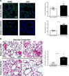
Thrombospondin 1 (TSP1) augments pulmonary oxidative damage and promotes vascular congestion in chimeric mice with a sickle erythropoiesis. A: lung tissue sections from chimeras were stained for 4-hydroxynonenal (4-HNE) or 3-nitrotyrosine (3-NT) and examined by immunofluorescence to determine whether in vivo sickle cell disease-mediated pulmonary reactive oxygen species production (4-HNE) and secondary reactive oxygen species-mediated protein (3-NT) were modified by the absence of TSP1-CD47 signaling. 3-NT is shown as green fluorescence; DAPI stained blue (n = 3 per group, 2 sections per animal). Representative 4-HNE deposition is also shown by green fluorescent immunohistochemical staining; DAPI stained blue (n = 2 mice per group, 3 sections per animal). Fluorescence signal was quantified using ImageJ software and reported as 3-NT or 4-HNE intensity per cell (DAPI signal). Vascular congestion on hematoxylin-eosin-stained slides from chimeras (n = 3 mice per group) was scored by 3 blinded, independent readers, who used a semiquantitative, relative scale of 0–4, where 0 = absence of red blood cells in the lumens of pulmonary blood vessels. BERK, Berkeley; CD47KO, CD47 knockout. B: increasing lumen congestion and number of affected vessels scored on a scale of 1–4. Images were taken using a Nikon A1 confocal microscope or Nikon 90i upright microscope at ×20. Scale bars = 50 µm. Results are shown as representative slides. Values are means ± SE. *P < 0.05, ****P < 0.0001 (by unpaired t-test).

References

    1. Al Ghouleh I, Meijles DN, Mutchler S, Zhang Q, Sahoo S, Gorelova A, Henrich Amaral J, Rodríguez AI, Mamonova T, Song GJ, Bisello A, Friedman PA, Cifuentes-Pagano ME, Pagano PJ. Binding of EBP50 to Nox organizing subunit p47phox is pivotal to cellular reactive species generation and altered vascular phenotype. Proc Natl Acad Sci USA 113: E5308–E5317, 2016. doi:10.1073/pnas.1514161113. - DOI - PMC - PubMed
    1. Aslan M, Freeman BA. Oxidases and oxygenases in regulation of vascular nitric oxide signaling and inflammatory responses. Immunol Res 26: 107–118, 2002. doi:10.1385/IR:26:1-3:107. - DOI - PubMed
    1. Aslan M, Ryan TM, Adler B, Townes TM, Parks DA, Thompson JA, Tousson A, Gladwin MT, Patel RP, Tarpey MM, Batinic-Haberle I, White CR, Freeman BA. Oxygen radical inhibition of nitric oxide-dependent vascular function in sickle cell disease. Proc Natl Acad Sci USA 98: 15215–15220, 2001. doi:10.1073/pnas.221292098. - DOI - PMC - PubMed
    1. Aslan M, Thornley-Brown D, Freeman BA. Reactive species in sickle cell disease. Ann NY Acad Sci 899: 375–391, 2000. doi:10.1111/j.1749-6632.2000.tb06201.x. - DOI - PubMed
    1. Bakeer N, James J, Roy S, Wansapura J, Shanmukhappa SK, Lorenz JN, Osinska H, Backer K, Huby AC, Shrestha A, Niss O, Fleck R, Quinn CT, Taylor MD, Purevjav E, Aronow BJ, Towbin JA, Malik P. Sickle cell anemia mice develop a unique cardiomyopathy with restrictive physiology. Proc Natl Acad Sci USA 113: E5182–E5191, 2016. doi:10.1073/pnas.1600311113. - DOI - PMC - PubMed

MeSH terms