. 2019 Mar;8(2):10.1128/ecosalplus.ESP-0037-2018.
doi: 10.1128/ecosalplus.ESP-0037-2018.

Curli Biogenesis: Bacterial Amyloid Assembly by the Type VIII Secretion Pathway

Affiliations

Curli Biogenesis: Bacterial Amyloid Assembly by the Type VIII Secretion Pathway

Sujeet Bhoite et al. EcoSal Plus. 2019 Mar.

Abstract

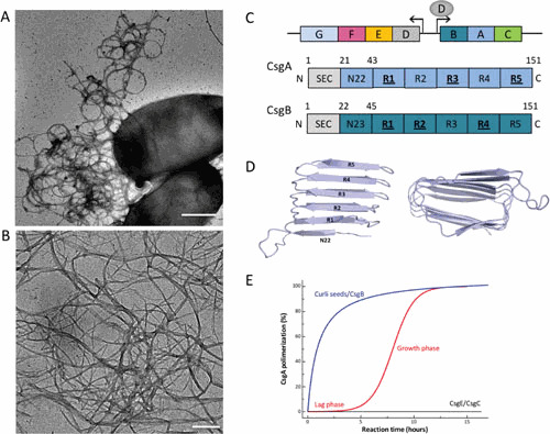
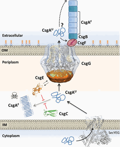
In 1989, Normark and coworkers reported on fibrous surface structures called curli on strains of Escherichia coli that were suspected of causing bovine mastitis. Subsequent work by many groups has revealed an elegant and highly regulated curli biogenesis pathway also referred to as the type VIII secretion system. Curli biogenesis is governed by two divergently transcribed operons, csgBAC and csgDEFG. The csgBAC operon encodes the structural subunits of curli, CsgA and CsgB, along with a chaperone-like protein, CsgC. The csgDEFG operon encodes the accessory proteins required for efficient transcription, secretion, and assembly of the curli fiber. CsgA and CsgB are secreted as largely unstructured proteins and transition to β-rich structures that aggregate into regular fibers at the cell surface. Since both of these proteins have been shown to be amyloidogenic in nature, the correct spatiotemporal synthesis of the curli fiber is of paramount importance for proper functioning and viability. Gram-negative bacteria have evolved an elegant machinery for the safe handling, secretion, and extracellular assembly of these amyloidogenic proteins.


Figures

Figure 1
Figure 1
Curli composition and structure. (A and B) Transmission electron micrographs of individual E. coli cells producing curli fibers (A) and curli-like fibers grown in vitro from purified CsgA (B). Scale bars: 500 nm and 200 nm, respectively. (C) Schematic organization of the csgDEFG and csgBAC curli gene clusters and architecture of the curli subunits CsgA (blue) and CsgB (dark blue). Subunits comprise an N-terminal signal sequence (SEC) that is cleaved upon export into the periplasm. The mature proteins contain curlin pseudorepeat regions (N22, R1 to R5) that guide substrate specificity in the secretion pathway and form the amyloidogenic core of the curli subunits. Repeats that efficiently self-polymerize in vitro are underscored. (D) Theoretical model of CsgA predicted based on amino acid covariation analysis (42). The predictions point to a right- or left-handed β-helix made up from stacked curlin repeats (labeled R1 to R5). (E) Representation of typical in vitro CsgA polymerization profiles in the absence (red) or presence (blue) of preformed fibers or the CsgB nucleator. In the presence of CsgE (1:1 ratio) or CsgC (1:500 ratio), no CsgA polymerization is observed (black curve).
Figure 2
Figure 2
Integrated model for curli subunit secretion. Curli subunits enter the periplasm via the SecYEG translocon, from where they progress to the cell surface as unfolded polypeptides via the curli transporter CsgG. Premature folding and polymerization of CsgA in the periplasm (right dotted line) are inhibited by CsgE and CsgC. CsgE binds and targets subunits to the secretion channel, while CsgC provides a safeguard against runaway polymerization, likely by the binding and neutralization of early assembly intermediates and/or nascent fibers. CsgG forms a nonameric complex that acts as a peptide diffusion channel and cooperates with the periplasmic factor CsgE, which binds the channel and forms a capping structure to the secretion complex. Recruitment and (partial) enclosure of CsgA in the secretion complex are proposed to create an entropy gradient over the channel that favors CsgA’s outward diffusion as an unfolded, soluble polypeptide. Once secreted, curli fiber formation and elongation are templated by CsgB, in a CsgF-dependent manner. CsgF is likely to be in contact or close proximity to the CsgG channel. The exact role of CsgF and whether fibers extend from the proximal or distal end (dashed arrows) are presently unknown. Abbreviations: IM, inner membrane; OM, outer membrane.

References

    1. Olsén A, Jonsson A, Normark S. 1989. Fibronectin binding mediated by a novel class of surface organelles on Escherichia coli. Nature 338:652–655. 10.1038/338652a0. [PubMed] - DOI - PubMed
    1. Dueholm MS, Albertsen M, Otzen D, Nielsen PH. 2012. Curli functional amyloid systems are phylogenetically widespread and display large diversity in operon and protein structure. PLoS One 7:e51274. 10.1371/journal.pone.0051274. [PubMed] - DOI - PMC - PubMed
    1. Barnhart MM, Chapman MR. 2006. Curli biogenesis and function. Annu Rev Microbiol 60:131–147. 10.1146/annurev.micro.60.080805.142106. [PubMed] - DOI - PMC - PubMed
    1. Collinson SK, Doig PC, Doran JL, Clouthier S, Trust TJ, Kay WW. 1993. Thin, aggregative fimbriae mediate binding of Salmonella enteritidis to fibronectin. J Bacteriol 175:12–18. 10.1128/jb.175.1.12-18.1993. [PubMed] - DOI - PMC - PubMed
    1. Römling U, Sierralta WD, Eriksson K, Normark S. 1998. Multicellular and aggregative behaviour of Salmonella typhimurium strains is controlled by mutations in the agfD promoter. Mol Microbiol 28:249–264. 10.1046/j.1365-2958.1998.00791.x. [PubMed] - DOI - PubMed

Publication types

MeSH terms