

Apoptotic bodies from endplate chondrocytes enhance the oxidative stress-induced mineralization by regulating PPi metabolism
- PMID: 30892812
- PMCID: PMC6484318
- DOI: 10.1111/jcmm.14268
Apoptotic bodies from endplate chondrocytes enhance the oxidative stress-induced mineralization by regulating PPi metabolism
Abstract
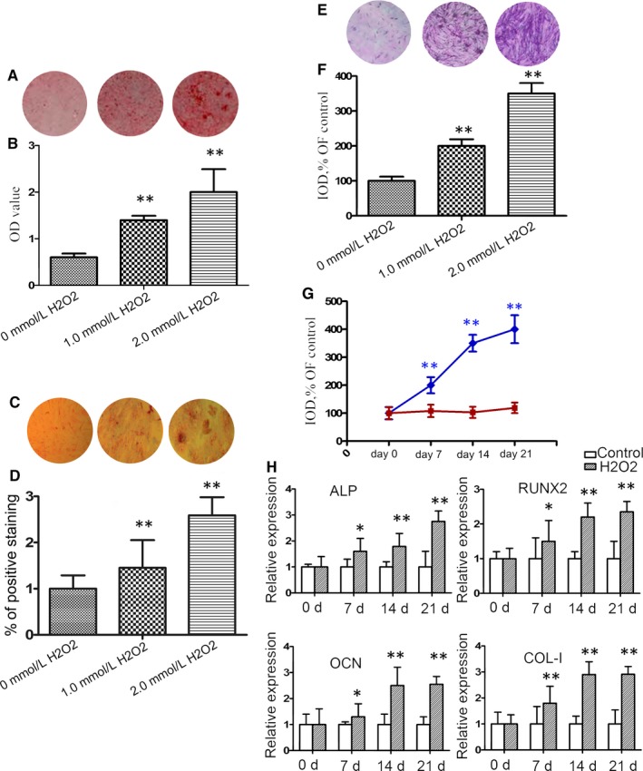
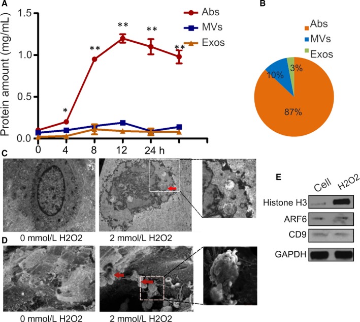
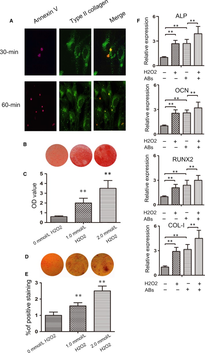
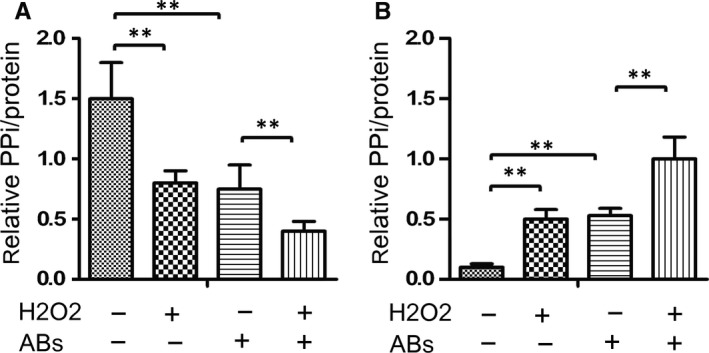
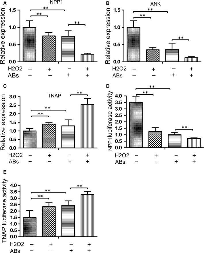
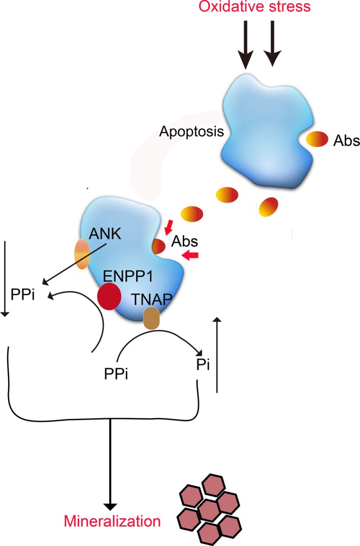
This study aimed to investigate the role of apoptotic bodies (Abs) from the oxidative stressed endplate chondrocytes in regulating mineralization and potential mechanisms. Endplate chondrocytes were isolated from rats and treated with H2O2 to induce oxidative stress. The calcium deposition for matrix mineralization in the cells was examined by histological staining. The expression levels of calcification-related genes in individual groups of cells were determined by quantitative real time-PCR (qRT-PCR). Subsequently, extracellular vesicles (EVs) were purified and characterized. The effect of treatment with H2O2 and/or Abs on the mineralization, extracellular PPi metabolism and related gene expression were determined. Oxidative stress significantly increased the mineralization and promoted the generation of main Abs from endplate chondrocytes. Abs were effectively endocytosed by endplate chondrocytes and co-localized with collagen (COL)-II in the cytoplasm, which enhanced the mineralization, alkaline phosphatase (ALP), osteocalcin (OCN), Runt-related transcription factor 2 (RUNX2) and COL-I expression in endplate chondrocytes. Furthermore, treatment either H2O2 or Abs significantly decreased PPi, but increased Pi production and treatment with both further enhancing the changes in endplate chondrocytes. Similarly, treatment either H2O2 or Abs significantly decreased the ectonucleotide pyrophosphatase/phosphodiesterase 1 (ENPP1), and ankylosis protein (ANK) expression and ENPP1 promoter activity, but increased the tissue-nonspecific alkaline phosphatase (TNAP) expression and TNAP promoter activity in endplate chondrocytes. Oxidative stress promoted the generation of Abs, which might enhance the oxidative stress-mediated mineralization in endplate chondrocytes by regulating the PPi metabolism.
Keywords: apoptotic bodies; endplate chondrocytes; intervertebral disc degeneration; mineralization.
© 2019 The Authors. Journal of Cellular and Molecular Medicine published by John Wiley & Sons Ltd and Foundation for Cellular and Molecular Medicine.
Conflict of interest statement
The auths declare that there are no conflicts of interest associated with this study.
Figures
Similar articles
-
[Expression and significance of ENPP1, TNAP and ANK proteins in the degeneration of endplate chondrocytes in rats].Zhonghua Yi Xue Za Zhi. 2011 Jan 18;91(3):189-92. Zhonghua Yi Xue Za Zhi. 2011. PMID: 21418901 Chinese.
-
Effects of mechanical strain on ANK, ENPP1 and TGF-β1 expression in rat endplate chondrocytes in vitro.Mol Med Rep. 2011 Sep-Oct;4(5):831-5. doi: 10.3892/mmr.2011.508. Epub 2011 Jun 14. Mol Med Rep. 2011. PMID: 21674130
-
Activation of nuclear factor-kappa B by TNF promotes nucleus pulposus mineralization through inhibition of ANKH and ENPP1.Sci Rep. 2021 Apr 15;11(1):8271. doi: 10.1038/s41598-021-87665-2. Sci Rep. 2021. PMID: 33859255 Free PMC article.
-
The role of phosphatases in the initiation of skeletal mineralization.Calcif Tissue Int. 2013 Oct;93(4):299-306. doi: 10.1007/s00223-012-9672-8. Epub 2012 Nov 27. Calcif Tissue Int. 2013. PMID: 23183786 Free PMC article. Review.
-
Inorganic pyrophosphate (PPI) in pathologic calcification of articular cartilage.Front Biosci. 2005 Jan 1;10:988-97. doi: 10.2741/1593. Print 2005 Jan 1. Front Biosci. 2005. PMID: 15569637 Review.
Cited by
-
Mechanisms and clinical implications of intervertebral disc calcification.Nat Rev Rheumatol. 2022 Jun;18(6):352-362. doi: 10.1038/s41584-022-00783-7. Epub 2022 May 9. Nat Rev Rheumatol. 2022. PMID: 35534553 Free PMC article. Review.
-
The pathogenesis and targeted therapies of intervertebral disc degeneration induced by cartilage endplate inflammation.Front Cell Dev Biol. 2024 Dec 2;12:1492870. doi: 10.3389/fcell.2024.1492870. eCollection 2024. Front Cell Dev Biol. 2024. PMID: 39687521 Free PMC article. Review.
-
Achilles' Heel-The Significance of Maintaining Microenvironmental Homeostasis in the Nucleus Pulposus for Intervertebral Discs.Int J Mol Sci. 2023 Nov 22;24(23):16592. doi: 10.3390/ijms242316592. Int J Mol Sci. 2023. PMID: 38068915 Free PMC article. Review.
-
Col9a2 gene deletion accelerates the degeneration of intervertebral discs.Exp Ther Med. 2022 Mar;23(3):207. doi: 10.3892/etm.2022.11130. Epub 2022 Jan 7. Exp Ther Med. 2022. PMID: 35126710 Free PMC article.
-
Roles of organokines in intervertebral disc homeostasis and degeneration.Front Endocrinol (Lausanne). 2024 Mar 12;15:1340625. doi: 10.3389/fendo.2024.1340625. eCollection 2024. Front Endocrinol (Lausanne). 2024. PMID: 38532900 Free PMC article. Review.
References
-
- Borenstein DG. Epidemiology, etiology, diagnostic evaluation, and treatment of low back pain. Curr Opin Rheumatol. 2001;13(2):128‐134. - PubMed
-
- Kelsey JL, White AA III. Epidemiology and impact of low‐back pain. Spine (Phila Pa 1976). 1980;5(2):133‐142. - PubMed
-
- Grunhagen T, Shirazi‐Adl A, Fairbank JC, Urban JP. Intervertebral disk nutrition: a review of factors influencing concentrations of nutrients and metabolites. Orthop Clin North Am. 2011;42(4):465‐477, vii. - PubMed
Publication types
MeSH terms
Substances
LinkOut - more resources
Full Text Sources
Research Materials
Miscellaneous
