. 2019 May 1;316(5):R651-R665.
doi: 10.1152/ajpregu.00393.2018. Epub 2019 Mar 20.

Angiotensin type 1a receptors in the median preoptic nucleus support intermittent hypoxia-induced hypertension

Affiliations

Angiotensin type 1a receptors in the median preoptic nucleus support intermittent hypoxia-induced hypertension

Brent Shell et al. Am J Physiol Regul Integr Comp Physiol. .

Abstract

Chronic intermittent hypoxia (CIH) is a model of the hypoxemia from sleep apnea that causes a sustained increase in blood pressure. Inhibition of the central renin-angiotensin system or FosB in the median preoptic nucleus (MnPO) prevents the sustained hypertensive response to CIH. We tested the hypothesis that angiotensin type 1a (AT1a) receptors in the MnPO, which are upregulated by CIH, contribute to this hypertension. In preliminary experiments, retrograde tract tracing studies showed AT1a receptor expression in MnPO neurons projecting to the paraventricular nucleus. Adult male rats were exposed to 7 days of intermittent hypoxia (cycling between 21% and 10% O2 every 6 min, 8 h/day during light phase). Seven days of CIH was associated with a FosB-dependent increase in AT1a receptor mRNA without changes in the permeability of the blood-brain barrier in the MnPO. Separate groups of rats were injected in the MnPO with an adeno-associated virus containing short hairpin (sh)RNA against AT1a receptors to test their role in intermittent hypoxia hypertension. Injections of shRNA against AT1a in MnPO blocked the increase in mRNA associated with CIH, prevented the sustained component of the hypertension during normoxia, and reduced circulating advanced oxidation protein products, an indicator of oxidative stress. Rats injected with shRNA against AT1a and exposed to CIH had less FosB staining in MnPO and the rostral ventrolateral medulla after intermittent hypoxia than rats injected with the control vector that were exposed to CIH. Our results indicate AT1a receptors in the MnPO contribute to the sustained blood pressure increase to intermittent hypoxia.

Keywords: angiotensin; hypertension; sleep apnea.


Conflict of interest statement

No conflicts of interest, financial or otherwise, are declared by the authors.

Figures
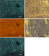
Fig. 1.
Fig. 1.
Expression of angiotensin type 1a (AT1a) receptor in median preoptic nucleus (MnPO) neurons that project to the paraventricular nucleus (PVN). A: digital image of neurons on the dorsal MnPO retrogradely labeled with FluoroGold injected in PVN. B: the same section showing in situ hybridization for the AT1a receptor in the dorsal MnPO. C: digitally merged image of A and B, with high-magnification inset showing retrograde labeling of a neuron that is positive for AT1a receptor. D: representative image showing expression of AT1a mRNA (black punctate dots) and the presence of astrocytes [yellow, glia fibrillary acid protein-positive cells] in MnPO. Digital image of dorsal MnPO with high-magnification inset showing that hybridization signal is not present in astrocytes. E: digital image of colocalization in ventral MnPO. Magenta arrow, astrocyte positive for AT1a mRNA; red arrows, astrocytes with adjacent AT1a mRNA signals; blue arrows, astrocytes that lack AT1a mRNA.
Fig. 2.
Fig. 2.
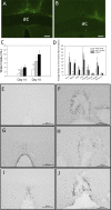
Median preoptic nucleus (MnPO) knockdown of angiotensin type 1a (AT1a) receptors and responses to intracerebroventricular ANG II. AAV, adeno-associated virus; sh, short hairpin RNA. A: digital image of green fluorescent protein (GFP) staining in MnPO from AAV-shAT1a injection. B: image from a different subject showing the same vector injected lateral to the MnPO. ac, anterior commissure. C: drinking responses to ANG II (2 ng/1 µl icv) in rats injected with AAV-shAT1a that included the MnPO (open bars, n = 9), AAV-shAT1a injections that missed (shAT1a Miss) MnPO (gray bars, n = 7), and rats injected in MnPO with AAV-shRNA-scrambled (SCR; black bars, n = 7). *Significantly different from all other groups. D: summary of effects of AT1a knockdown in MnPO on cFos staining in the organum vasculosum lamina terminalis (OVLT; n = 5–8), MnPO (n = 7–8), supraoptic nucleus (SON; n = 7–8), paraventricular nucleus (PVN; n = 7–8), and subregions of the PVN (n = 7–8). *Significantly different from all other groups [Student-Newman-Keuls (SNK), P < 0.05; †significantly different from shAT1a Miss group (SNK, P < 0.05)]. EJ: digital images of cFos staining in MnPO (right column) and OVLT (left column) in rats from the AAV-shAT1a group (E and F), AAV-shAT1a Miss group (G and H), and rats injected in MnPO with AAV-shSCR (I and J).
Fig. 3.
Fig. 3.
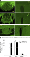
A: digital images of IgG staining in subfornical organ (SFO; left) and median preoptic nucleus (MnPO; right) from unstained controls (bottom), normoxic control (middle), and a rat exposed to chronic intermittent hypoxia (CIH; top). B: calculated integrated density of IgG staining in organum vasculosum lamina terminalis (OVLT), SFO, and MnPO from normoxic control rats and rats exposed to CIH. There were no differences in the density of IgG staining between normoxia (n = 6) and CIH (n = 6) for any of the 3 regions (all unpaired t tests).
Fig. 4.
Fig. 4.
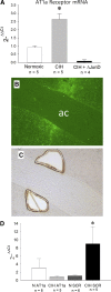
A: virally mediated expression of the ΔJunD dominant negative construct prevents elevation in angiotensin type 1a (AT1a) receptor message vs. controls and chronic intermittent hypoxia (CIH) animals (*P < 0.05). B: representative image of virally induced green fluorescent protein (GFP) expression in dorsal and ventral median preoptic nucleus (MnPO) Hit. ac, anterior commissure. C: the same section from B shown in bright field after removal of dorsal and ventral MnPO by laser capture microdissection. D: injections of shAT1a prevent the increase of AT1a expression after 7 days of CIH. qRT-PCR from laser-captured MnPOs demonstrate ability of shAT1a to prevent an increase in AT1a receptor message after 7 days of CIH vs. CIH scramble (SCR) animals (*P < 0.05). Expression of AT1a in CIH shAT1a was comparable to that in normoxic (N) counterparts (P > 0.05).
Fig. 5.
Fig. 5.
Knockdown of angiotensin type 1a (AT1a) receptors in median preoptic nucleus (MnPO) prevents the sustained component of hypertension from chronic intermittent hypoxia (CIH). A: changes in mean arterial pressure (MAP) recording from 0800 to 1600 h during light phase when rats were exposed to CIH or room air. AAV, adeno-associated virus; sh, small hairpin RNA. CIH scramble (CIH SCR) and AAV-AT1a not including MnPO (CIH shAT1a Miss) animals had significantly elevated blood pressure during CIH vs. normoxic (Norm) controls (*P < 0.05). AAV-shAT1a rats exposed to CIH were not significantly different from the other CIH groups. BL, baseline; IH, intermittent hypoxia. B: changes in MAP from the same rats recorded during dark phase from 1800 to 0600. During dark phase, increases in MAP of AAV-shSCR-injected and AAV-shAT1a Miss groups exposed to CIH were significantly greater than the 2 normoxic groups or (*P < 0.05) the CIH AAV-AT1a group (†P < 0.05). Symbols represent the following groups: ●, rats injected in MnPO with AAV-shAT1a and exposed to CIH (CIH shAT1a, n = 7); ○, CIH-treated rats with AAV-shAT1a injections that did not include MnPO (CIH AT1a Miss, n = 13); ▲, CIH-treated rats injected in MnPO with AAV-SCR (CIH SCR, n = 7), △, rats exposed to normoxia injected in MnPO with AAV-AT1a (Norm shAT1a, n = 8); ■, rats exposed to normoxia and injected in MnPO with AAV-SCR (Norm SCR, n = 8). C: average daily increases in MAP in CIH-treated AAV-shSCR injected group (CIH SCR), and CIH treated shAT1a Miss group (CIH AT1a Miss) were significantly greater than in normoxic groups [Student-Newman-Keuls (SNK), †P < 0.05] but not different from CIH-treated rats injected in MnPO with AAV-AT1a (CHI shAT1a). D: changes in MAP during dark phase from 1600 to 0800 h, when all rats were exposed to room air. Increased MAP from CIH SCR and CIH AT1a Miss groups were significantly greater that the other 3 groups (SNK, *P < 0.05). Numbers of rats are the same as in A and B.
Fig. 6.
Fig. 6.
Daily changes from baseline in heart rate (HR) and respiratory rate (RR). A: changes in HR recorded from 0800 to 1600 h when the rats were exposed to intermittent hypoxia (IH) during the light phase. There were no differences in HR among the groups. B: changes in HR recording during the normoxic (Norm) dark phase from 1800 to 0600 h. There was a significant difference between chronic intermittent hypoxia (CIH) shAT1a animals and their normoxic counterparts during normoxic dark phase (‡P > 0.05). C and D: changes in RR recorded during 0800–1600 when the rats were exposed to IH (C) and from 1800–0600 during the normoxic dark phase (D). There were no differences among the groups. AT1a, angiotensin type 1a; sh, short hairpin RNA; Miss, not including ventral median preoptic nucleus (MnPO); SCR, scramble. Norm shAT1a, n = 8; CIH shAT1a, n = 7; CIH shAT1a Miss, n = 13; Norm SCR, n = 8; CIH SCR, n = 7. Symbols and abbreviations represent the same groups as described in Fig. 5.
Fig. 7.
Fig. 7.
AD: representative examples of FosB/ΔFosB staining in ventral median preoptic nucleus (MnPO). AT1a, angiotensin type 1a; norm, normoxic; sh, short hairpin RNA; CIH, chronic intermittent hypoxia; AAV, adeno-associated virus; SCR, scramble. A: normoxic control injected with AAV-shAT1a. B: CIH and AAV-shAT1a injected. C: normoxic control injected with AAV-SCR. D: CIH and injected with AAV-SCR. E: summary data showing average FosB staining in the organum vasculosum of the lamina terminalis (OVLT), MnPO, and supraoptic nucleus (SON) in normoxic control injected with AAV-shAT1a (Norm shAT1a, open bars; n = 4–6), CIH and AAV-shAT1a injected (CIH shAT1a, light gray bars; n = 4–8), normoxic control injected with AAV-SCR (Norm SCR, dark gray bars; n = 6–7), and CIH and injected with AAV-SCR (CIH SCR, black bars; n = 6–7). No significant effects were observed for the SON. FI: representative examples of FosB/ΔFosB staining in paraventricular nucleus (PVN). A: normoxic control injected with AAV-shAT1a. G: CIH and AAV-shAT1a injected. H: normoxic control injected with AAV-SCR. I: CIH and injected with AAV-SCR. J: summary of FosB/ΔFosB staining in total PVN and specific subregions in normoxic control injected with AAV-shAT1a (Norm shAT1a, open bars; n = 6), CIH and AAV-shAT1a injected (CIH shAT1a, light gray bars; n = 8), normoxic control injected with AAV-SCR (Norm SCR, dark gray bars; n = 7), and CIH and injected with AAV-SCR (CIH SCR, black bars; n = 7). *Significantly different from both normoxic exposed groups (P < 0.05); †significantly different from all other groups for that area (P < 0.05). Subregions of PVN: PVN-DP, dorsal parvocellular; PVN-MP, medial parvocellular; PVN-VLP, ventrolateral parvocellular; PVN-PM, posterior magnocellular.
Fig. 8.
Fig. 8.
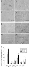
Effects of angiotensin type 1a (AT1a) knockdown in median preoptic nucleus (MnPO) on FosB/ΔFosB staining in hindbrain. Norm, normoxic; sh, short hairpin RNA; CIH, chronic intermittent hypoxia; AAV, adeno-associated virus; SCR, scramble. Representative images of FosB staining in the MnPO (AD) and subcommissural nucleus tractus solitarius (NTS; EH). Images are from A and E: normoxic control injected with AAV-shAT1a; B and F: CIH and AAV-shAT1a injected. C and G: normoxic control injected with AAV-SCR. D and H: CIH and injected with AAV-SCR. I: summary data of FosB/ΔFosB staining in NTS: precommissural NTS (NTS-PC), commissural NTS (NTS-C), subpostremal NTS (NTS-SP), caudal ventrolateral medulla (CVLM), and ventrolateral medulla (RVLM) in normoxic control injected with AAV-shAT1a (Norm shAT1a, open bars; n = 6), CIH and AAV-shAT1a injected (CIH shAT1a, light gray bars; n = 8), normoxic control injected with AAV-SCR (Norm SCR, dark gray bars; n = 7), and CIH and injected with AAV-SCR (CIH SCR, black bars; n = 7). *Significantly different from both normoxic groups (P < 0.05); †significantly different from all other groups (P < 0.05).
Fig. 9.
Fig. 9.
Effects of angiotensin type 1a (AT1a) knockdown in median preoptic nucleus (MnPO) on circulating advanced oxidation protein products (AOPP) in rats exposed to normoxia (Norm) or chronic intermittent hypoxia (CIH) after radio telemetry surgery alone (Uninj), or radio telemetry surgery followed by MnPO injections with AAV-SCR (shSCR) or adeno-associated virus (AAV)-shAT1a (shAT1a). *Significantly different from normoxic control group within the same surgical condition [Student-Newman-Keuls (SNK), P < 0.05]; +significantly lower than the other 2 normoxic groups (SNK, P < 0.05). Norm Uninjected, n = 5; CIH Uninjected, n = 6; Norm shSCR, n = 7; CIH shSCR, n = 9; Norm shAT1a, n = 6; CIH shAT1a, n = 6.

References

    1. Aihara N, Tanno H, Hall JJ, Pitts LH, Noble LJ. Immunocytochemical localization of immunoglobulins in the rat brain: relationship to the blood-brain barrier. J Comp Neurol 342: 481–496, 1994. doi:10.1002/cne.903420402. - DOI - PubMed
    1. Allen AM. Angiotensin AT1 receptor-mediated excitation of rat carotid body chemoreceptor afferent activity. J Physiol 510: 773–781, 1998. doi:10.1111/j.1469-7793.1998.773bj.x. - DOI - PMC - PubMed
    1. Allen AM, Moeller I, Jenkins TA, Zhuo J, Aldred GP, Chai SY, Mendelsohn FA. Angiotensin receptors in the nervous system. Brain Res Bull 47: 17–28, 1998. doi:10.1016/S0361-9230(98)00039-2. - DOI - PubMed
    1. Bathina CS, Rajulapati A, Franzke M, Yamamoto K, Cunningham JT, Mifflin S. Knockdown of tyrosine hydroxylase in the nucleus of the solitary tract reduces elevated blood pressure during chronic intermittent hypoxia. Am J Physiol Regul Integr Comp Physiol 305: R1031–R1039, 2013. doi:10.1152/ajpregu.00260.2013. - DOI - PMC - PubMed
    1. Biancardi VC, Son SJ, Ahmadi S, Filosa JA, Stern JE. Circulating angiotensin II gains access to the hypothalamus and brain stem during hypertension via breakdown of the blood-brain barrier. Hypertension 63: 572–579, 2014. doi:10.1161/HYPERTENSIONAHA.113.01743. - DOI - PMC - PubMed

Publication types

MeSH terms