

Radiological and clinical patterns of myeloid sarcoma
- PMID: 30893056
- PMCID: PMC6572499
- DOI: 10.2478/raon-2019-0014
Radiological and clinical patterns of myeloid sarcoma
Abstract
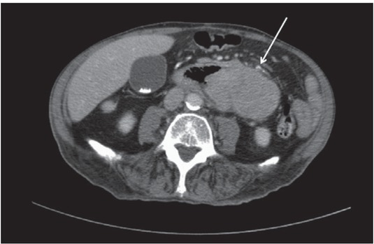
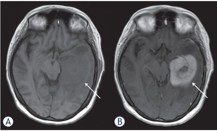
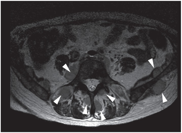
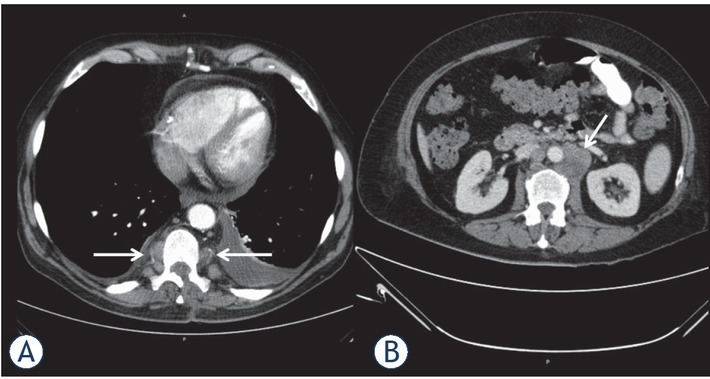
Background Myeloid sarcoma (MS), also known as granulocytic sarcoma or chloroma, is a solid tumor of extramedullary localization composed of malignant primitive myeloid cells. The purpose of the study was to identify clinical and imaging features in a large patient sample. Patients and methods Overall, 71 cases (34 females (47.9%) and 37 males (52.1%) with a median age of 56 (± 16 years) of histopathologically confirmed myeloid sarcoma were included into this study. The underlying hematological disease, occurrence, localizations and clinical symptoms as well as imaging features on computed tomography and magnetic resonance imaging were investigated. Results In 4 cases (5.63%) the manifestation of MS preceded the systemic hematological disease by a mean value of 3.8 ± 2.1 months. In 13 cases, first presentation of MS occurred simultaneously with the initial diagnosis of leukemia, and 51 patients presented MS after the initial diagnosis of the underlying malignancy with a mean latency of 39.8 ± 44.9 SD months. The visceral soft tissue was affected in 26 cases, followed by the cutis/subcutis was affected in 21 cases. Further localizations were bones (n = 13), central nervous system (n = 9), lymph nodes (n = 4) and visceral organs (n = 9). Conclusions MS is a rare complication of several hematological malignancies, predominantly of acute myeloid leukemia, which can affect any part of the body. In most cases it occurs after the diagnosis of the underlying malignancy, and affects frequently the cutis and subcutis.
Keywords: acute myeloid leukaemia; chloroma; granulocytic sarcoma; myeloid sarcoma.
Figures
References
-
- Liu PI, Ishimaru T, McGregor DH, Okada H, Steer Al. Autopsy study of granulocytic sarcoma (chloroma) in patients with myelogenous leukemia, Hiroshima-Nagasaki 1949-1969. Cancer. 1973;31:948. –. - PubMed
-
- Burns A. Observations of surgical anatomy in head and neck. London: Royce; 1811. p. 364.
-
- King A. A case of chloroma. Monthly J Med. 1853;17:17.
MeSH terms
LinkOut - more resources
Full Text Sources
Medical
