

Conserved Residues Control the T1R3-Specific Allosteric Signaling Pathway of the Mammalian Sweet-Taste Receptor
- PMID: 30893427
- PMCID: PMC6538948
- DOI: 10.1093/chemse/bjz015
Conserved Residues Control the T1R3-Specific Allosteric Signaling Pathway of the Mammalian Sweet-Taste Receptor
Erratum in
-
Corrigendum: Conserved Residues Control the T1R3-Specific Allosteric Signaling Pathway of the Mammalian Sweet-Taste Receptor.Chem Senses. 2019 Oct 17;44(8):649-650. doi: 10.1093/chemse/bjz038. Chem Senses. 2019. PMID: 31420679 Free PMC article. No abstract available.
Abstract
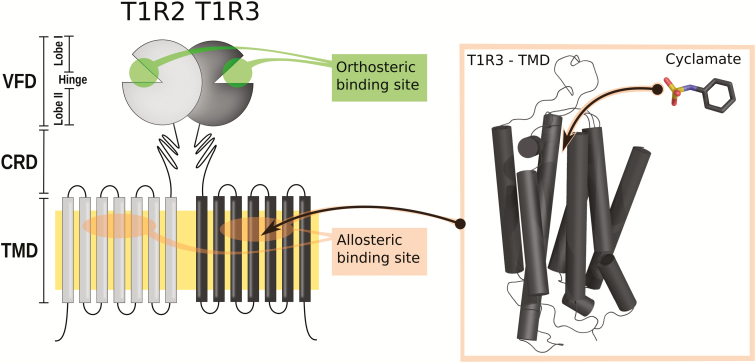
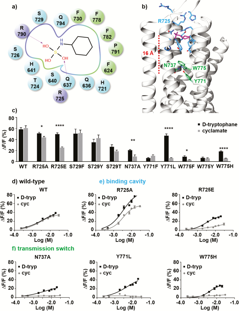
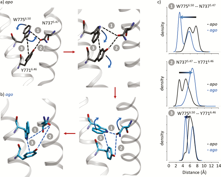
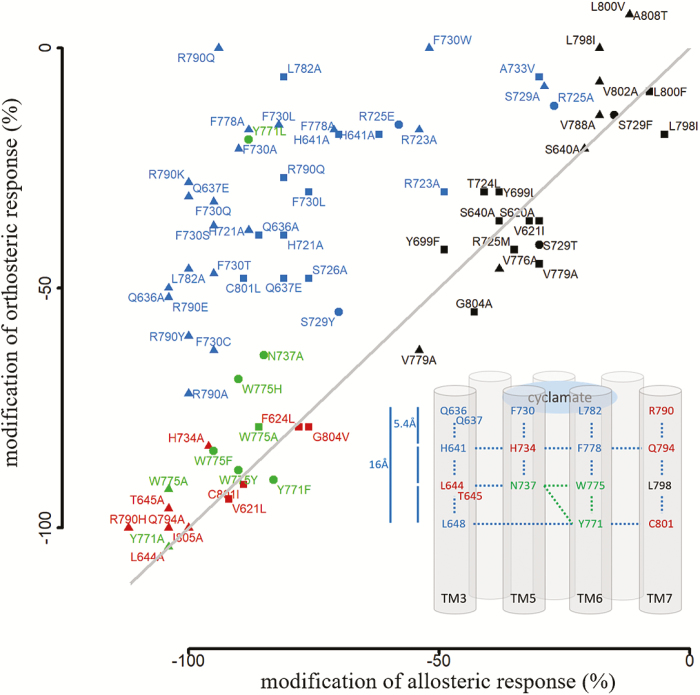
Mammalian sensory systems detect sweet taste through the activation of a single heteromeric T1R2/T1R3 receptor belonging to class C G-protein-coupled receptors. Allosteric ligands are known to interact within the transmembrane domain, yet a complete view of receptor activation remains elusive. By combining site-directed mutagenesis with computational modeling, we investigate the structure and dynamics of the allosteric binding pocket of the T1R3 sweet-taste receptor in its apo form, and in the presence of an allosteric ligand, cyclamate. A novel positively charged residue at the extracellular loop 2 is shown to interact with the ligand. Molecular dynamics simulations capture significant differences in the behavior of a network of conserved residues with and without cyclamate, although they do not directly interact with the allosteric ligand. Structural models show that they adopt alternate conformations, associated with a conformational change in the transmembrane region. Site-directed mutagenesis confirms that these residues are unequivocally involved in the receptor function and the allosteric signaling mechanism of the sweet-taste receptor. Similar to a large portion of the transmembrane domain, they are highly conserved among mammals, suggesting an activation mechanism that is evolutionarily conserved. This work provides a structural basis for describing the dynamics of the receptor, and for the rational design of new sweet-taste modulators.
Keywords: allosteric binding site; class C GPCR; cyclamate; mammalian; sweet-taste receptor; taste modulator.
© The Author(s) 2019. Published by Oxford University Press. All rights reserved. For permissions, please e-mail: journals.permissions@oup.com.
Figures
References
-
- Case DA, Babin V, Berryman JT, Betz RM, Cai Q, Cerutti DS, Cheatham TE, Darden TA, Duke RE, Gohlke H, et al. . 2014. Amber 14. San Francisco (CA): University of California.
-
- Chéron JB, Golebiowski J, Antonczak S, Fiorucci S. 2017. The anatomy of mammalian sweet taste receptors. Proteins. 85:332–341. - PubMed
-
- Doré AS, Okrasa K, Patel JC, Serrano-Vega M, Bennett K, Cooke RM, Errey JC, Jazayeri A, Khan S, Tehan B, et al. . 2014. Structure of class C GPCR metabotropic glutamate receptor 5 transmembrane domain. Nature. 511:557–562. - PubMed
-
- DuBois GE. 2016. Molecular mechanism of sweetness sensation. Physiol Behav. 164:453–463. - PubMed
Publication types
MeSH terms
Substances
LinkOut - more resources
Full Text Sources
