Review
. 2019 Jun 11;42(6):zsz073.
doi: 10.1093/sleep/zsz073.

Role of the L-PGDS-PGD2-DP1 receptor axis in sleep regulation and neurologic outcomes

Affiliations
Review

Role of the L-PGDS-PGD2-DP1 receptor axis in sleep regulation and neurologic outcomes

Abdullah Shafique Ahmad et al. Sleep. .

Abstract

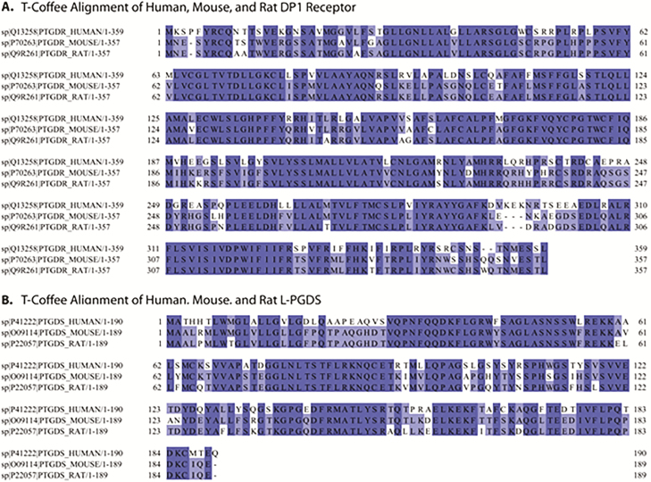
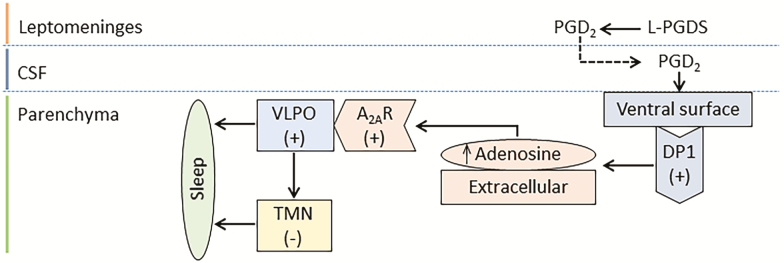
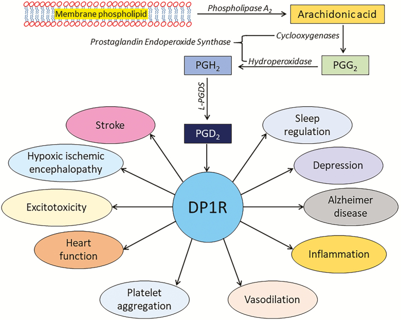
To meet the new challenges of modern lifestyles, we often compromise a good night's sleep. In preclinical models as well as in humans, a chronic lack of sleep is reported to be among the leading causes of various physiologic, psychologic, and neurocognitive deficits. Thus far, various endogenous mediators have been implicated in inter-regulatory networks that collectively influence the sleep-wake cycle. One such mediator is the lipocalin-type prostaglandin D2 synthase (L-PGDS)-Prostaglandin D2 (PGD2)-DP1 receptor (L-PGDS-PGD2-DP1R) axis. Findings in preclinical models confirm that DP1R are predominantly expressed in the sleep-regulating centers. This finding led to the discovery that the L-PGDS-PGD2-DP1R axis is involved in sleep regulation. Furthermore, we showed that the L-PGDS-PGD2-DP1R axis is beneficial in protecting the brain from ischemic stroke. Protein sequence homology was also performed, and it was found that L-PGDS and DP1R share a high degree of homology between humans and rodents. Based on the preclinical and clinical data thus far pertaining to the role of the L-PGDS-PGD2-DP1R axis in sleep regulation and neurologic conditions, there is optimism that this axis may have a high translational potential in human therapeutics. Therefore, here the focus is to review the regulation of the homeostatic component of the sleep process with a special focus on the L-PGDS-PGD2-DP1R axis and the consequences of sleep deprivation on health outcomes. Furthermore, we discuss whether the pharmacological regulation of this axis could represent a tool to prevent sleep disturbances and potentially improve outcomes, especially in patients with acute brain injuries.

Keywords: DP1 receptor; animal models; brain injuries; eicosanoids; inflammation; outcome assessments; poststroke sleep disturbance; prostaglandin D2; prostaglandin receptors; sleep apnea.


Figures

Figure 1.
Figure 1.
Electroencephalographic signature of sleep stages. This figure represents only EEG recordings with leads placed at different locations. All epochs demonstrate at least 15 s of EEG recording captured with high-pass filter at 1 Hz, low-pass filter at 70 Hz, sensitivity at 7 µV/mm, Notch filter “off,” paper speed of 30 mm/s, and displayed in longitudinal bipolar montage. (A) Drowsiness (N1). Note the roving eye movements (red box; generated due to the positive polarity of the cornea in relation to the retina) and vertex waves (red arrow). (B) Stage 2 (N2). Note the K complexes (red box) and sleep spindles (red arrows). (C) Slow-wave sleep (N3). Note the abundance of high-amplitude delta waveforms (red arrow) admixed with sleep architecture that resembles K complexes and spindles. (D) REM. Note the abundant rapid eye movement sharp waveforms generated by the difference in polarity between the cornea and the retina (red boxes). There is no EMG electrode displayed in this picture, but if it was there, one would see lack of fast activity generated by muscle tone during this sleep stage. The abbreviations on the left side of the recordings show the location. Fp1-F7/F7-T7/T7-P7/P7-O1: left fronto-temporal-parietal-occipital chains; Fp1-F3/F3-C3/C3-P3/P3-O1: left fronto-central-parietal-occipital chains; Fz-Cz/Cz-Pz: midline leads; Fp2-F4/F4-C4/C4-P4/P4-O2: right fronto-central-parietal-occipital chains; Fp2-F8/F8-T8/T8-P8/P8-O2: right fronto-temporal-parietal-occipital chains.
Figure 2.
Figure 2.
Multiple sequence alignment of human, mouse, and rat DP1R (A) and L-PGDS (B) using T-Coffee (version 11.00.d625267). Results were visualized using Jalview (version 2.10.3b1) and colored according to the percent identity among the proteins.
Figure 3.
Figure 3.
Role of L-PGDS-PGD2-DP1R signaling in sleep regulation: The sleep-regulating L-PGDS is abundantly present in leptomeninges and produces PGD2 in the arachnoid trabecular cells. The PGD2 then enters the CSF circulation and activates the DP1R on the ventral surface from the basal forebrain to the hypothalamus. This increases the extracellular level of adenosine which then activates the adenosine A2A receptors (A2AR). The activated A2AR excites the ventrolateral preoptic (VLPO) neurons and promote sleep initiation. Furthermore, the excited VLPO neurons subsequently send inhibitory signals to the wake-regulating histaminergic TMN. Thus, the activation of VLPO and inhibition of TMN area also results in sleep initiation through the “flip-flop” mechanism. The signs + or – were used to described activation or inhibition, respectively.
Figure 4.
Figure 4.
Roles of L-PGDS-PGD2-DP1R system reported in CNS: Phospholipase A2 (PLA2) acts on membrane phospholipids and generates arachidonic acid (AA). The AA is then converted into the intermediate PGG2 and PGH2 by the action of cyclooxygenase and hydroperoxidase, respectively (collectively known as prostaglandin endoperoxide synthase). The unstable PGH2 is then converted to prostaglandin D2 (PGD2) by lipocalin-type PGD2 synthase (L-PGDS). PGD2 then activates the DP1R, which then affects the outcomes of various physiologic and neurologic conditions. Please note that another type of PGDS, hematopoietic-type PGDS (H-PGDS), is also reported. Moreover, another receptor for PGD2, known as CRTH2 or DP2R, is also reported. However, it has been shown that DP2R is not needed for the action of PGD2, and DP2R receptor is on a different branch in the phylogenetic tree and has a different homology than DP1R. Therefore, because the focus of this review is on L-PGDS-PGD2-DP1R system, the H-PGDS and DP2R receptor are not shown in the illustration.

Similar articles

Cited by

References

    1. Blum ID, et al. . Time for bed: genetic mechanisms mediating the circadian regulation of sleep. Trends Genet. 2018;34(5):379–388. - PMC - PubMed
    1. Tononi G, et al. . Sleep and the price of plasticity: from synaptic and cellular homeostasis to memory consolidation and integration. Neuron. 2014;81(1):12–34. - PMC - PubMed
    1. Wang G, et al. . Synaptic plasticity in sleep: learning, homeostasis and disease. Trends Neurosci. 2011;34(9):452–463. - PMC - PubMed
    1. Saper CB, et al. . Sleep state switching. Neuron. 2010;68(6):1023–1042. - PMC - PubMed
    1. Brown RE, et al. . Control of sleep and wakefulness. Physiol Rev. 2012;92(3):1087–1187. - PMC - PubMed

Publication types