. 2019 Mar 19;26(12):3298-3312.e4.
doi: 10.1016/j.celrep.2019.02.080.

Ndfip Proteins Target Robo Receptors for Degradation and Allow Commissural Axons to Cross the Midline in the Developing Spinal Cord

Affiliations

Ndfip Proteins Target Robo Receptors for Degradation and Allow Commissural Axons to Cross the Midline in the Developing Spinal Cord

Madhavi Gorla et al. Cell Rep. .

Abstract

Commissural axons initially respond to attractive signals at the midline, but once they cross, they become sensitive to repulsive cues. This switch prevents axons from re-entering the midline. In insects and mammals, negative regulation of Roundabout (Robo) receptors prevents premature response to the midline repellant Slit. In Drosophila, the endosomal protein Commissureless (Comm) prevents Robo1 surface expression before midline crossing by diverting Robo1 into late endosomes. Notably, Comm is not conserved in vertebrates. We identified two Nedd-4-interacting proteins, Ndfip1 and Ndfip2, that act analogously to Comm to localize Robo1 to endosomes. Ndfip proteins recruit Nedd4 E3 ubiquitin ligases to promote Robo1 ubiquitylation and degradation. Ndfip proteins are expressed in commissural axons in the developing spinal cord and removal of Ndfip proteins results in increased Robo1 expression and reduced midline crossing. Our results define a conserved Robo1 intracellular sorting mechanism between flies and mammals to avoid premature responsiveness to Slit.

Keywords: Commissureless; E3 ubiquitin ligase; Ndfip; Nedd4; Robo; Slit; axon guidance; midline; repulsion; spinal cord.


Conflict of interest statement

DECLARATION OF INTERESTS

The authors declare no competing interests.

Figures

Figure 1.
Figure 1.. Ndfip1 and Ndfip2 Trigger Robo1 Re-localization and Degradation In Vitro
(A) Sequence alignment showing the conservation of the PY (PPxY and LPxY) motifs between Drosophila Comm and mammalian Ndfip proteins. (B and C) Cos-7 cells were transiently co-transfected with Myc-Robo1 (0.5 μg) and with N-terminally HA-tagged Ndfip1 (B) or Ndfip2 (C) (0.5 μg) expression constructs. 48 h after transfection, cell extracts were prepared and analyzed using western blotting with anti-Myc and anti-HA antibodies. Robo1 levels are strongly reduced in cells transfected with either Ndfip1 or Ndfip2 (Ndfip1: 0.56 ± 0.09, p = 0.013; Ndfip2: 0.194 ± 0.06, p = 0.002). (D and E) HeLa cells were transiently transfected with either HA-tagged Ndfip1 (D) or Ndfip2 (E) (0.5 μg) expression constructs, and the levels of endogenous Robo1 protein were analyzed using anti-Robo1 antibody. Endogenous Robo1 levels were reduced in both Ndfip1- and Ndfip2-transfected cells (Ndfip1: 0.62 ± 0.04, p = 0.005; Ndfip2: 0.50 ± 0.02, p = 0.0009), but integrin beta-1 receptor levels are unaltered. An anti-Tubulin antibody was used to control for equal protein loading. (F and G) Quantitative representations of band intensities of Myc-tagged Robo1 (F) or endogenous Robo1 (G) levels in Ndfip1- and Ndfip2-transfected cells. (H–J) Confocal micrographs of COS-7 cells expressing Myc-tagged Robo1 and HA-tagged Ndfip1 or Ndfip2. (H) In cells that were transfected with Myc-Robo1 alone, Robo1 was mainly at the plasma membrane and Golgi apparatus. (I and J) Co-transfection of Myc-Robo1 (in green) either with HA-Ndfip1 (I) or HA-Ndfip2 (J) (in red) results in redistribution of Robo1 into endosomes and reduced plasma membrane staining. (K) Cell lysates from COS-7 cells expressing Myc-hRobo1 and HA-tagged Ndfip proteins immunoprecipitated with anti-Myc antibody and analyzed using western blot. Immunoprecipitates were probed with anti-HA, and the inputs (10% of total cell lysate used in the immunoprecipitation step) were analyzed using the indicated antibodies. Both Ndfip1 and Ndfip2 are detected in Robo1 immunoprecipitates in Cos cell lysates. Error bars represent SEM. Significance was assessed using Student’s t test (*p < 0.05 and **p < 0.01). Scale bar represents 10 μm.
Figure 2.
Figure 2.. Ectopic Expression of Ndfip1 or Ndfip2 Decreases Robo1 Surface Levels In Vitro
(A–C′) Confocal micrographs of COS-7 cells expressing N-Myc-Robo1 with empty HA-vector (A and A′) or with Ndfip1-HA (B and B′) or with Ndfip2-HA (C and C′). Surface expression of Robo1 was visualized by staining the N-terminal Myc tag before fixation and permeabilization (A–C, green). The HA staining reveals the expression of Ndfip1 (B′, red) and Ndfip2 (C′, red). DRAQ-5 is a nuclear marker. Co-expression of Ndfip1 or Ndfip2 with Robo1 leads to a significant decrease in Robo1 at the cell surface. (D) The fluorescent intensity of surface Robo1 is measured as a mean gray value. Error bars represent SEM. Control, n = 8; Ndfip1-HA, n = 10; Ndfip2-HA, n = 12 (n, number of cells scored for each transfection) (Ndfip1, 29.8 ± 5.74; Ndfip2, 17.6 ± 1.33; p < 0.001). Significance was assessed using Student’s t test (**p < 0.001). (E) HeLa cells transiently transfected with Myc-Robo1 and Ndfip1-HA or Ndfip2-HA plasmids. 48 h after transfection, cell surface proteins isolated using biotinylation were analyzed using western blot using anti-Myc antibody (top panel). Levels of total Robo1 and the expression of Ndfip proteins were analyzed using western blot using anti-Myc and anti-HA antibodies, respectively. An anti-Tubulin antibody was used to control for equal protein loading. Biotinylated surface Robo1 levels are strongly reduced in Ndfip1 and Ndfip2 transfected cells (Ndfip1: 0.60 ± 0.103, p = 0.022; Ndfip2: 0.006 ± 0.004; p = 0.0014). (F) Quantitative representations for biotinylated surface Robo1 band intensities in control vector and Ndfip1-HA- and Ndfip2-HA-transfected cells. Data were normalized to control. Error bars represent SEM. Significance was assessed using Student’s t test (*p < 0.05 and **p = 0.001). Scale bar represents 10 μm.
Figure 3.
Figure 3.. Ndfip PY Motifs and E3 Ligase Activity Are Required for Robo1 Degradation
(A–C) Confocal micrographs of COS-7 cells expressing Myc-tagged Robo1 and HA-tagged Ndfip1 or Ndfip2 with mutations in the PY motifs. (A) Robo1 (in green) is localized mainly at the plasma membrane and Golgi apparatus in cells that co-expressed a vector control. (B and C) Co-transfection of Robo1 with either Ndfip1 (B) or Ndfip2 (C) (red) re-localizes Robo1 into endosomes. (D and E) Co-transfection of Robo1 with either Ndfip1PY (D) or Ndfip2PY (E) does not alter Robo1 localization. (F and G) COS-7 cells were transfected with plasmids expressing Myc-tagged Robo1 and either HA-tagged Ndfip1 or Ndfip1PY (F), or HA-tagged Ndfip2 or Ndfip2PY (G) as indicated. PY mutant indicates Ndfip versions in which each PY motif was mutated from PxY to PAG. 48 h after transfection, cell surface proteins isolated using biotinylation were analyzed using western blot using anti-Myc antibody. Co-expression of Ndfip1 or Ndfip2 strongly reduces both surface and total Robo1 protein levels, but co-expression of either Ndfip1 PY or Ndfip2PY does not (Ndfip1: 0.3 ± 0.01, p < 0.001; and Ndfip1-PY: 0.94 ± 0.19, p < 0.05; Ndfip2: 0.33 ± 0.10, p = 0.012; Ndfip2-PY: 0.75 ± 0.05, p < 0.05). (H and I) COS-7 cells were transiently co-transfected with Myc-Robo1, FLAG-Ub, HA-Ndfip1, and HA-Ndfip2 expression constructs as indicated. After 48 h of transfection, cells were treated with 100 μM Heclin for 2 h. (H) Cell lysates were immunoprecipitated with anti-Myc antibody, and immunoprecipitates were western-blotted with anti-FLAG antibody. Ubiquitylated Robo1 is strongly reduced upon Heclin treatment. Ubiquitylated forms appear as smears. (I) Robo1 protein is stabilized in Ndfip1 and Ndfip2 transfected cells that were treated with Heclin (Ndfip1 with Heclin: 0.57 ± 0.004 versus Ndfip1: 0.3 ± 0.01, p < 0.05; Ndfip2 with Heclin: 0.63 ± 0.01 versus Ndfip2: 0.33 ± 0.10, p < 0.05). The expression levels of both Ndfip proteins and Robo1 were analyzed using western blot using anti-HA and anti-Myc antibodies. An anti-Tubulin antibody was used to control for equal protein loading. (J and K) Quantification of total Robo protein levels in cells expressing Ndfip1 (J) or Ndfip2 (K) proteins with mutations in the PY motifs or in cells treated with Heclin. Data were normalized to tubulin levels. Error bars represent SEM. Significance was assessed using Student’s t test (*p < 0.05 and **p < 0.001). (L and M) Schematic illustrations demonstrating the mechanism and the effect of PY mutations in Ndfip proteins (L) or Heclin treatment (M) on Robo protein levels. Scale bars in (A)–(E) represent 10 μm.
Figure 4.
Figure 4.. Ndfip1 and Ndfip2 Expression in the Developing Spinal Cord
(A and B) mRNA in situ hybridization reveals clear expression of Ndfip1(A) and Ndfip2 (B) in E10.5 and E11.5 mouse spinal cord. mRNA probes to the sense strand serve as controls for the specificity of Ndfip1 (A) and Ndfip2 (B) expression. Yellow arrows in the E11.5 images show expression in regions of dorsal commissural axon cell bodies. (C) Representative confocal images of transverse sections of wild-type mouse spinal cord from E10.5 to E12.5 labeled with anti-Ndfip1 antibody. Ndfip1 is expressed at the floor plate, in the motor column, and in DRGs. (D) Anti-GFP immunostaining of E10, E10.5, and E11.5 of embryos reveals the pattern of Ndfip2 expression. Embryos are heterozygous for an allele of Ndfip2 where the coding sequence has been replaced by a GFP reporter. Commissural axons are clearly labeled by E11.5. (E–G) Higher magnification images of E10.5 and E11.5 spinal cord sections illustrate co-labeling of Ndfip1 and TAG1 (E and F) or Robo1 (G) in the ventral commissure. Co-localization of Ndfip1 with TAG1-positive commissural axons demonstrates the commissural axonal expression of Ndfip1. (H–J) Higher magnification of anti-GFP immunostaining of E10.5 and E11.5 of Ndfip2-GFP heterozygous embryos reveals co-labeling of Ndfip2 and TAG1 (H and I) or DCC (J) in the ventral commissure. (K and L) Double immunostaining of Ndfip1 (K and L, green) and DCC (K, red) or TAG1 (L, red) in dissociated commissural neurons showing the expression of Ndfip1 in the cell body, axon and growth cone of commissural neurons. Scale bars represent 50 μm in (A)–(D), 20 μm in (E)–(J), and 10 μm in (K) and (L).
Figure 5.
Figure 5.. Ndfip1 and Ndfip2 Mutant Embryos Have Defects in Midline Crossing
(A–D) Representative confocal images of E10.5 transverse spinal cord sections that were taken from Ndfip1 or Ndfip2 heterozygous or mutant littermate mouse embryos. All sections were processed for immunohistochemistry for TAG1 and Robo3. (A′–D′) Bottom rows show the ventral commissure bundle at higher magnification. (A and C) Ndfip1 (A) and Ndfip2 (C) mutant embryos have a much reduced or no TAG1-positive ventral commissure at E10.5. (B and D) Cross sections of E10.5 heterozygous or mutant Ndfip1 (B) and Ndfip2 (D) embryos stained with Robo3. Ndfip1 and Ndfip2 mutant embryos have a reduced Robo3-positive ventral commissure at E10.5. Robo3-positive axons are defasciculated at E10.5 (arrows) with a few axons observed in the motor column in E10.5 Ndfip1 mutant embryos. (E and F) Quantification of TAG1-positive (E) and Robo3-positive (F) commissure thickness at E10.5. The thickness of the axon bundle at the ventral midline is represented as commissure size in wild-type and Ndfip1 or Ndfip2 mutant embryos. In order to control for any variation in size of the embryos, the values of commissure thickness were normalized with the length of the spinal cord (distance between the floor plate and roof plate using ImageJ). Data were normalized to sibling controls. There was a significant reduction in either TAG1- or Robo3-positive commissural axon bundle thickness at the ventral midline at E10.5. The quantifications show the mean and SEM of five to eight sections per embryo, with n = 3 embryos for Ndfip1 heterozygotes and mutants, n = 3 embryos for Ndfip2 heterozygotes, and n = 4 for Ndfip2 mutants. (A) Ndfip1 mutant, TAG1+ (0.5 ± 0.003, p = 0.0024); (B) Ndfip1 mutant Robo3+ (0.50 ± 0.006 p = 0.0058); (C) Ndfip2 mutant, TAG1+ (0.72 ± 0.008, p = 0.032); and (D) Ndfip2 mutant, Robo3+ (0.72 ± 0.012, p = 0.015). Significance was assessed using Student’s t test (**p < 0.01 and *p < 0.05). Scale bars represent 50 μm in (A)–(D) and 20 μm in (A′)–(D′).
Figure 6.
Figure 6.. Commissural Axon Guidance Defects in Ndfip Double Mutants
(A and B) Representative confocal images of E11.5 transverse spinal cord sections that were taken from Ndfip2+/− or Ndfip2−/− or Ndfip1−/−; Ndfip2−/− mouse embryos. All sections were processed for immunohistochemistry for Robo3 (A) and TAG1 (B). Bottom rows show the ventral commissure bundle at higher magnification. Ndfip1−/−; Ndfip2−/− mutant embryos exhibit significant reduction in ventral commissure thickness compared with Ndfip2−/− and Ndfip2+/− embryos. (C and D) Quantification of Robo3-positive (C) and TAG1-positive (D) commissure thickness normalized with the length of the spinal cord at E11.5 in Ndfip2+/− or Ndfip2−/− or Ndfip1−/−; Ndfip2−/− mouse embryos. Robo3+ commissure thickness in Ndfip2−/− (0.8 ± 0.013, p = 0.0015) and Ndfip1−/−; Ndfip2−/− (0.6 ± 0.014, p < 0.0001) and TAG1 + commissure thickness in Ndfip2−/− (0.73 ± 0.012, p = 0.0004) and Ndfip1−/−; Ndfip2−/− (0.55 ± 0.008, p < 0.0001). The quantifications show the mean and SEM of five to eight sections per embryo, with n = 3 embryos were analyzed for each indicated genotype. (E–L) Confocal images of Dil injections in E12.5 spinal cord open-book preparations labeling commissural axons. The majority of axons in open-book preparations of wild-type embryos cross the floor plate and turn anteriorly on the contralateral side (E and F). In contrast, labeled axons in Ndfip2−/− spinal cords frequently stop short and fail to make the correct anterior turn (G). In a few embryos, we also observed that some axons take an abnormal posterior turn in Ndfip2 mutant spinal cords (denoted with asterisk in H). (I–L) In Ndfip1−/−; Ndfip2−/−, these phenotypes are significantly stronger than those observed in the Ndfip2 single-mutant cords (G and H). In addition to stalling phenotypes, we sometimes observe ipsilateral mis projections in Ndfip1−/−; Ndfip2−/− spinal cords (denoted with asterisk in L). (M) The graph represents the percentage of the axons with the indicated phenotype. The percentage of axons that turned anteriorly is significantly decreased in Ndfip2−/− and Ndfip1−/−; Ndfip2−/− mouse embryos compared with wild-type control. Percentage of axons that turned anteriorly in Ndfip2−/− (50% ± 0.40, p = 0.0016) and in Ndfip1−/−; Ndfip2−/− (18% ± 0.34, p < 0.0001). Wild-type; n = 4 with number of injection sites 17, Ndfip2−/−; n = 5 with number of injection sites 22, Ndfip1−/−; Ndfip2−/−; n = 3 with number of injection sites 11 (n, number of embryos analyzed for each genotype). Significance was assessed using Student’s t test (**p < 0.0001 and *p < 0.01). FP, floor plate. Scale bars represent 50 μm in (A) and (B); higher magnification images in (A) and (B) are 20 μm and 20 μm in (E)–(L).
Figure 7.
Figure 7.. Robo1 Expression Is Increased in Ndfip Mutants
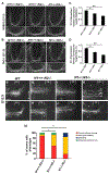
(A–C′) Immunohistochemistry against Robo1 protein labels post-crossing axons and pre-crossing (arrow mark) and crossing commissural axons (arrowhead). At E11.5, in Ndfip1 mutant spinal cord, Robo1 levels are increased in pre-crossing commissural axons (B and C, arrow with asterisk) compared with wild-type (A, arrow). Robo1-positive axons are observed crossing the midline in Ndfip1 mutant embryos (B′ and C′, arrowhead with asterisk) (n = 4; n, number of embryos). The variability of Robo1 expression in Ndfip1 mutant embryos is represented in (B) and (C). (B′ and C′) Higher magnification images at the floor plate region. (D) Quantitative representation of Robo1 pixel intensity at the commissure in control, Ndfip1−/−, Ndfip2−/−, and Ndfip1−/−; Ndfip2−/− spinal cord sections. (E and F) Brain extracts (E) and spinal cord extracts (F) from wild-type and Ndfip1 mutant adult mice were immunoblotted with anti-Robo1 and anti-Robo2 antibodies. Anti-Tubulin antibody was used as a loading control. Robo1 levels are increased in both Ndfip1 mutant brain or spinal cord lysates compared with wild-type, whereas Robo2 levels are unaltered. (G and H) Quantitative representation of band intensities of Robo1, Robo2, and DCC in brain lysates (G) or Robo1 and Robo2 in spinal cord lysates (H) that were normalized with tubulin levels. Error bars represent SEM. Significance was assessed using Student’s t test (*p < 0.05 and **p < 0.01); ns, non-significant. Scale bars represent 50 μm in (A)–(C) and 20 μm in (A′)–(C′).

Similar articles

Cited by

References

    1. Ambrozkiewicz MC, and Kawabe H (2015). HECT-type E3 ubiquitin ligases in nerve cell development and synapse physiology. FEBS Lett 589, 1635–1643. - PubMed
    1. Bai G, Chivatakarn O, Bonanomi D, Lettieri K, Franco L, Xia C, Stein E, Ma L, Lewcock JW, and Pfaff SL (2011). Presenilin-dependent receptor processing is required for axon guidance. Cell 144, 106–118. - PMC - PubMed
    1. Blockus H, and Chédotal A (2014). The multifaceted roles of Slits and Robos in cortical circuits: from proliferation to axon guidance and neurological diseases. Curr. Opin. Neurobiol 27, 82–88. - PubMed
    1. Brose K, Bland KS, Wang KH, Arnott D, Henzel W, Goodman CS, Tessier-Lavigne M, and Kidd T (1999). Slit proteins bind Robo receptors and have an evolutionarily conserved role in repulsive axon guidance. Cell 96, 795–806. - PubMed
    1. Cao XR, Lill NL, Boase N, Shi PP, Croucher DR, Shan H, Qu J, Sweezer EM, Place T, Kirby PA, et al. (2008). Nedd4 controls animal growth by regulating IGF-1 signaling. Sci. Signal 1, ra5. - PMC - PubMed

Publication types

MeSH terms