. 2019 Mar 19;20(6):1367.
doi: 10.3390/ijms20061367.

In Vitro Induction of T Helper 17 Cells by Synergistic Activation of Human Monocyte-Derived Langerhans Cell-Like Cells with Bacterial Agonists

Affiliations

In Vitro Induction of T Helper 17 Cells by Synergistic Activation of Human Monocyte-Derived Langerhans Cell-Like Cells with Bacterial Agonists

Robert Gramlich et al. Int J Mol Sci. .

Abstract

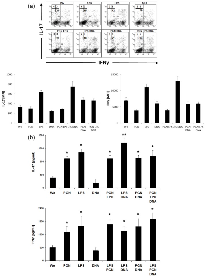
In the case of epidermal barrier disruption, pathogens encounter skin-resident Langerhans cells (LCs) and are recognized by pathogen recognition receptors such as Toll-like receptors (TLRs). As the majority of microorganisms exhibit more than one TLR ligand, the mechanisms of subsequent T cell differentiation are complex and far from clear. In this study, we investigated combinatory effects on Th cell polarization by bacterial cell wall compounds peptidoglycan (PGN) and lipopolysaccharide (LPS) and by bacterial nucleic acid (DNA). Expression of maturation markers CD40, CD80, HLA-DR and CCR7 and the release of IL-1β, IL-6 and IL-23 was strongly enhanced by simultaneous exposure to PGN, LPS and DNA in LCs. As all these factors were potential Th17 driving cytokines, we investigated the potency of combinatory TLR stimuli to induce Th17 cells via LC activation. High amounts of IL-17A and IL-22, key cytokines of Th17 cells, were detected. By intracellular costaining of IL-17⁺T cells, IL-22- (Th17) and IL-22⁺ (immature Th17) cells were identified. Interestingly, one population of LPS stimulated cells skewed into IL-9⁺Th cells, and LPS synergized with PGN while inducing high IL-22. In conclusion, our data indicates that when mediated by a fine-tuned signal integration via LCs, bacterial TLR agonists synergize and induce Th17 differentiation.

Keywords: IL-17; IL-22; IL-9; T helper cell; Th17; Toll-like receptor; dendritic cells; skin immune system.


Conflict of interest statement

The authors declare no conflict of interest.

Figures

Figure 1
Figure 1
PGN and CD40 ligand synergize in upregulating maturation molecules on MoLCs. Derived from the same PBMC, MoDCs and MoLCs were stimulated for 48 h by TLR2 agonist 20 µg/mL PGN and 1 µg/mL trimer of CD40 ligand or remained without stimulus (Wo). (a) Expression of CD86 and HLA-DR and (b) expression of CD80 and CD83 was detected by flow cytometry on the surface of MoLCs (upper panel) and MoDCs (lower panel). Quadrants indicate staining with isotype controls, numbers percent positive cells. Dot plots shown are representative for four independent experiments with different donors.
Figure 2
Figure 2
Triple TLR agonist activation increases LC-surface markers. MoLCs were activated by combinatory TLR2, TLR4 and TLR9 agonists: 20 µg/mL PGN, 1 µg/mL LPS, and 5 µg/mL DNA, respectively. (a) Dot plots show coexpression of CD80 and HLA-DR after 48 h. (b) Dot plots show coexpression of CD40 and CCR7 after 48 h. Quadrants indicate isotype controls, numbers percent positive cells. (c) Surface expression of TLR2, TLR4 and TLR9 in MoLCs. FACS staining of cells from one donor represents experiments from five donors with similar results.
Figure 3
Figure 3
Triple TLR agonist activation increases crucial LC cytokines. MoLCs were activated by combinatory TLR2, TLR4 and TLR9 agonists: 20 µg/mL PGN, 1 µg/mL LPS, and 5 µg/mL DNA, respectively. The amounts of IL-1β, IL-6, and IL-23 were detected in supernatant after 48 h. ELISA bars show mean of cytokine values from four donors. Differences of values for cytokines after stimulation were analyzed for statistical significance and compared to control (without, wo); * p ≤ 0.05, ** p ≤ 0.01.
Figure 4
Figure 4
Analogs of nucleic acids elevate LPS-induced IL-1β and IL-12p70. MoLC were challenged with combinations of polyI:C (pIC, 5 µg/mL) and polyU (pU, 5 µg/mL) with LPS (1 µg/mL). Release of IL-1β, IL-6 and IL-12p70 was measured by ELISA after 48 h. Bars represent inter-experimental means ±SD of protein release in the media of MoLC from 4 different donors. Differences of values for cytokines after stimulation were analyzed for statistical significance and compared to control (without, wo); * p ≤ 0.05, ** p ≤ 0.01.
Figure 5
Figure 5
Intracellular and supernatant-derived IL-17 and IFN-γ in CD4+T cells from TLR-stimulated cocultures. MoLC were stimulated with single and combinations of TLR agonists 20 µg/mL PGN, 1 µg/mL LPS and 5 µg/mL DNA for 48 h. The stimuli were removed by washing and LCs further cocultured with allogeneic CD4+T cells. (a) Intracellular costaining of IL-17 and IFN-γ by flow cytometry in 5 days-cocultured cells. One experiment out of four with different donors and similar results is shown. Numbers represent the percentage positive cytokine expression among CD4+T cells; Bars indicate mean fluorescence intensity (MFI) ± SD from intracellular FACS for IL-17 and IFN-γ. (b) ELISA detects IL-17 and IFN-γ after 5 days in the supernatants. Bars represent results of experiments with four different donors (mean±SD) and include data from the same donor as used in flow cytometry (a). Differences of values for cytokines after stimulation were analyzed for statistical significance and compared to control (without, wo); * p ≤ 0.05, ** p ≤ 0.01.
Figure 6
Figure 6
LPS, PGN and DNA synergize in inducing IL-22 and IL-9. After 48 h stimulation with 20 µg/mL PGN, 1 µg/mL LPS and 5 µg/mL DNA, stimuli were removed by washing and LCs further cocultured with allogeneic CD4+T cells. (a) Coexpression of IL-22 with IL-9 in cells cultured for 5 days. Shown is one experiment representing a total of tree with different donors, numbers indicate the percentage of positive cells, means of fluorescence intensity (mean FI) were analyzed from the dotplots. Bars indicate mean fluorescence intensity (MFI) ± SD from intracellular FACS for IL-9 and IL-22. (b) After additional 5 days, supernatants were analyzed for IL-9 and IL-22 by ELISA. Bars represent amounts of protein as inter-experimental means±SD of cytokine secretion in the supernatants of T cells from experiments using 5 independent MoLC and T cell donors. Differences of values for cytokines after stimulation were analyzed for statistical significance and compared to control (without, wo); * p ≤ 0.05, ** p ≤ 0.01.

References

    1. Mosmann T.R., Cherwinski H., Bond M.W., Giedlin M.A., Coffman R.L. Two types of murine helper T cell clone. I. Definition according to profiles of lymphokine activities and secreted proteins. J. Immunol. 1986;136:2348–2357. - PubMed
    1. O’Shea J.J., Paul W.E. Mechanisms underlying lineage commitment and plasticity of helper CD4+ T cells. Science. 2010;327:1098–1102. doi: 10.1126/science.1178334. - DOI - PMC - PubMed
    1. Malik S., Awasthi A. Transcriptional Control of Th9 Cells: Role of Foxo1 in Interleukin-9 Induction. Front. Immunol. 2018;9:995. doi: 10.3389/fimmu.2018.00995. - DOI - PMC - PubMed
    1. Miossec P., Korn T., Kuchroo V.K. Interleukin-17 and type 17 helper T cells. N. Engl. J. Med. 2009;361:888–898. doi: 10.1056/NEJMra0707449. - DOI - PubMed
    1. Plank M.W., Kaiko G.E., Maltby S., Weaver J., Tay H.L., Shen W., Wilson M.S., Durum S.K., Foster P.S. Th22 Cells Form a Distinct Th Lineage from Th17 Cells In Vitro with Unique Transcriptional Properties and Tbet-Dependent Th1 Plasticity. J. Immunol. 2017;198:2182–2190. doi: 10.4049/jimmunol.1601480. - DOI - PMC - PubMed

MeSH terms