Review
. 2019 Mar 19;11(3):274.
doi: 10.3390/v11030274.

Ebola Virus Entry: From Molecular Characterization to Drug Discovery

Affiliations
Review

Ebola Virus Entry: From Molecular Characterization to Drug Discovery

Cristiano Salata et al. Viruses. .

Abstract

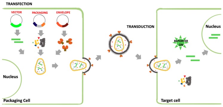
Ebola Virus Disease (EVD) is one of the most lethal transmissible infections, characterized by a high fatality rate, and caused by a member of the Filoviridae family. The recent large outbreak of EVD in Western Africa (2013⁻2016) highlighted the worldwide threat represented by the disease and its impact on global public health and the economy. The development of highly needed anti-Ebola virus antivirals has been so far hampered by the shortage of tools to study their life cycle in vitro, allowing to screen for potential active compounds outside a biosafety level-4 (BSL-4) containment. Importantly, the development of surrogate models to study Ebola virus entry in a BSL-2 setting, such as viral pseudotypes and Ebola virus-like particles, tremendously boosted both our knowledge of the viral life cycle and the identification of promising antiviral compounds interfering with viral entry. In this context, the combination of such surrogate systems with large-scale small molecule compounds and haploid genetic screenings, as well as rational drug design and drug repurposing approaches will prove priceless in our quest for the development of a treatment for EVD.

Keywords: Ebola virus; Filoviridae; VSV; antivirals; pseudovirus; retroviral vectors; small molecules; viral entry; virus-like particles.


Conflict of interest statement

The authors declare no conflict of interest.

Figures

Figure 1
Figure 1
Schematic representation of EBOV entry. Following interaction with attachment factors (1), the virion is internalized by the macropinocytosis (2). Inside the membrane-bound vesicle, GP is cleaved by cysteine proteases to activate its fusogenic potential (3). Cleaved GP is then able to interact with the specific NPC1 viral receptor (4). Such event, in addition to the activity of the TPC2 calcium channel (5), helps triggering the fusion between the viral envelope and the endosomal/lysosomal membrane (6), leading to viral genome release followed by transcription and replication (7).
Figure 2
Figure 2
Recovery, growth and pseudotyping of rVSV-ΔG-GFP. The system is based on a plasmid encoding the viral genome, containing a reporter gene (GFP) instead of the native gene coding for the glycoprotein G, and four plasmids providing the packaging system (matrix M, polymerase L, phosphoprotein P and G). At the beginning, cells are cotransfected with the pVSV-ΔG-GFP plasmid along with the four packaging plasmids to recover the G-complemented rVSV-ΔG-GFP. To express the viral genome for the first viral rescue, a plasmid encoding the T7 RNA polymerase is also required (not shown). This virus can be used for the generation of a pseudotyped rVSV-ΔG-GFP by transducing cells preventively transfected with a plasmid encoding for the heterologous glycoproteins. Then, the pseudotyped virus can be used to transduce target cells.
Figure 3
Figure 3
Schematic representation of the production of a pseudotyped retroviral vector. This system is based on a plasmid encoding for the retroviral vector (cis-acting sequences, reporter gene), and constructs expressing the packaging system factors and the heterologous envelope glycoprotein. Packaging cells are cotransfected with the different plasmids to recover pseudotyped retroviral particles in the supernatant. Pseudotyped particles can be used to transduce target cells.
Figure 4
Figure 4
Transcription- and replication-competent eVLP (tr-eVLP). This system is based on a minigenome, encoding for a reporter gene, the viral proteins VP40, GP, and in some cases p24, co-transfected with the constructs expressing RNP proteins (N, VP35, VP30, and L). Inside the producer cells, VP40 drives the formation of eVLPs that harbor minigenome-containing nucleocapsids. These tr-eVLPs can transduce target cells and deliver the minigenome that undergoes primary transcription mediated by RNP proteins brought into the target cells within the tr-eVLPs (in the form of nucleocapsids), resulting into the expression of the reporter gene. If target cells are pre-transfected with plasmids encoding for RNPs, the minigenome is replicated and undergoes a secondary transcription (with the expression of the reporter gene) mediated by RNP proteins provided in trans from expression constructs. Furthermore, a new progeny of infectious tr-eVLPs is produced and can be used to transduce new target cells.

References

    1. Feldmann H., Geisbert T.W. Ebola haemorrhagic fever. Lancet. 2011;377:849–862. doi: 10.1016/S0140-6736(10)60667-8. - DOI - PMC - PubMed
    1. Goldstein T., Anthony S.J., Gbakima A., Bird B.H., Bangura J., Tremeau-Bravard A., Belaganahalli M.N., Wells H.L., Dhanota J.K., Liang E., et al. The discovery of Bombali virus adds further support for bats as hosts of ebolaviruses. Nat. Microbiol. 2018;3:1084–1089. doi: 10.1038/s41564-018-0227-2. - DOI - PMC - PubMed
    1. Yang X.-L., Tan C.W., Anderson D.E., Jiang R.-D., Li B., Zhang W., Zhu Y., Lim X.F., Zhou P., Liu X.-L., et al. Characterization of a filovirus (Měnglà virus) from Rousettus bats in China. Nat. Microbiol. 2019;4:390–395. doi: 10.1038/s41564-018-0328-y. - DOI - PubMed
    1. Subissi L., Keita M., Mesfin S., Rezza G., Diallo B., Van Gucht S., Musa E.O., Yoti Z., Keita S., Djingarey M.H., et al. Ebola Virus Transmission Caused by Persistently Infected Survivors of the 2014–2016 Outbreak in West Africa. J. Infect. Dis. 2018;218:S287–S291. doi: 10.1093/infdis/jiy280. - DOI - PMC - PubMed
    1. Burk R., Bollinger L., Johnson J.C., Wada J., Radoshitzky S.R., Palacios G., Bavari S., Jahrling P.B., Kuhn J.H. Neglected filoviruses. FEMS Microbiol. Rev. 2016;40:494–519. doi: 10.1093/femsre/fuw010. - DOI - PMC - PubMed

Publication types

Substances