. 2019 Mar 19;11(3):167.
doi: 10.3390/toxins11030167.

The Dual Prey-Inactivation Strategy of Spiders-In-Depth Venomic Analysis of Cupiennius salei

Affiliations

The Dual Prey-Inactivation Strategy of Spiders-In-Depth Venomic Analysis of Cupiennius salei

Lucia Kuhn-Nentwig et al. Toxins (Basel). .

Abstract

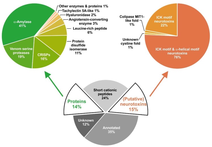
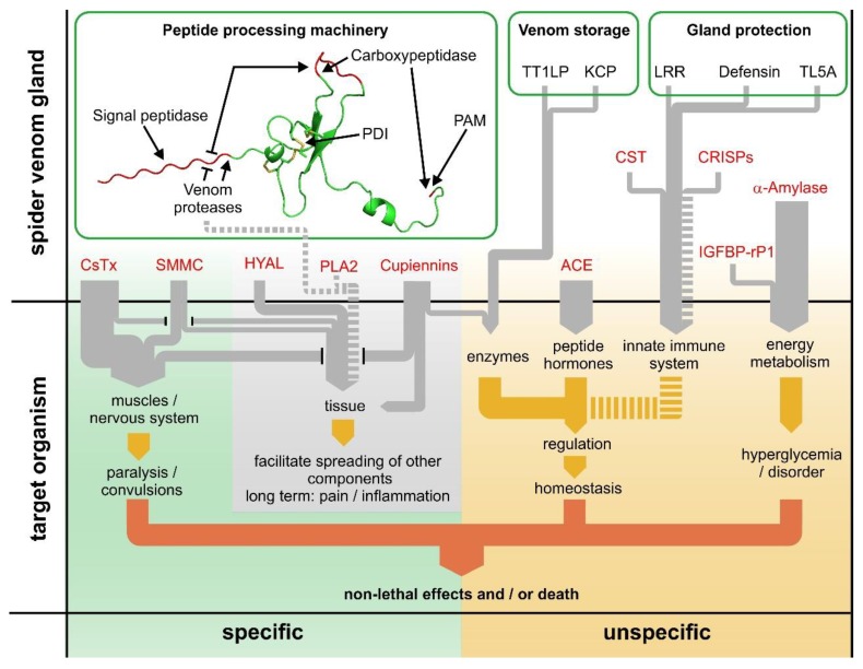
Most knowledge of spider venom concerns neurotoxins acting on ion channels, whereas proteins and their significance for the envenomation process are neglected. The here presented comprehensive analysis of the venom gland transcriptome and proteome of Cupiennius salei focusses on proteins and cysteine-containing peptides and offers new insight into the structure and function of spider venom, here described as the dual prey-inactivation strategy. After venom injection, many enzymes and proteins, dominated by α-amylase, angiotensin-converting enzyme, and cysteine-rich secretory proteins, interact with main metabolic pathways, leading to a major disturbance of the cellular homeostasis. Hyaluronidase and cytolytic peptides destroy tissue and membranes, thus supporting the spread of other venom compounds. We detected 81 transcripts of neurotoxins from 13 peptide families, whereof two families comprise 93.7% of all cysteine-containing peptides. This raises the question of the importance of the other low-expressed peptide families. The identification of a venom gland-specific defensin-like peptide and an aga-toxin-like peptide in the hemocytes offers an important clue on the recruitment and neofunctionalization of body proteins and peptides as the origin of toxins.

Keywords: enzymes; in depth transcriptomics; neurotoxins; proteomics; venom; α-amylase.


Conflict of interest statement

The authors declare no conflict of interest.

Figures

Figure 1
Figure 1
Functional profile of venom gland-specific transcripts of proteins and (putative) neurotoxins of C. salei. Contigs were blasted against UniProt KB (E = 10−5) and searched against Pfam-A.hmm [16]. The relative abundancy of functional groups is given in % of normalized read counts per contig (TPM), and specified for proteins (green) and cysteine-containing (putative) neurotoxins (orange). Proteins and neurotoxin-like structures are annotated based on their similarity to known proteins or structural motifs (α-helical motif, inhibitor cystine knot motif (ICK), and colipase MIT1-like fold).
Figure 2
Figure 2
Classification and characterization of high- and low-expressed (putative) neurotoxins identified in the transcriptome of C. salei. Toxin names, following the CsTx numbering system, are indicated by numbers in the slices and slice size represents the expression level as estimated by full read counts of relevant contigs. Structural motifs (detailed in the figure legend) and family annotations of toxins according to reference [79] are detailed in black and colored lines on the outer side of the respective slices.
Figure 3
Figure 3
Multiple sequence alignment of mature peptides belonging to the SN_19 family. Amino acid residue differences within the different subgroups are given in red characters, possible C-terminal α-helical parts are marked in blue and italic, glycine residues for C-terminal amidation are highlighted in green and in brackets, and cysteines are highlighted in black. Post-translational removal of amino acid residues is given in yellow. Not shown are identical amino acid sequences with silent mutations and visible mutations within signal and pro-peptide. CsTx-11b * was sequenced by Edman degradation [13].
Figure 4
Figure 4
Multiple sequence alignment of mature peptides belonging to the SN_02 family. Amino acid residue differences within the different subgroups are given in red characters, C-terminal amidation is highlighted in green, and cysteines are highlighted in black. Not shown are identical amino acid sequences with silent and visible mutations within the signal and pro-peptide. CsTx-18b* was sequenced by Edman degradation [13].
Figure 5
Figure 5
Sequence relationship of peptides of the SN_02 family. Sequence relationship was calculated using Bayesian inference methods. Sequences derived from venom gland transcriptomes are highlighted in dark gray, sequences from transcriptomes of other tissues or genomes in light gray. Nodes are labeled with posterior probability values. The peptide sequences used for analysis are accessible online with the following sequence identifiers: C. salei HC_agaLP_CUPSA (MH754628), Amblyomma triste (tr|A0A023G4R1|), Ixodes ricinus (tr|A0A131YAX3|), Nephila clavipes (tr|A0A2P6L7U6|), Xibalbanus tulumensis (contig00124SPETU, [99]), C. salei CsTx-17 (MH754572), C. salei CsTx-25 (MH754577), C. salei CsTx-31a (MH754573), Superstitionia donensis (tr|A0A1V1WBV1|), Hadogenes troglodytes (tr|A0A1B3IJ31|), Viridasius fasciatus (tr|A0A1V0FWF9|), Apis mellifera (XP_003249808).
Figure 6
Figure 6
Multiple sequence alignment of mature peptides belonging to different low abundant peptide families. Amino acid residue differences within the different subgroups are given in red characters, C-terminal amidation is highlighted in green, post-translational removal of amino acid residues is given in yellow, and cysteines are highlighted in black. Not shown are identical amino acid sequences with silent and visible mutations within the signal and pro-peptide.
Figure 7
Figure 7
Identification of different pro-peptide cleavage motifs in one precursor. (A) Pro-peptide cleavage motifs of CsTx-33, CsTx-35, and other peptides featuring a dibasic motif. The sequence parts shown include the ends of pro-peptides and the beginning of mature peptides. The start of the mature peptide sequences is indicated with a bold red line. Amino acids of the mature peptide are in bold. Potential protease cleavage motifs are displayed in dashed boxes. Red boxes indicate the cleavage motif, which directly locates the N-terminal of the experimentally found start of the mature peptide. (B) Sequence comparison of transcripts coding for CsTx-33 and other C. salei peptides with a similar N-terminal sequence of the mature peptide. Nucleotides differing between sequences are highlighted in red.
Figure 8
Figure 8
Multiple sequence alignment of mature peptides belonging to the SN_20 and SN_32 peptide families. Amino acid residue differences within the different subgroups are given in red characters, and cysteines are highlighted in black. Not shown are identical amino acid sequences with silent and visible mutations within the signal and pro-peptide.
Figure 9
Figure 9
Multiple sequence alignment of mature defensin-like peptides from different arachnids and types of tissue. Amino acid residue differences of defensins within different tissue subgroups (VG, venom gland; HE, hemocytes; * venom glands, ** salivary glands) are given in red characters and cysteines are highlighted in black. Sequences belong to the spiders C. salei (defensin2_VG_CUPSA, (CSG_98_27980_30560); defensin1_HE_CUPSA [121]), Cupiennius getazi (defensin2_VG_CUPGE, (DN21681_c1_g2_i3_4); defensin1_HE_CUPGE, (DN14943_c0_g1_i1_6)), Polybetes pythagoricus (defensin_HE_POLPY [121]), Eratigena atrica (defensin_HE_ERAAT [121]), Phoneutria reidyi (defensin_HE_PHORE [121]), the scorpions Mesobuthus martensii (BmKDfsin4_MESMA [122]), Centruroides hentzi (tr|A0A2I9LNX0_defensin_CENHE [123]), Androctonus bicolor (tr|A0A0K0LBV1_defensin1_ANDBI [124]), and the ticks Ixodes scapularis (tr|V5IGJ0_defensin2_IXORI [125]), Ixodes ricinus (tr|Q5Q979_defensin_IXOSC), and Amblyomma cajennense (tr|A0A023FQT7_defensin1_AMBCA [120]).
Figure 10
Figure 10
Dual prey-inactivation strategy of the venom of C. salei based on specific (left) and unspecific (right) venom pathways. Main interactions of the major venom components are shown in the venom gland (upper half) and, after venom injection, in the target organism (lower half). The specific pathway, mainly based on neurotoxins and other compounds, usually leads to death. The unspecific or metabolic pathway, based on a variety of regulatory elements, disturbs homeostasis or leads to hyperglycemia. The thickness of the gray arrows indicates the estimated impact on the prey. Dashed lines represent vague or uninvestigated connections (for further details compare text). ACE, angiotensin-converting enzyme; CRISPs, Cysteine-rich secretory proteins; CST, cystatin; CsTx, Cupiennius salei toxins; HYAL, hyaluronidase; IGFBP-rP1, insulin-like growth factor-binding protein-related protein 1; KCP, Kunitz domain-containing protein; LRR, leucine-rich repeat domain-containing protein; PAM, peptidylglycine α-amidating monooxygenase; PDI, protein disulfide isomerase; PLA2, phospholipase A2; SMMC, small molecular mass compounds; TL5A, tachylectin 5A-like protein; TT1LP, thyroglobulin type-1 domain-like protein.

Similar articles

Cited by

References

    1. Natural History Museum Bern; [(accessed on 11 September 2018)]. World Spider Catalog. Version 19.5. Available online: http://wsc.nmbe.ch.
    1. Wise D.H. Spiders in Ecological Webs. Cambridge University Press; Cambridge, MA, USA: 1993.
    1. Kuhn-Nentwig L., Stöcklin R., Nentwig W. Venom composition and strategies in spiders: Is everything possible? Adv. Insect Physiol. 2011;40:1–86.
    1. Saez N.J., Senff S., Jensen J.E., Er S.Y., Herzig V., Rash L.D., King G.F. Spider-venom peptides as therapeutics. Toxins. 2010;2:2851–2871. doi: 10.3390/toxins2122851. - DOI - PMC - PubMed
    1. Smith J.J., Herzig V., King G.F., Alewood P.F. The insecticidal potential of venom peptides. Cell. Mol. Life Sci. 2013;70:3665–3693. doi: 10.1007/s00018-013-1315-3. - DOI - PMC - PubMed

Publication types