. 2019 Mar 19;7(1):21.
doi: 10.3390/biomedicines7010021.

Calcium Carbonate Nanoparticles Can Activate the Epithelial⁻Mesenchymal Transition in an Experimental Gastric Cancer Model

Affiliations

Calcium Carbonate Nanoparticles Can Activate the Epithelial⁻Mesenchymal Transition in an Experimental Gastric Cancer Model

Marina Senchukova et al. Biomedicines. .

Abstract

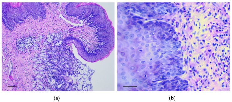
Previously, we have shown the possibility of intramucosal gastric carcinoma induction by the intragastric administration of a mixture of formaldehyde and hydrogen peroxide in rats. In this study, we report a sizable increase in carcinogenic properties of the mixture when a suspension containing calcium carbonate nanoparticles was added to it. This technique allowed us to reduce both the number of the carcinogen administrations from twelve to two and the time to the cancer induction from six to four months. Although the induced tumors were represented by the intramucosal carcinomas, they were characterized by the extensive invasion of individual tumor cells and their clusters into the muscle layer and serosa as well as into the omentum and blood vessels. Considering that the invasive tumor cells were positive for vimentin, Snail and TGF-β2, we concluded that their invasion was the result of the activation of epithelial⁻mesenchymal transition (EMT) mechanisms. Thus, taking into account the data obtained, it can be assumed that under the conditions of inflammation or carcinogenesis, the calcium carbonate nanoparticles may affect the activation of EMT mechanisms.

Keywords: calcium carbonate; carcinogenesis; epithelial–mesenchymal transition; gastric cancer; nanoparticles.


Conflict of interest statement

The authors declare no conflict of interest.

Figures

Figure 1
Figure 1
The pathological changes in the forestomach. (a) Area of the groove between the forestomach and glandular stomach. Mayer’s hematoxylin and eosin (H&E) staining, scale bar 200 μm; (b) Increasing thickness and number of epidermal rows in the basal and spinous layers of forestomach. H&E staining, scale bar 100 μm.
Figure 2
Figure 2
The pathological changes in stomach body. (a) Basal part of gastric glands with marked cytological and architectural atypia (arrows). In the box the selected area is shown. H&E staining, scale bar 200 μm (in box—100 μm). (b) Proliferations of atypical epithelium in the lumen of glandular-like structures (arrows). H&E staining, scale bar 100 μm. (cd) The marked Snail (c) and TGF-β2 (d) expression in gastric mucosa in rats of the experimental group is shown. Immunoperoxidase staining with antibody to Snail and TGF-β2, scale bar 100 μm. (ef) Very weak Snail (arrows) (e) and TGF-β2 (f) expression in gastric mucosa in rats of the control group is shown. Immunoperoxidase staining with antibody to Snail and TGF-β2, scale bar 100 μm.
Figure 2
Figure 2
The pathological changes in stomach body. (a) Basal part of gastric glands with marked cytological and architectural atypia (arrows). In the box the selected area is shown. H&E staining, scale bar 200 μm (in box—100 μm). (b) Proliferations of atypical epithelium in the lumen of glandular-like structures (arrows). H&E staining, scale bar 100 μm. (cd) The marked Snail (c) and TGF-β2 (d) expression in gastric mucosa in rats of the experimental group is shown. Immunoperoxidase staining with antibody to Snail and TGF-β2, scale bar 100 μm. (ef) Very weak Snail (arrows) (e) and TGF-β2 (f) expression in gastric mucosa in rats of the control group is shown. Immunoperoxidase staining with antibody to Snail and TGF-β2, scale bar 100 μm.
Figure 3
Figure 3
The pathological changes in the pyloric stomach. (a) The dysplastic changes in the pyloric glands of stomach. H&E staining, scale bar 100 μm. In the box the polymorphic prismatic cells with large nuclei, having irregular hyperchromatism and irregular nucleoli in generative zone of pyloric glands are shown. H&E staining, scale bar 100 μm. (b) Positive reaction to Snail in cells lined the pyloric glands resembling signet ring cells. In the box the selected area with signet ring cells (arrows) is shown. Immunoperoxidase staining with antibody to Snail, scale bar 100 μm.
Figure 4
Figure 4
Invasion of tumor cells. H&E staining. (a) Tumor embolus in the vessel, scale bar 100 μm. (b) Invasion of tumor cells in muscle layer (arrows), scale bar 100 μm. (c) Invasion of tumor cells in serosa layer (arrow). In the box the marked area is shown at magnification, scale bar 200 μm. (d) Invasion of tumor cells in omentum. In the box the selected area is shown at higher magnification, scale bar 200 μm. (e) The clusters of tumor cells in the muscle layer consisting of fibroblast-like cells with a curved shape (1-black arrows) and cells with finely dispersed oxyphilic inclusions in the cytoplasm (2-red arrows), scale bar 100 μm. (f) The cells with large, polymorphic, hyperchromic nuclei and with a markedly increased nuclear/cytoplasmic ratio in the muscle layer of stomach, scale bar 100 μm.
Figure 4
Figure 4
Invasion of tumor cells. H&E staining. (a) Tumor embolus in the vessel, scale bar 100 μm. (b) Invasion of tumor cells in muscle layer (arrows), scale bar 100 μm. (c) Invasion of tumor cells in serosa layer (arrow). In the box the marked area is shown at magnification, scale bar 200 μm. (d) Invasion of tumor cells in omentum. In the box the selected area is shown at higher magnification, scale bar 200 μm. (e) The clusters of tumor cells in the muscle layer consisting of fibroblast-like cells with a curved shape (1-black arrows) and cells with finely dispersed oxyphilic inclusions in the cytoplasm (2-red arrows), scale bar 100 μm. (f) The cells with large, polymorphic, hyperchromic nuclei and with a markedly increased nuclear/cytoplasmic ratio in the muscle layer of stomach, scale bar 100 μm.
Figure 5
Figure 5
Invasion of tumor cells in the muscle layer (a,c,d) and in omentum (b,d,f). Positive reaction to vimentin (a,b), Snail (c,d) and TGF-β2 (e,f). Immunoperoxidase staining with antibody to vimentin, Snail and TGF-β2, scale bar 100 μm.
Figure 6
Figure 6
Well-differentiated adenocarcinoma of cecum. H&E staining: (a) 200×, scale bar 200 μm, (b) 800×, scale bar 100 μm.
Figure 7
Figure 7
Detection and characterization of calcium carbonate nanoparticles in the carcinogenic suspension and microcalcifications in the gastric mucosa. (a) Multiple microcalcifications in the gastric mucosa of experimental rats. von Kossa staining, scale bar 100 μm. (b) The absence of microcalcifications in the gastric mucosa of control rats. von Kossa staining, scale bar 100 μm. (c) The imaging of aggregates (nanoparticles) obtained by the atomic force microscopy. The scan size is 7.8 × 5.6 μm. (d) The histogram of calcium carbonate nanoparticles distribution in carcinogenic suspension according to their hydrodynamic radii, obtained by the dynamic light scattering method.

Similar articles

Cited by

References

    1. Ferlay J., Soerjomataram I., Dikshit R., Eser S., Mathers C., Rebelo M., Parkin D.M., Forman D., Bray F. Cancer incidence and mortality worldwide: Sources, methods and major patterns in GLOBOCAN 2012. Int. J. Cancer. 2015;136:359–386. doi: 10.1002/ijc.29210. - DOI - PubMed
    1. Torre L.A., Bray F., Siegel R.L., Ferlay J., Lortet-Tieulent J., Jemal A. Global cancer statistics, 2012. CA Cancer J. Clin. 2015;65:87–108. doi: 10.3322/caac.21262. - DOI - PubMed
    1. Chen X.L., Zhao L.Y., Xue L., Xu Y.H., Zhang W.H., Liu K., Chen X.Z., Yang K., Zhang B., Chen Z.X., et al. Prognostic significance and the role in TNM stage of extranodal metastasis within regional lymph nodes station in gastric carcinoma. Oncotarget. 2016;7:67047–67060. doi: 10.18632/oncotarget.11478. - DOI - PMC - PubMed
    1. Wei S., Wang L., Zhang L., Li B., Li Z., Zhang Q., Wang J., Chen L., Sun G., Li Q., et al. ZNF143 enhances metastasis of gastric cancer by promoting the process of EMT through PI3K/AKT signaling pathway. Tumour Biol. 2016;37:12813–12821. doi: 10.1007/s13277-016-5239-z. - DOI - PubMed
    1. Zhao W., Jia L., Kuai X., Tang Q., Huang X., Yang T., Qiu Z., Zhu J., Huang J., Huang W., et al. The role and molecular mechanism of Trop2 induced epithelial-mesenchymal transition through mediated β-catenin in gastric cancer. Cancer Med. 2019 doi: 10.1002/cam4.1934. - DOI - PMC - PubMed