. 2019 Mar 19;9(3):53.
doi: 10.3390/metabo9030053.

The Effect of Exercise Training on Myocardial and Skeletal Muscle Metabolism by MR Spectroscopy in Rats with Heart Failure

Affiliations

The Effect of Exercise Training on Myocardial and Skeletal Muscle Metabolism by MR Spectroscopy in Rats with Heart Failure

Mingshu Shi et al. Metabolites. .

Abstract

The metabolism and performance of myocardial and skeletal muscle are impaired in heart failure (HF) patients. Exercise training improves the performance and benefits the quality of life in HF patients. The purpose of the present study was to determine the metabolic profiles in myocardial and skeletal muscle in HF and exercise training using MRS, and thus to identify targets for clinical MRS in vivo. After surgically establishing HF in rats, we randomized the rats to exercise training programs of different intensities. After the final training session, rats were sacrificed and tissues from the myocardial and skeletal muscle were extracted. Magnetic resonance spectra were acquired from these extracts, and principal component and metabolic enrichment analysis were used to assess the differences in metabolic profiles. The results indicated that HF affected myocardial metabolism by changing multiple metabolites, whereas it had a limited effect on skeletal muscle metabolism. Moreover, exercise training mainly altered the metabolite distribution in skeletal muscle, indicating regulation of metabolic pathways of taurine and hypotaurine metabolism and carnitine synthesis.

Keywords: MRS; cardiac metabolism; magnetic resonance spectroscopy; metabolomics.


Conflict of interest statement

The authors declare no conflict of interest.

Figures

Figure 1
Figure 1
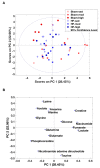
Principle component analysis (PCA) of myocardial metabolites. (A) The score plot from the PCA of the myocardial metabolites demonstrates a significant difference in the metabolite distribution between HF and Sham based on the first principal component (PC1), which explains 34% of the total variation. (B) The loading plot showing the contribution of the individual metabolites to the model.
Figure 2
Figure 2
The scatter and bar plots show the relative metabolite levels (AUC) of the 10 metabolites that are significantly different in the ANOVA model (Table 1). Pairwise comparison within each group was performed using Tukey’s Post Hoc Test. In total 25 samples were distributed into 6 subgroups: Sham-sed, n = 5; Sham-mod, n = 3; Sham-high, n = 4; HF-sed, n = 4; HF-mod, n = 4; HF-high, n = 5. “#”above the HF bars donates that the exercise training subgroup was significantly different compared to its Sham counterparts (e.g., Sham-sed vs. HF-sed). #, ##, ### denote p values < 0.05, 0.01, and 0.001, respectively. Five metabolites (glutamine, acetate, lactate, succinate, and aspartate) were significantly different in the ANOVA model but showed no significant difference in the pairwise comparison.
Figure 3
Figure 3
Enrichment analysis of the myocardial tissue. The pathways shown were significantly different between sham-sed and HF-sed, sorted by fold enrichment and p value. False Discovery Rate (FDR) > 0.05 was considered as statistically different.
Figure 4
Figure 4
PCA of skeletal muscle metabolites. (A) The score plot from PCA within all skeletal muscle metabolites. Principal components 1 and 2 (PC1 and PC2) explain the model with 28% and 22% values, respectively. However, no obvious separation between HF and Sham was visually observed. (B) The contribution of the individual metabolites to the model.
Figure 5
Figure 5
The skeletal muscle metabolic profile. The scatter and bar plots show relative metabolite levels (AUC) of the seven metabolites that are significantly different in the ANOVA model (Table 2). Pairwise comparison within each group was performed using Tukey’s Post Hoc Test. In total, 48 samples were distributed into 6 subgroups: Sham-sed, n = 7; Sham-mod, n = 7; Sham-high, n = 7; HF-sed, n = 10; HF-mod, n = 8; HF-high, n = 9. “#”above the HF bars denotes that the exercise training subgroup is significantly different (p < 0.05) compared to its Sham counterpart (e.g., Sham-sed vs. HF-sed). “*” denotes a significant difference between different exercise groups which underwent the same surgical process (e.g., Sham-sed vs. Sham-high). *, **, *** denotes p value < 0.05, 0.01, and 0.001, respectively. Although alanine was recognized as significantly different in the ANOVA model, the pairwise comparison under the exercise training or surgical factor did not result in a significant difference.
Figure 6
Figure 6
Enrichment analysis of skeletal muscle tissue. The pathways shown were significantly different between (A) Sham-sed and HF-sed; (B) Sham-sed and Sham-high; and (C) HF-sed and HF-mod, sorted by fold enrichment and p value. FDR > 0.05 was considered as statistically different.

References

    1. White H.D., Chew D.P. Acute myocardial infarction. Lancet. 2008;372:570–584. doi: 10.1016/S0140-6736(08)61237-4. - DOI - PubMed
    1. McMurray J.J., Pfeffer M.A. Heart failure. Lancet. 2005;365:1877–1889. doi: 10.1016/S0140-6736(05)66621-4. - DOI - PubMed
    1. James S., Barton D., O’Connell E., Voon V., Murtagh G., Watson C., Murphy T., Prendiville B., Brennan D., Hensey M., et al. Life expectancy for community-based patients with heart failure from time of diagnosis. Int. J. Cardiol. 2015;178:268–274. doi: 10.1016/j.ijcard.2014.09.131. - DOI - PubMed
    1. Savage P.A., Shaw A.O., Miller M.S., VanBuren P., LeWinter M.M., Ades P.A., Toth M.J. Effect of resistance training on physical disability in chronic heart failure. Med. Sci. Sports Exerc. 2011;43:1379–1386. doi: 10.1249/MSS.0b013e31820eeea1. - DOI - PMC - PubMed
    1. Fukushima A., Milner K., Gupta A., Lopaschuk G.D. Myocardial Energy Substrate Metabolism in Heart Failure: From Pathways to Therapeutic Targets. Curr. Pharm. Des. 2015;21:3654–3664. doi: 10.2174/1381612821666150710150445. - DOI - PubMed