. 2019 Mar 19;11(3):385.
doi: 10.3390/cancers11030385.

Perilipin 5 and Lipocalin 2 Expression in Hepatocellular Carcinoma

Affiliations

Perilipin 5 and Lipocalin 2 Expression in Hepatocellular Carcinoma

Anastasia Asimakopoulou et al. Cancers (Basel). .

Abstract

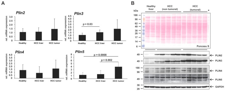
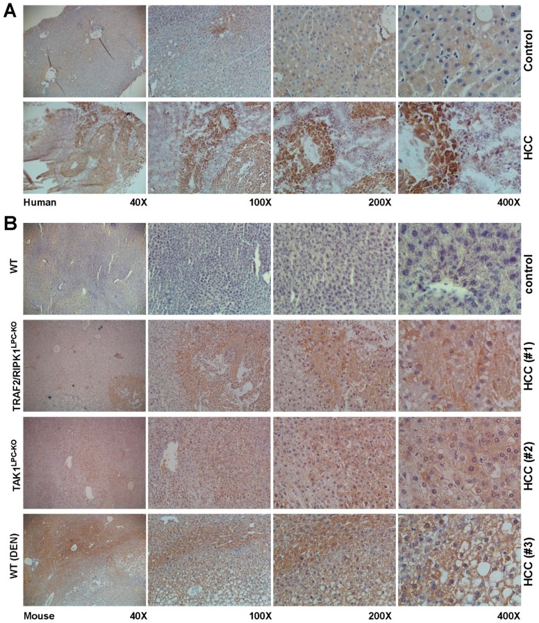
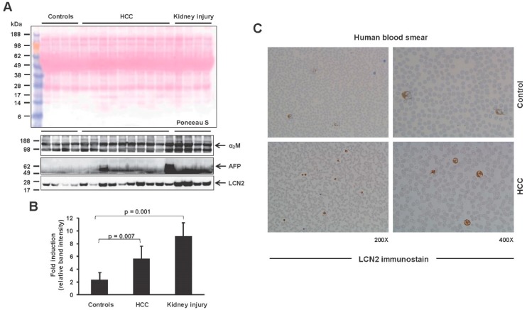
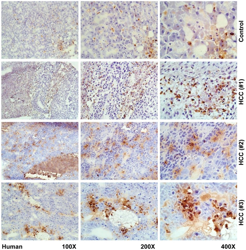
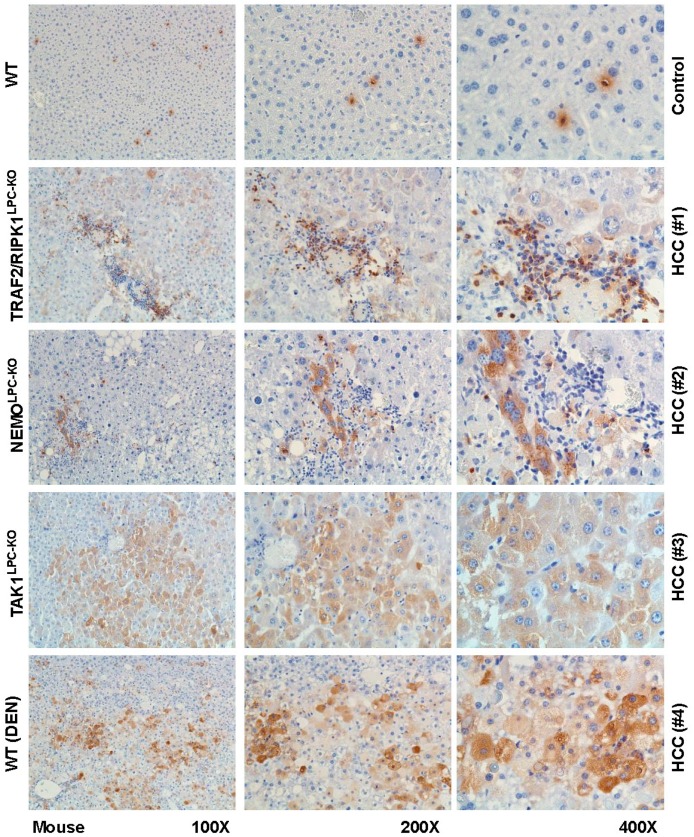
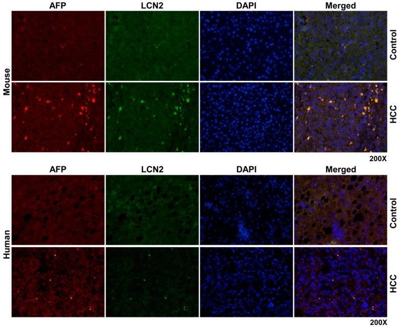
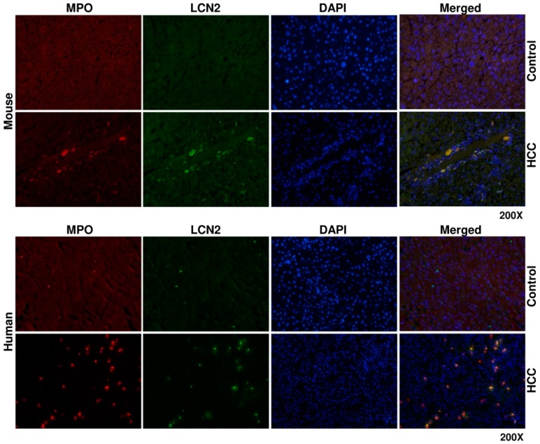
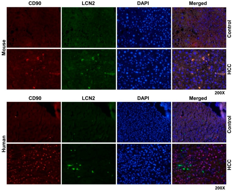
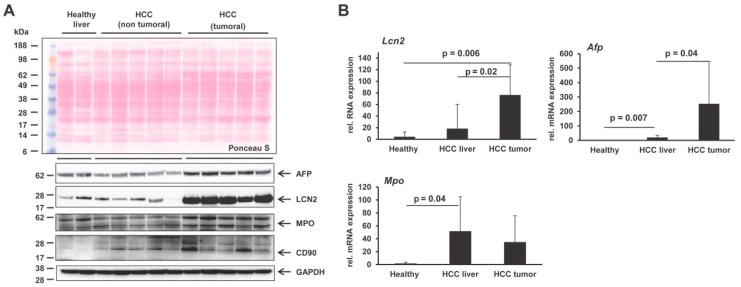
Hepatocellular carcinoma (HCC) is one of the most prevalent and deadly cancers worldwide. Therefore, current global research focuses on molecular tools for early diagnosis of HCC, which can lead to effective treatment at an early stage. Perilipin 5 (PLIN5) has been studied as one of the main proteins of the perilipin family, whose role is to maintain lipid homeostasis by inhibiting lipolysis. In this study, we show for the first time that PLIN5 is strongly expressed in tumors of human patients with HCC as well as in mouse livers, in which HCC was genetically or experimentally induced by treatment with the genotoxic agent diethylnitrosamine. Moreover, the secreted acute phase glycoprotein Lipocalin 2 (LCN2) established as a biomarker of acute kidney injury, is also proven to indicate liver injury with upregulated expression in numerous cases of hepatic damage, including steatohepatitis. LCN2 has been studied in various cancers, and it has been assigned roles in multiple cellular processes such as the suppression of the invasion of HCC cells and their metastatic abilities. The presence of this protein in blood and urine, in combination with the presence of α -Fetoprotein (AFP), is hypothesized to serve as a biomarker of early stages of HCC. In the current study, we show in humans and mice that LCN2 is secreted into the serum from liver cancer tissue. We also show that AFP-positive hepatocytes represent the main source for the massive expression of LCN2 in tumoral tissue. Thus, the strong presence of PLIN5 and LCN2 in HCC and understanding their roles could establish them as markers for diagnosis or as treatment targets against HCC.

Keywords: AFP; HCC; LCN2; PLIN5; cancer; liver.


Conflict of interest statement

The authors declare no conflict of interest.

Figures

Figure 1
Figure 1
Expression of the perilipin family in liver and hepatocellular carcinoma (HCC). (A) Quantitative real-time PCR analysis of hepatic Plin2, Plin3, Plin4 and Plin5 expression in healthy mice (n = 4), non-tumoral liver (n = 8) and tumoral liver extracts (n = 8) of HCC mice. The quantity of mRNA in healthy mouse livers was set to 1, and expression levels in the HCC groups were expressed as relative values. All measurements were normalized to β-Actin expression. Primers are listed in Table 2. Statistical analysis was performed by Student t-test and standard deviations below 0.05 were considered significant. (B) Confirmation of differential expression of the perilipin proteins by Western blot analysis. Western blots were performed to detect PLIN2, PLIN3, PLIN4 and PLIN5 proteins in liver protein extracts from healthy mice (n = 3) and mice with HCC on tumoral and non-tumoral liver extracts (n = 8 animals/group). An extract of primary mouse hepatocytes (marked with asterisk), transient overexpressing PLIN5, was used to define the protein band of PLIN5 as it is lowly expressed in liver. Glycerinaldehyd-3-phosphate dehydrogenase (GAPDH) was used as a loading control. The antibodies used are listed in Table 1.
Figure 2
Figure 2
Immunohistochemical localization of PLIN5 in human livers of HCC patients and experimental mouse HCC models. Liver cryosections of (A) human hepatic biopsies and (B) paraffin sections of HCC mouse models were stained with an antibody against PLIN5. The depicted results are representative of all samples analyzed. As negative controls, either non-HCC human patient biopsies or hepatic sections from healthy mice were used. Magnifications presented are of 40×, 100×, 200× and 400×, respectively.
Figure 3
Figure 3
Lipocalin 2 (LCN2) expression in blood serum and blood smears withdrawn from HCC patients. (A) LCN2 protein secretion in the blood serum of human HCC patients. α-Fetoprotein (AFP) was shown as a known marker elevated in HCC patients, while α2-Macroglobulin (α2M) was used as a loading control. (B) Densitometry of LCN2 results depicted in (A). Statistical analysis was performed by Student t-test and standard deviations below 0.05 were considered significant. (C) Blood smears were stained for LCN2 by routine immunocytochemistry procedure to visualize LCN2 positive cells. Serum from healthy subjects was used as a control. Magnifications presented are 200× and 400×.
Figure 4
Figure 4
Immunohistochemical localization of LCN2 in human livers of HCC patients. Liver cryosections of human hepatic biopsies were stained with antibody against LCN2. Representative stains of three HCC patients and one control are depicted. As negative controls non-HCC human patient biopsies were used. Magnifications are 100×, 200× and 400×, respectively.
Figure 5
Figure 5
Immunohistochemical localization of LCN2 in livers of HCC mice. Liver paraffin sections of healthy and HCC mice were stained with antibody against LCN2. Representative images of one control and four HCC samples taken from different HCC models are depicted. Magnifications shown are 100×, 200× and 400×, respectively.
Figure 6
Figure 6
Double fluorescent Immunohistochemical staining of LCN2 and AFP in livers of HCC mice and human HCC liver biopsies. Liver paraffin sections of healthy and HCC mice as well as human liver biopsies of HCC or non-HCC patients were stained with antibodies against LCN2 and AFP. Alexa Fluor-conjugated secondary antibodies were used for visualization (green, LCN2; red, AFP). Nuclei were counterstained with DAPI (blue). Representative images are depicted. Magnification: 200×.
Figure 7
Figure 7
Double fluorescent immunohistochemical staining of LCN2 and MPO in livers of HCC mice and human HCC liver biopsies. Liver paraffin sections of healthy and HCC mice, as well as human liver biopsies of HCC or non-HCC patients were stained with antibodies against LCN2 and Myeloperoxidase (MPO). Alexa Fluor-conjugated secondary antibodies were used for visualization (green, LCN2; red, MPO) and nuclei were counterstained with DAPI (blue). Magnification: 200×.
Figure 8
Figure 8
Double fluorescent immunohistochemical staining of LCN2 and CD90 in livers of HCC mice and human HCC liver biopsies. Liver paraffin sections of healthy and HCC mice as well as human liver biopsies of HCC or non-HCC patients were stained with antibodies against LCN2 and CD90. Alexa Fluor-conjugated secondary antibodies secondary antibodies were used for visualization (green, LCN2; red, CD90) and nuclei were counterstained with DAPI (blue). Magnification: 200×.
Figure 9
Figure 9
Differential expression of LCN2, AFP, MPO and CD90 in HCC mouse livers. (A) Western blots were performed to detect LCN2, AFP, MPO and CD90 in liver protein extracts from healthy mice (n = 3) and mice with HCC on tumoral and non-tumoral liver extracts (n = 8 animals/group). GAPDH was used as a loading control. The antibodies used are listed in Table 1. (B) Quantitative real-time PCR analysis of Lcn2, Afp and Mpo mRNA levels in healthy mice (n = 4), non-tumoral liver (n = 8), and tumoral liver extracts of HCC mice (n = 8). The quantity of mRNA in healthy mouse liver was set to 1, and expression levels in the HCC groups were expressed as relative values. All measurements were normalized to β-Actin expression. Primers are listed in Table 2. Statistical analysis was performed by the Student t-test and standard deviations below 0.05 were considered significant.

Similar articles

Cited by

References

    1. Kimmel A.R., Sztalryd C. The perilipins: Major cytosolic lipid droplet-associated proteins and their roles in cellular lipid storage, mobilization, and systemic homeostasis. Annu. Rev. Nutr. 2016;36:471–509. doi: 10.1146/annurev-nutr-071813-105410. - DOI - PubMed
    1. Sztalryd C., Brasaemle D.L. The perilipin family of lipid droplet proteins: Gatekeepers of intracellular lipolysis. Biochim. Biophys. Acta Mol. Cell Biol. Lipids. 2017;1862:1221–1232. doi: 10.1016/j.bbalip.2017.07.009. - DOI - PMC - PubMed
    1. Wolins N.E., Quaynor B.K., Skinner J.R., Tzekov A., Croce M.A., Gropler M.C., Varma V., Yao-Borengasser A., Rasouli N., Kern P.A., et al. OXPAT/PAT-1 is a PPAR-induced lipid droplet protein that promotes fatty acid utilization. Diabetes. 2006;55:3418–3428. doi: 10.2337/db06-0399. - DOI - PubMed
    1. Hashani M., Witzel H.R., Pawella L.M., Lehmann-Koch J., Schumacher J., Mechtersheimer G., Schnölzer M., Schirmacher P., Roth W., Straub B.K. Widespread expression of perilipin 5 in normal human tissues and in diseases is restricted to distinct lipid droplet subpopulations. Cell Tissue Res. 2018;374:121–136. doi: 10.1007/s00441-018-2845-7. - DOI - PubMed
    1. Dalen K.T., Dahl T., Holter E., Arntsen B., Londos C., Sztalryd C., Nebb H.I. LSDP5 is a PAT protein specifically expressed in fatty acid oxidizing tissues. Biochim. Biophys. Acta. 2007;1771:210–227. doi: 10.1016/j.bbalip.2006.11.011. - DOI - PubMed

LinkOut - more resources