. 2019 Jul 1:194:272-282.
doi: 10.1016/j.neuroimage.2019.03.021. Epub 2019 Mar 17.

Thalamus Optimized Multi Atlas Segmentation (THOMAS): fast, fully automated segmentation of thalamic nuclei from structural MRI

Affiliations

Thalamus Optimized Multi Atlas Segmentation (THOMAS): fast, fully automated segmentation of thalamic nuclei from structural MRI

Jason H Su et al. Neuroimage. .

Abstract

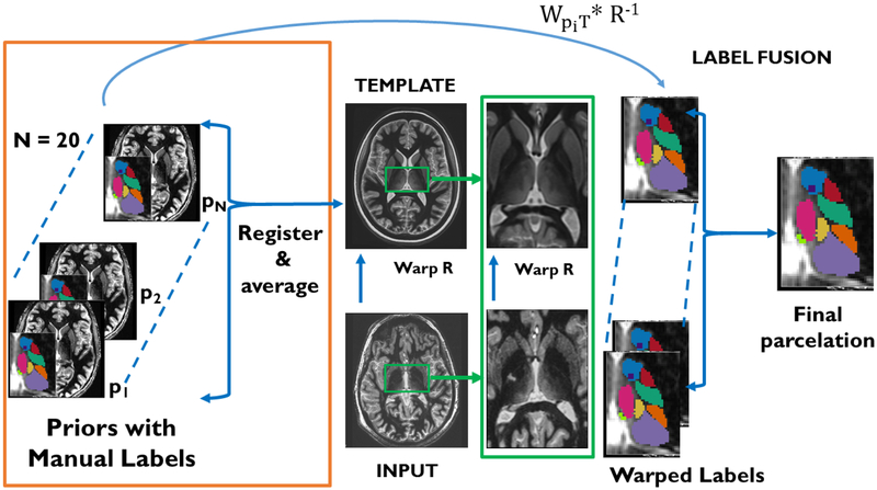
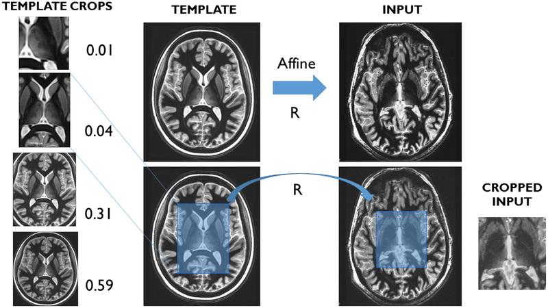
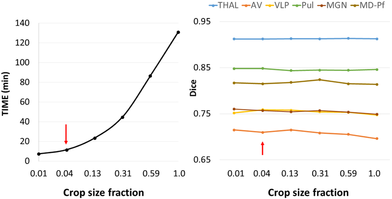
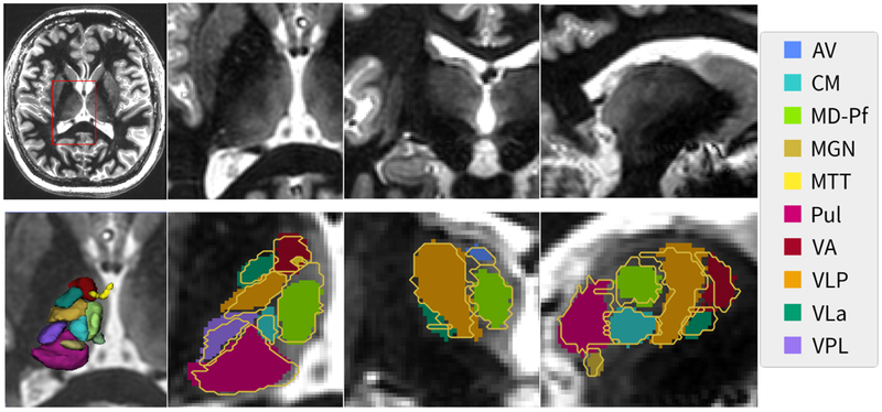
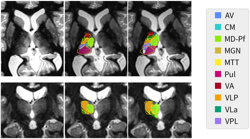
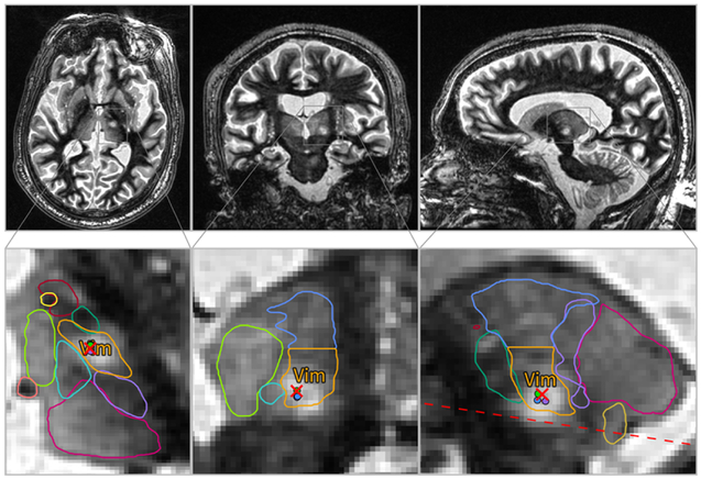
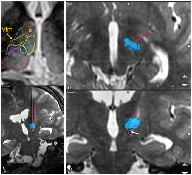
The thalamus and its nuclei are largely indistinguishable on standard T1 or T2 weighted MRI. While diffusion tensor imaging based methods have been proposed to segment the thalamic nuclei based on the angular orientation of the principal diffusion tensor, these are based on echo planar imaging which is inherently limited in spatial resolution and suffers from distortion. We present a multi-atlas segmentation technique based on white-matter-nulled MP-RAGE imaging that segments the thalamus into 12 nuclei with computation times on the order of 10 min on a desktop PC; we call this method THOMAS (THalamus Optimized Multi Atlas Segmentation). THOMAS was rigorously evaluated on 7T MRI data acquired from healthy volunteers and patients with multiple sclerosis by comparing against manual segmentations delineated by a neuroradiologist, guided by the Morel atlas. Segmentation accuracy was very high, with uniformly high Dice indices: at least 0.85 for large nuclei like the pulvinar and mediodorsal nuclei and at least 0.7 even for small structures such as the habenular, centromedian, and lateral and medial geniculate nuclei. Volume similarity indices ranged from 0.82 for the smaller nuclei to 0.97 for the larger nuclei. Volumetry revealed that the volumes of the right anteroventral, right ventral posterior lateral, and both right and left pulvinar nuclei were significantly lower in MS patients compared to controls, after adjusting for age, sex and intracranial volume. Lastly, we evaluated the potential of this method for targeting the Vim nucleus for deep brain surgery and focused ultrasound thalamotomy by overlaying the Vim nucleus segmented from pre-operative data on post-operative data. The locations of the ablated region and active DBS contact corresponded well with the segmented Vim nucleus. Our fast, direct structural MRI based segmentation method opens the door for MRI guided intra-operative procedures like thalamotomy and asleep DBS electrode placement as well as for accurate quantification of thalamic nuclear volumes to follow progression of neurological disorders.

Keywords: Multi-atlas segmentation; Thalamic nuclei; Thalamic parcellation; White matter nulled MP-RAGE.


Figures

Figure 1.
Figure 1.
THOMAS workflow- The input image is nonlinearly registered to the average brain template. Precomputed warps from each of the 20 priors to the template is then combined with the template-input warp (R−1) to generate 20 candidate labels, which are then fused to get the final parcellation.
Figure 2.
Figure 2.
Automated cropping scheme for THOMAS- the mean template is cropped manually with a desired crop mask (ranging in size from unilateral and bilateral thalamus to the whole brain). An affine registration between the template and the input is performed and the crop mask warped to the input space to crop generate a cropped input image.
Figure 3.
Figure 3.
Comparable slices from a conventional CSF-nulled MP-RAGE sequence (left), a white-matter-nulled MP-RAGE sequence (middle) and the mean brain template (right) generated from the white-matter-nulled MP-RAGE prior datasets. Note the lack of boundaries and poor inter nuclear contrast in the conventional MP-RAGE compared to the white-matter-nulled MP-RAGE sequence. Note also the improved SNR of the mean template as well as preservation of fine structures such as the MTT (white arrows).
Figure 4.
Figure 4.
Processing times and Dice coefficients as a function of crop size fraction for THOMAS obtained from a small subset of 5 datasets. While the crop size fractions of 0.01 and 0.04 are both ideal from a processing time standpoint, 0.04 corresponds to a bilateral thalamus crop and was chosen. See Figure 2 for the visualization. Note the relative insensitivity of Dice to crop fraction while the processing time increases exponentially with size.
Figure 5.
Figure 5.
A representative slice from a WMn MP-RAGE dataset (top left) along with the zoomed insets in three orthogonal planes (top right panels). THOMAS segmentation results (solid color) are shown in the bottom right panels with the manual segmentation shown as yellow outlines. A 3D rendering of the segmentation is shown in the bottom left panel. Note that even small structures like the MTT (yellow) are clearly visualized.
Figure 6.
Figure 6.
Comparison of THOMAS (middle column) and FreeSurfer (right column) segmentation results on a representative slice shown in axial (top row) and coronal (bottom row) planes with the manual segmentation based on the Morel atlas overlaid as gold outline. Native WMn MP-RAGE images are shown in the left column. Note that the THOMAS segmentation clearly follows the thalamic boundaries demarcated in the WMn MP-RAGE images. FreeSurfer used both conventional and WMn MP-RAGE images for this segmentation.
Figure 7.
Figure 7.
Application of THOMAS to focused ultrasound thalamotomy- the top 3 panels show orthogonal planes of post-operative 3T WMn MP-RAGE images obtained on a patient undergoing ablation of the Vim nucleus for treatment of essential tremor. Pre-operative 7T WMn MP-RAGE data acquired on the patient was segmented using THOMAS and warped to the post-operative space along with the labels. The lower panels show zoomed insets with thalamic nuclei boundaries overlaid on top. The ablation region (white edematous region) is clearly within the Vim nucleus (yellow). The red cross is the targeted ablation hot spot and the dotted line is the AC-PC axis.
Figure 8.
Figure 8.
Application to DBS electrode placement- pre-operative 3T WMn MP-RAGE images acquired on a patient who underwent traditional DBS surgery using awake physiologic guidance were segmented using THOMAS (top left). The right top and bottom panels show axial and coronal planes of post-operative 1.5T T2 Cube images with the volume rendered Vim label overlaid after registration (blue). The segmented DBS electrode is shown in pink with the bottom left image showing the full path (“electrode” view). The active contact point (second from the end) is depicted using the white arrow and is at the inferior margin of the Vim.

References

    1. Abosch A, Yacoub E, Ugurbil K, Harel N, 2010. An assessment of current brain targets for deep brain stimulation surgery with susceptibility-weighted imaging at 7 tesla. Neurosurgery 67, 1745–1756; discussion 1756. 10.1227/NEU.0b013e3181f74105 - DOI - PMC - PubMed
    1. Andreasen NC, 1997. The role of the thalamus in schizophrenia. Can J Psychiatry 42, 27–33. 10.1177/070674379704200104 - DOI - PubMed
    1. Artaechevarria X, Muñoz-Barrutia A, Ortiz-de-Solórzano C, 2008. Efficient classifier generation and weighted voting for atlas-based segmentation: two small steps faster and closer to the combination oracle, in: Reinhardt JM, Pluim JPW (Eds.), . p. 69141W 10.1117/12.769401 - DOI
    1. Arts NJ, Walvoort SJ, Kessels RP, 2017. Korsakoff’s syndrome: a critical review. Neuropsychiatr Dis Treat 13, 2875–2890. 10.2147/NDT.S130078 - DOI - PMC - PubMed
    1. Battistella G, Najdenovska E, Maeder P, Ghazaleh N, Daducci A, Thiran J-P, Jacquemont S, Tuleasca C, Levivier M, Bach Cuadra M, Fornari E, 2017. Robust thalamic nuclei segmentation method based on local diffusion magnetic resonance properties. Brain Struct Funct 222, 2203–2216. 10.1007/s00429-016-1336-4 - DOI - PMC - PubMed

Publication types