. 2019 May 1;79(9):2314-2326.
doi: 10.1158/0008-5472.CAN-18-3668. Epub 2019 Mar 20.

Inhibition of miR-328-3p Impairs Cancer Stem Cell Function and Prevents Metastasis in Ovarian Cancer

Affiliations

Inhibition of miR-328-3p Impairs Cancer Stem Cell Function and Prevents Metastasis in Ovarian Cancer

Amit K Srivastava et al. Cancer Res. .

Abstract

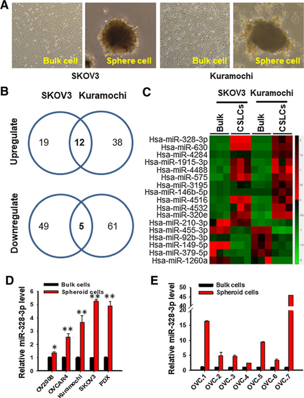
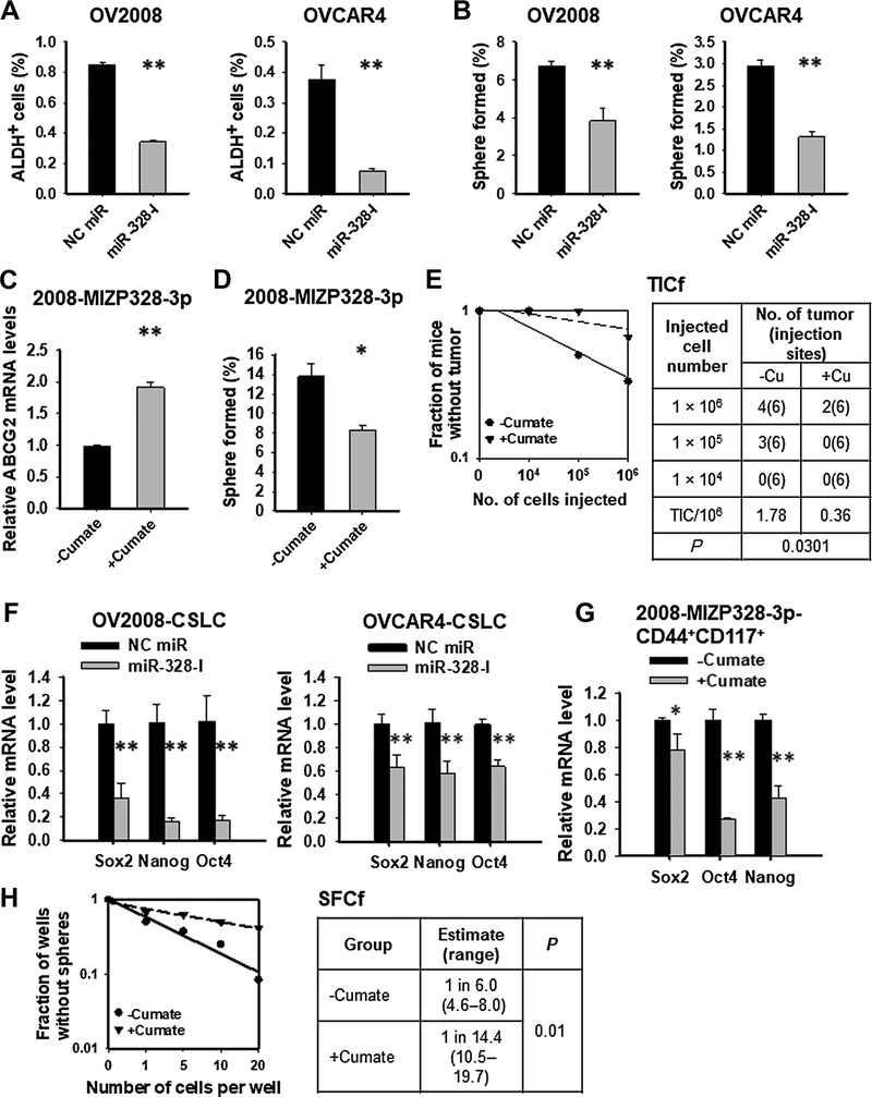
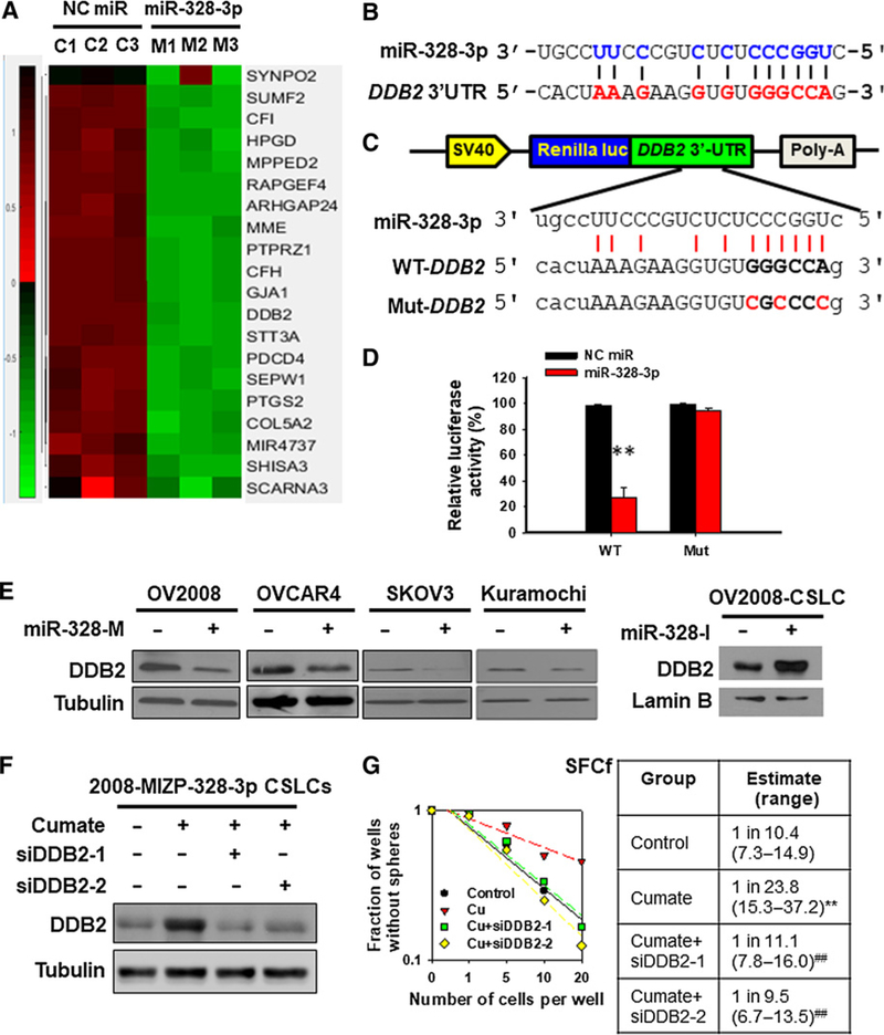
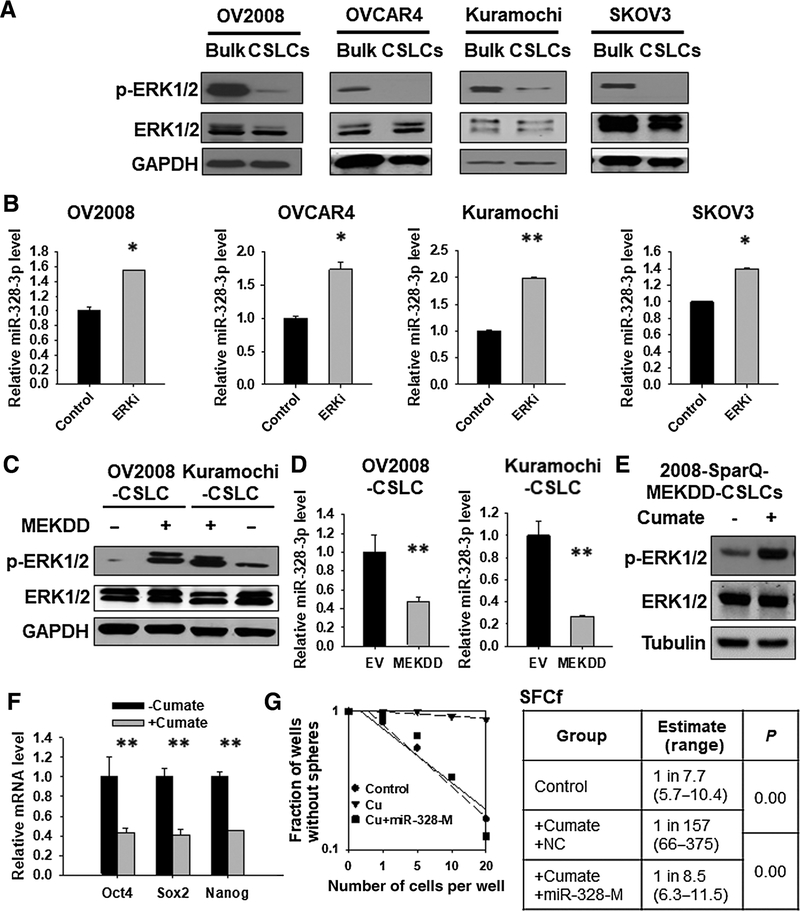
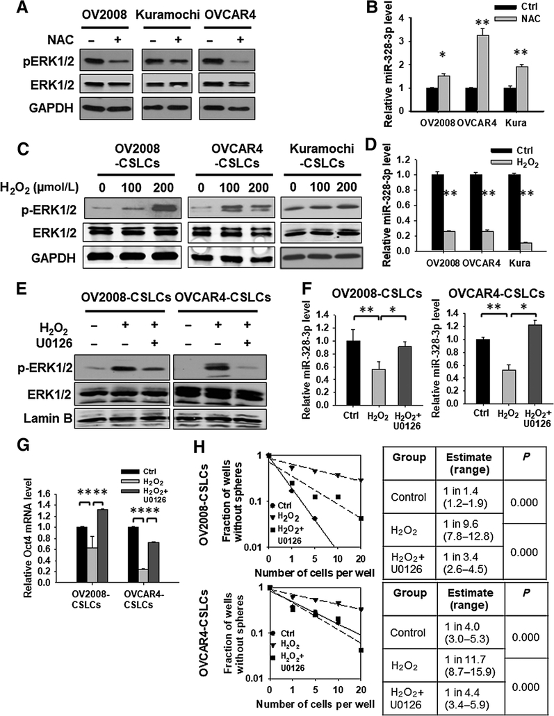
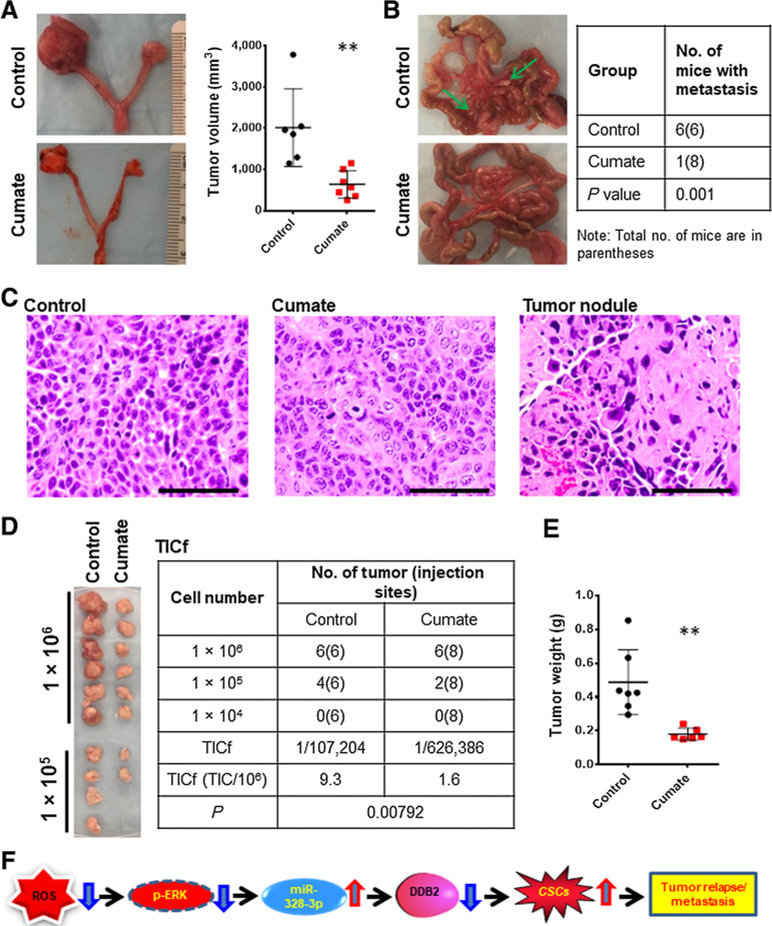
Cancer stem cells (CSC) play a central role in cancer metastasis and development of drug resistance. miRNA are important in regulating CSC properties and are considered potential therapeutic targets. Here we report that miR-328-3p (miR-328) is significantly upregulated in ovarian CSC. High expression of miR-328 maintained CSC properties by directly targeting DNA damage binding protein 2, which has been shown previously to inhibit ovarian CSC. Reduced activity of ERK signaling in ovarian CSC, mainly due to a low level of reactive oxygen species, contributed to the enhanced expression of miR-328 and maintenance of CSC. Inhibition of miR-328 in mouse orthotopic ovarian xenografts impeded tumor growth and prevented tumor metastasis. In summary, our findings provide a novel mechanism underlying maintenance of the CSC population in ovarian cancer and suggest that targeted inhibition of miR-328 could be exploited for the eradication of CSC and aversion of tumor metastasis in ovarian cancer. SIGNIFICANCE: These findings present inhibition of miR-328 as a novel strategy for efficient elimination of CSC to prevent tumor metastasis and recurrence in patients with epithelial ovarian cancer.


Conflict of interest statement

Disclosure of Potential Conflicts of Interest

No potential conflicts of interest were disclosed.

Figures

Figure 1.
Figure 1.
Ovarian CSCs possess highly expressed miR-328. A–C, NanoString nCounter analysis revealed 12 upregulated and 5 downregulated miRNAs in spheroid cells compared with their corresponding bulk cancer cells. EOC cell lines Kuramochi and SKOV3 were cultured in Ultra-Low Attachment dishes with CSC culture medium as described in Materials and Methods for 12 days to enrich CSCs (A). The spheres formed under these culture conditions (CSLCs), as well as monolayer cells cultured under the regular culture conditions (bulk cells), were harvested; RNA was isolated and subjected to miRNA screening using NanoString nCounter. B, Venn diagram shows the number of altered miRNA in CSLCs derived from Kuramochi and SKOV3 cell lines. C, Heat map of altered miRNA in CSLCs derived from both cell lines was generated. D, Enhanced expression of miR-328 was confirmed in CSLCs isolated from four EOC cell lines and one PDX. Spheroid (CSLCs) and adherent (Bulk) cells were harvested from various EOC cell lines and one ovarian tumor PDX; miR-328 was quantified using qRT-PCR. n = 3; Bar, SD; *, P < 0.05; **, P < 0.001. E, Enhanced expression of miR-328 was confirmed in spheroid cells isolated from primary HGSOC samples. Tumor cells were isolated from seven freshly removed primary HGSOC samples, cultured under sphere culture conditions (Spheroid) and adherent culture conditions (Bulk). The miR-328 level was quantified using qRT-PCR. n = 3; bar, SD. Analysis by the linear mixed model indicates that miR-328 expression increased significantly in spheroid cells compared with bulk cancer cells (P < 0.0001).
Figure 2.
Figure 2.
miR-328 is critical to maintaining the CSC population in EOC cells. A, Inhibition of miR-328 (miR-328-I) decreased the abundance of ALDH+ cells in various EOC cell lines. OV2008 and OVCAR4 cells were transiently transfected with either NC miR or miR-328-I for 48 hours. The proportion of ALDH+ cells was analyzed with ALDEFLUOR assay using FACS. N = 3; bar, SD. B, Inhibition of miR-328 reduced the sphere formation capability of EOC cell lines. OV2008 and OVCAR4 cells were transiently transfected with either NC miR or miR-328-I for 48 hours. Semisolid colony formation assay was used to determine their sphere formation capacity. N = 3; bar, SD. C–E, Inhibition of miR-328 reduced the tumorigenic potential of the EOC cells. OV2008 cells possessing cumate-inducible anti-miR-328 expression vectors (2008-MIZP328–3p) were treated with or without cumate for 3 days. The expression level of ABCG2 (a direct target of miR-328) was determined to assess the inhibition of miR-328 (C). Semisolid colony formation assay was used to determine their sphere formation capacity (D). Xenotransplantation limiting dilution assay was used to estimate the TICf (E). N = 3; bar, SD. F–H, Inhibition of miR-328 in ovarian CSLCs reduces the CSC properties. CSLCs were isolated from OV2008 and OVCAR4 cells, transfected with either NC miR or miR-328-I for 48 hours, and the expression of stem cell–specific genes was determined using qRT-PCR (F). CSLCs were isolated from 2008-MIZP328–3p cells, treated with or without cumate for 3 days. The expression of stem cell–specific genes was determined (G). Cells were plated in a limiting dilution manner in 96-well Ultra-Low Attachment plates with CSC culture medium, and the number of wells containing spheres was calculated after 12 days to calculate the SFCf (H). N = 3; bar, SD; *, P < 0.05; **, P < 0.01.
Figure 3.
Figure 3.
DDB2 is a direct target of miR-328 and mediates the function of miR-328 in the regulation of CSC properties. A, Gene expression heatmap from microarray analysis of miR-328–overexpressing ovarian cancer cells. OV2008 cells were transfected with miR-328 mimics or NC miR for 2 days. Total RNA was isolated, and the differentially expressed genes were screened using GeneChip Human Transcriptome Array 2.0. B, Predicted binding of miR-328 to the 3′-UTR of DDB2. C and D, Dual luciferase reporter assay shows the interaction of miR-328 and its targeting sequence in the DDB2 3′-UTR. OV2008 cells were transfected with psiCHECK-2 containing the wild-type (WT) or mutated (Mut) target site of the DDB2 3′-UTR (C) plus miR-328-M or NC miR for 48 hours. The luciferase activity was determined and normalized to the Firefly activity and presented as relative activity to the corresponding NC miR (D). N = 3; bar, SD; **, P < 0.01. E, miR-328 downregulates the DDB2 protein level. miR-328-M was transfected into various EOC cell lines for 48 hours, and miR-328-I was transfected into OV2008-CSLCs for 48 hours; immunoblotting analyses were conducted to show the protein level of DDB2 in these cells. F and G, DDB2 downregulation antagonizes anti-miR-328-reduced sphere formation capability of ovarian CSLCs. The 2008-MIZP328–3p cells were treated with cumate to induce miR-328 inhibition. Meanwhile, cells were also transfected with two different siDDB2 for 48 hours, and DDB2 expression was determined (F). Cells were plated in Ultra-Low Attachment 96-well plates in a limiting dilution manner, and the number of wells containing spheres was counted after 12 days to generate the SFCf (G). **, P < 0.01 compared with control; ##, P < 0.01 compared with cumate.
Figure 4.
Figure 4.
Reduced ERK signaling activity contributes to the upregulation of miR-328 expression and maintenance of CSC properties in ovarian CSLCs. A, Expression of p-ERK1/2 in CSLCs isolated from various ovarian cancer cell lines. Spheroid (CSLCs) and adherent (Bulk) cells were harvested from various EOC cell lines. p-ERK1/2 was determined using immunoblotting; ERK1/2 and GAPDH were also determined to serve as loading controls. B, Inhibition of the ERK signaling enhanced miR-328 expression. EOC cell lines were treated with the ERK inhibitor U0126 for 24 hours, RNA was isolated, and the miR-328 level was determined using qRT-PCR. N = 3; bar, SD; *, P < 0.05; **, P < 0.01. C and D, Activation of the ERK signaling reduced the expression of miR-328. OV2008 and Kuramochi CSLCs were transiently transfected with either empty vector or MEKDD expressing vector for 48 hours; immunoblotting was used to determine the expression of p-ERK1/2 (C), and qRT-PCR was used to determine the level of miR-328 (D). N = 3; bar, SD; **, P < 0.01. E, Cumate induced ERK1/2 phosphorylation in 2008-SparQ-MEKDD CSLCs. F, Activation of the ERK signaling by treating cells with cumate reduced the expression level of stem cell–specific genes in 2008-SparQ-MEKDD CSLCs. N = 3; bar, SD; **, P < 0.01. G, Overexpression of miR-328 antagonized the decrease of SFCf induced by ERK activation. The 2008-SparQ-MEKDD CSLCs were treated with or without cumate. Meanwhile, cells were transfected with miR-328-M. After 3 days, the SFCf was determined using the sphere formation assay in a limiting dilution manner.
Figure 5.
Figure 5.
Low ROS level contributes to the reduced ERK activity and enhanced miR-328 expression in ovarian CSLCs. A and B, Depleting ROS inhibits the ERK1/2 phosphorylation and increases miR-328 expression in ovarian cancer cell lines. Various EOC cell lines were treated with the ROS inhibitor N-acetyl-l-cysteine (NAC, 10 mmol/L) for 12 hours, p-ERK1/2 was detected using Western blotting (A), and miR-328 expression was determined using qRT-PCR (B). N = 3; bar, SD; *, P < 0.05; **, P < 0.01. C and D, Oxidative stress enhances ERK phosphorylation and reduces miR-328 expression in ovarian CSLCs. CSLCs enriched from various EOC cell lines were treated with H2O2 for 24 hours. p-ERK1/2 was detected using Western blotting (C), and miR-328 expression was determined using qRT-PCR (D). N = 3; bar, SD; **, P < 0.01. E–H, ERK signaling inhibition antagonizes H2O2-induced reduction of miR-328 expression and CSC properties. OV2008-CSLCs and OVCAR4-CSLCs were treated with H2O2 and/or the ERK inhibitor U0126 for 24 hours, and the p-ERK1/2 level was determined using immunoblotting (E). F, The miR-328 level was determined using qRT-PCR. N = 3; bar, SD; *, P < 0.05; **, P < 0.01. G, Expression of the stem cell–specific gene Oct4 was determined using qRT-PCR. N = 3; bar, SD; **, P < 0.01. The SFCf in these cells were determined using the sphere formation assay with limiting dilution (H).
Figure 6.
Figure 6.
Inhibition of miR-328 impedes tumor growth and metastasis in the orthotopic ovarian xenograft model. A, Representative images of orthotopic ovarian xenografts generated by injecting 2008-MIZP328–3p cells into the ovary of nude mice. Volumes of xenografts in mice treated or untreated with cumate were measured. **, P < 0.01. B, Macroscopic appearances and counts of ovarian metastasis nodules in peritoneal cavity. C, Representative H&E-stained images of the xenograft tumors and metastasis nodules. Bar, 100 μm. D and E, Xenotransplantation limiting dilution assay was used to estimate TICs in these xenografts. Tumor cells were isolated from xenografts and injected into NOD/SCID mice subcutaneously in a limiting dilution manner. Images of tumors after 2 weeks of injection were presented, and the number of xenografts was counted to generate TICf using the online algorithm described in Materials and Methods (D). Weight of tumors generated with 1 × 10⁶ xenograft cells isolated from control and cumate-treated mice bearing orthotopic xenografts (E). F, Model of the mechanism by which miR-328 regulates the maintenance of CSC properties and tumor metastasis.

Similar articles

Cited by

References

    1. Zhang S, Balch C, Chan MW, Lai HC, Matei D, Schilder JM, et al. Identification and characterization of ovarian cancer-initiating cells from primary human tumors. Cancer Res 2008;68:4311–20. - PMC - PubMed
    1. Baba T, Convery PA, Matsumura N, Whitaker RS, Kondoh E, Perry T, et al. Epigenetic regulation of CD133 and tumorigenicity of CD133+ ovarian cancer cells. Oncogene 2009;28:209–18. - PubMed
    1. Bapat SA, Mali AM, Koppikar CB, Kurrey NK. Stem and progenitor-like cells contribute to the aggressive behavior of human epithelial ovarian cancer. Cancer Res 2005;65:3025–9. - PubMed
    1. Marcato P, Dean CA, Giacomantonio CA, Lee PW. Aldehyde dehydrogenase: its role as a cancer stem cell marker comes down to the specific isoform. Cell Cycle 2011;10:1378–84. - PubMed
    1. Weiswald LB, Bellet D, Dangles-Marie V. Spherical cancer models in tumor biology. Neoplasia 2015;17:1–15. - PMC - PubMed

Publication types

MeSH terms