. 2019 May 6;218(5):1670-1685.
doi: 10.1083/jcb.201809161. Epub 2019 Mar 20.

Tks5 and Dynamin-2 enhance actin bundle rigidity in invadosomes to promote myoblast fusion

Affiliations

Tks5 and Dynamin-2 enhance actin bundle rigidity in invadosomes to promote myoblast fusion

Mei-Chun Chuang et al. J Cell Biol. .

Abstract

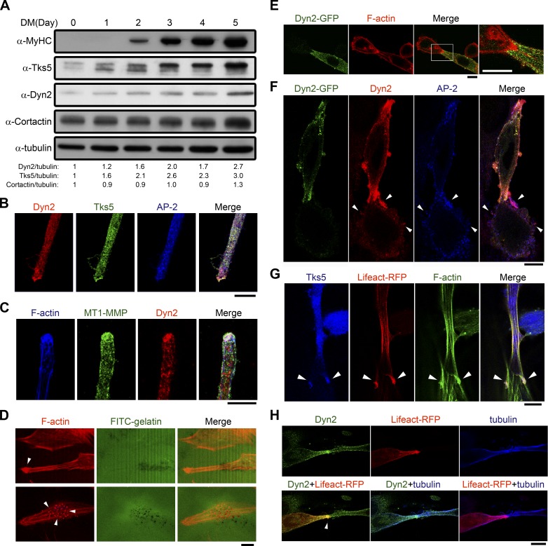
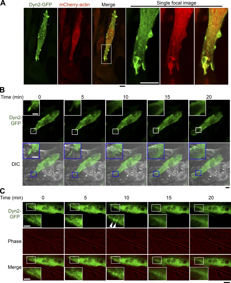
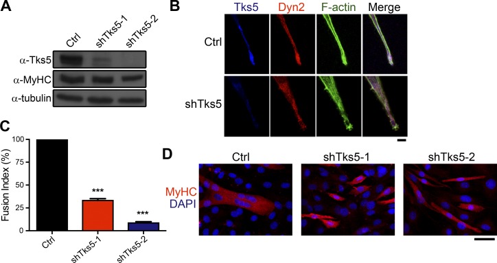
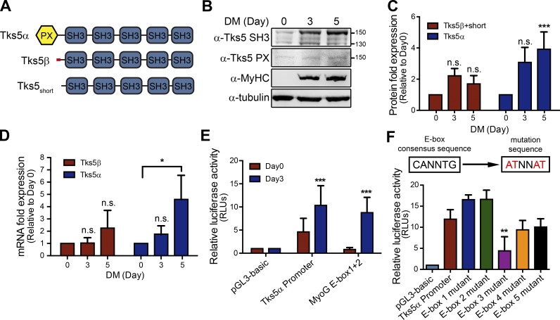
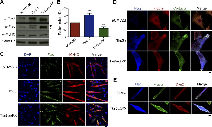
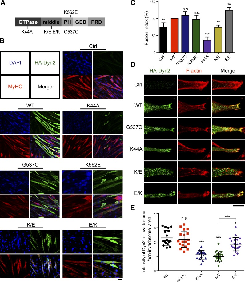
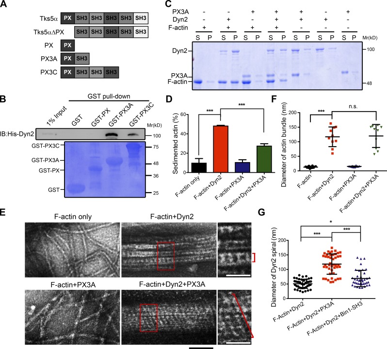
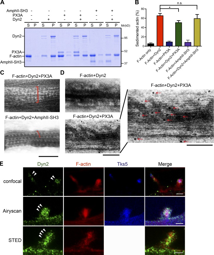
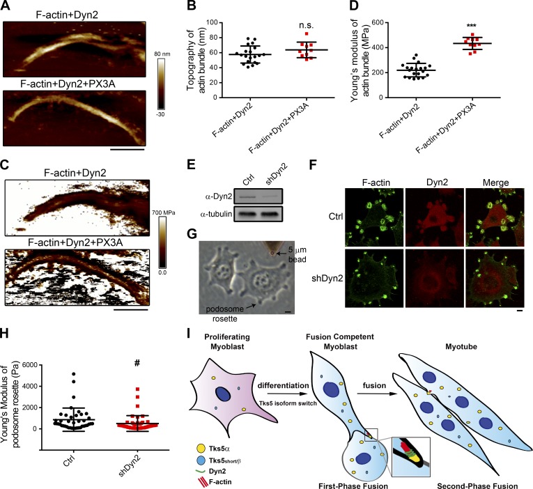
Skeletal muscle development requires the cell-cell fusion of differentiated myoblasts to form muscle fibers. The actin cytoskeleton is known to be the main driving force for myoblast fusion; however, how actin is organized to direct intercellular fusion remains unclear. Here we show that an actin- and dynamin-2-enriched protrusive structure, the invadosome, is required for the fusion process of myogenesis. Upon differentiation, myoblasts acquire the ability to form invadosomes through isoform switching of a critical invadosome scaffold protein, Tks5. Tks5 directly interacts with and recruits dynamin-2 to the invadosome and regulates its assembly around actin filaments to strengthen the stiffness of dynamin-actin bundles and invadosomes. These findings provide a mechanistic framework for the acquisition of myogenic fusion machinery during myogenesis and reveal a novel structural function for Tks5 and dynamin-2 in organizing actin filaments in the invadosome to drive membrane fusion.


Figures

Figure 1.
Figure 1.
Invadosome forms and distributes asymmetrically in differentiated myoblasts. (A) Expression level of different invadosome components in myoblasts upon differentiation. Myoblast lysates derived from different days of DM treatment were immunoblotted with indicated antibodies. Numbers below indicate the fold-change of Dyn2, Tks5, and Cortactin compared with day 0 after normalization with tubulin. (B and C) Colocalization of invadosome components at the myoblast tip of differentiated myoblast. Day 3 differentiated C2C12 myoblasts were immunofluorescence stained to detect endogenous Tks5, Dyn2, MT1-MMP, F-actin, and AP-2. Images were acquired with z-stack confocal microscopy and shown as single focal planes. (D) Matrix degradation ability. Day 3 differentiated myoblasts were seeded onto an FITC-gelatin–coated coverslip and imaged after 24 h. Arrowheads indicate invadosomes. (E–H) Asymmetrical distribution of invadosome in fusing myoblasts. Two close-positioned, day 3 differentiated myoblasts with one cell labeled with Dyn2-GFP or Lifeact-RFP were stained for F-actin (E), Dyn2 and AP-2 (F), Tks5 (G), and tubulin (H). Arrowheads in F indicate the enriched Dyn2 and AP2 at the cell periphery of the receiving cell or the invadosome in attacking cells (G and H). Scale bars (both black and white), 10 µm.
Figure 2.
Figure 2.
Dyn2 enriches at myoblast fusion site. (A) Dyn2-GFP and actin-based finger-like structures in differentiated myoblasts. Differentiated C2C12-expressing Dyn2-GFP and mCherry-actin was fixed and imaged with z-stack confocal microscopy. Maximum-intensity projections are shown, while magnified insets are single focal images. Scale bars (black and white), 10 µm. (B) Dyn2-GFP enrichment at the site of myoblast fusion. The dynamic localization of Dyn2-GFP in day 3 differentiated C2C12 was monitored with time-lapse microscopy. Five frames right before and after myoblast fusion from a 5-h recording were extracted and shown. Boxed regions were enlarged and shown in insets. Scale bars (black and white), 10 µm. (C) Dyn2-rich membrane protrusion in second-phase myoblast fusion. Time-lapse microscopic imaging of Dyn2-GFP in day 4 differentiated C2C12. Phase-contrast image was pseudocolored to better observe the boundary between two myotubes. White arrowheads indicate the Dyn2-GFP foci at the fusion interface. Black bar, 20 µm. White bar, 10 µm.
Figure 3.
Figure 3.
Invadosome is required for myoblast fusion. (A and B) Differentiation and morphology of Tks5-depleted myoblasts. Tks5 was depleted by two lentiviral shRNAs and selected with puromycin for 3 d. After 3 d of DM treatment, cells were processed for immunoblotting (A) or immunostaining (B). Scale bar, 10 µm. (C and D) Myoblast fusion in Tks5-depleted myoblasts. Fusion efficiency was analyzed in day 4 DM-treated myoblasts by immunofluorescent staining and quantified as number of nuclei in multinuclear cell (nuclear number ≥3)/number of nuclei in MyHC-positive cell. Fusion efficiency is considered as 100% in control cells. Scale bar, 50 µm. All values reported in this study represent the mean ± SD of at least three independent experiments, and data were analyzed with one-way ANOVA. ***, P < 0.001.
Figure 4.
Figure 4.
Tks5 undergoes isoform switch during myoblast differentiation. (A) Domain structure of different Tks5 isoforms. Red box represents exon 6β, which is unique in Tks5β. (B–D) The expression of Tks5 isoforms during myogenesis. Expression of protein or mRNA levels of different Tks5 isoforms in myoblasts upon differentiation were examined with immunoblotting (B and C) or quantitative PCR (D). C was derived from the results using anti-SH3 antibody. (E) Promoter activity of Tks5α promoter upon myoblast differentiation. Dual luciferase reporter assay of Tks5α promoter was performed and compared among day 0 or 3 differentiated C2C12. The MyoG E-box serves as positive control. (F) Dual luciferase reporter assay of different E-box mutations of Tks5α promoter. Data were analyzed with one-way ANOVA (C, D, and F) or Student’s t test (E). *, P < 0.05; **, P < 0.01; ***, P < 0.001.
Figure 5.
Figure 5.
Isoform switch of Tks5 is critical for myoblast fusion. (A) Effects of ectopic expression of flag-tagged Tks5α or Tks5αΔPX in day 4 differentiated myoblasts. Cell lysates were immunoblotted with anti-Tks5 SH3 or anti-Flag antibodies, with arrow and arrowhead indicating Tks5α and Tks5αΔPX, respectively. Asterisk shows nonspecific signal of anti-Flag antibody. (B and C) Fusion efficiency of different Tks5 constructs expressing myoblasts. Scale bar, 30 µm. **, P < 0.01; ***, P < 0.001. (D) Effects of ectopic expression of flag-tagged Tks5 in undifferentiated myoblasts. After 48-h transfection, undifferentiated myoblasts were seeded on fibronectin-coated coverslips and stained for actin, Cortactin, and Flag. Maximum-intensity projections are shown. (E) Effects of Tks5αΔPX expression on invadosome formation. Day 3 differentiated myoblasts with Tks5α or Tks5αΔPX expression were seeded on fibronectin-coated coverslips and stained to detect endogenous Dyn2 and F-actin distribution. Scale bars, 10 µm.
Figure 6.
Figure 6.
Dyn2 is directly involved in myoblast fusion. (A) Dyn2 domain structure and the mutations used in this study. (B and C) Fusion efficiency of myoblasts infected with different Dyn2 mutants were imaged (B) and quantified (C). These Dyn2 mutants were tetracycline-regulated and were induced 2 d after differentiation to avoid their effects on differentiation. **, P < 0.01; ***, P < 0.001. (D and E) Distribution of Dyn2 mutants in day 3 differentiated myoblasts. After 2 d of differentiation, C2C12 myoblasts were replated into lower density on fibronectin-coated coverslips and infected with HA-Dyn2 mutants expressing adenoviruses. After 16-h induction, Dyn2 mutants and F-actin were stained and imaged with confocal microscopy. The enrichment of Dyn2 mutants in invadosomes was quantified by the intensity of Dyn2 in actin focus divided by the intensity outside the invadosome (E). 20 cells of each mutant were analyzed. Scale bars, 10 µm.
Figure 7.
Figure 7.
Tks5 interacts with Dyn2 and regulates its assembly around actin. (A) Domain structure of different Tks5 constructs used in this study. (B) Direct interaction between Dyn2 and Tks5. GST pulldown assay was performed using purified full-length Dyn2 and three GST-tagged Tks5 fragments, GST-PX, GST-PX3A, and PX3C. Bound His-Dyn2 was detected with immunoblotting, and GST-tagged proteins were shown with Coomassie Blue stain. (C and D) F-actin bundling assay. Prepolymerized filamentous actin was incubated with purified Dyn2 or Tks5 as indicated. After 30 min of incubation, bundled actin was sedimented into pellet (P), and the ratio of bundled actin (pellet/total) was quantified with SDS-PAGE, Coomassie Blue staining, and ImageJ (D). (E and G) Electron micrographs of Dyn2-mediated actin bundling with or without Tks5. Single Dyn2 spiral was indicated by red braces in the magnified panels, and the diameters of actin bundles or Dyn2 spirals in the presence or absence of Tks5 were quantified with ImageJ (F and G). Black bar, 100 nm. White bars, 50 nm. *, P < 0.05; ***, P < 0.001.
Figure 8.
Figure 8.
Tks5 regulates Dyn2 assembly around actin. (A–C) Effects of Amphiphysin II and Tks5 on Dyn2-actin bundle. SH3 domain from Tks5 (PX3A) or Amphiphysin II (AmphII-SH3) were added into Dyn2 and actin filament reactions. Bundled actin was sedimented, and the ratio of bundled actin (pellet/total) was analyzed and quantified with ImageJ (B) or imaged with negative stain TEM (C). (D) Immunogold staining for Tks5 on Dyn2-actin bundle. Tks5/GST-PX3A was labeled with anti-GST antibody and subsequently with 6 nm gold particle–conjugated anti-rabbit antibody. A magnified image was shown with red arrows indicating the gold particles. Scale bars, 100 nm. (E) Distribution of endogenous Dyn2 in invadosome. Differentiated myoblasts were stained with indicated antibodies and imaged with confocal, confocal with Airyscan detector, or STED microscopy. Single focal images are shown. Scale bars, 2 µm. *, P < 0.05.
Figure 9.
Figure 9.
Tks5 regulates the physical properties of Dyn2-actin bundles. (A–D) AFM topography and stiffness map (Young’s modulus) of Dyn2-actin bundles. Height (A) and corresponding stiffness (C) of Dyn2-actin bundles with or without GST-PX3A addition were recorded and quantified with Peakforce QNM. Quantification results are shown in C and D. n ≥ 10. Scale bars, 800 µm. ***, P < 0.001. (E and F) Dyn2 knockdown in c-SrcY527F–transformed NIH3T3 cells. Dyn2 was depleted by lentiviral shRNA and selected with puromycin for 3 d. Cells were processed for immunoblotting (E) or immunostaining (F). Scale bar, 10 µm. (G and H) AFM stiffness map (Young’s modulus) of podosome rosette. Podosome rosettes in control or Dyn2-depleted c-SrcY527F–transformed NIH3T3 cells with diameter ∼5 µm were analyzed with AFM probe equipped with a 5-µm bead in medium (G). Approximately 40 podosome rosettes in >10 cells of each condition were measured and quantified in H. #, P = 0.079. (I) Tks5 mediates invadosome maturation through regulating dynamin-actin organization to drive myoblast fusion. Upon myoblast differentiation, Tks5 is up-regulated and undergoes isoform switching to the long isoform Tks5α that encodes a membrane-interacting PX domain. Tks5α promotes the formation and maturation of an invadosome by dictating Dyn2 assembly, strengthening the protrusive actin-rich structure and perhaps coupling these force-generating filaments to the membrane. The invadosome thus functions as a molecular drill to propel both the first- and second-phase myoblast fusion.

Similar articles

Cited by

References

    1. Abmayr S.M., and Pavlath G.K.. 2012. Myoblast fusion: lessons from flies and mice. Development. 139:641–656. 10.1242/dev.068353 - DOI - PMC - PubMed
    1. Albiges-Rizo C., Destaing O., Fourcade B., Planus E., and Block M.R.. 2009. Actin machinery and mechanosensitivity in invadopodia, podosomes and focal adhesions. J. Cell Sci. 122:3037–3049. 10.1242/jcs.052704 - DOI - PMC - PubMed
    1. Almada A.E., and Wagers A.J.. 2016. Molecular circuitry of stem cell fate in skeletal muscle regeneration, ageing and disease. Nat. Rev. Mol. Cell Biol. 17:267–279. 10.1038/nrm.2016.7 - DOI - PMC - PubMed
    1. Antonny B., Burd C., De Camilli P., Chen E., Daumke O., Faelber K., Ford M., Frolov V.A., Frost A., Hinshaw J.E., et al. . 2016. Membrane fission by dynamin: what we know and what we need to know. EMBO J. 35:2270–2284. 10.15252/embj.201694613 - DOI - PMC - PubMed
    1. Blau H.M., Pavlath G.K., Hardeman E.C., Chiu C.P., Silberstein L., Webster S.G., Miller S.C., and Webster C.. 1985. Plasticity of the differentiated state. Science. 230:758–766. 10.1126/science.2414846 - DOI - PubMed

Publication types

LinkOut - more resources