. 2019 May 3;294(18):7419-7432.
doi: 10.1074/jbc.RA118.006672. Epub 2019 Mar 20.

Structural analysis of a plant fatty acid amide hydrolase provides insights into the evolutionary diversity of bioactive acylethanolamides

Affiliations

Structural analysis of a plant fatty acid amide hydrolase provides insights into the evolutionary diversity of bioactive acylethanolamides

Mina Aziz et al. J Biol Chem. .

Abstract

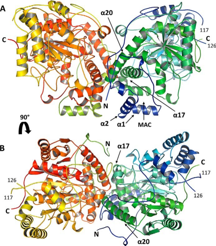
N-Acylethanolamines (NAEs) are fatty acid derivatives that in animal systems include the well-known bioactive metabolites of the endocannabinoid signaling pathway. Plants use NAE signaling as well, and these bioactive molecules often have oxygenated acyl moieties. Here, we report the three-dimensional crystal structures of the signal-terminating enzyme fatty acid amide hydrolase (FAAH) from Arabidopsis in its apo and ligand-bound forms at 2.1- and 3.2-Å resolutions, respectively. This plant FAAH structure revealed features distinct from those of the only other available FAAH structure (rat). The structures disclosed that although catalytic residues are conserved with the mammalian enzyme, AtFAAH has a more open substrate-binding pocket that is partially lined with polar residues. Fundamental differences in the organization of the membrane-binding "cap" and the membrane access channel also were evident. In accordance with the observed structural features of the substrate-binding pocket, kinetic analysis showed that AtFAAH efficiently uses both unsubstituted and oxygenated acylethanolamides as substrates. Moreover, comparison of the apo and ligand-bound AtFAAH structures identified three discrete sets of conformational changes that accompany ligand binding, suggesting a unique "squeeze and lock" substrate-binding mechanism. Using molecular dynamics simulations, we evaluated these conformational changes further and noted a partial unfolding of a random-coil helix within the region 531-537 in the apo structure but not in the ligand-bound form, indicating that this region likely confers plasticity to the substrate-binding pocket. We conclude that the structural divergence in bioactive acylethanolamides in plants is reflected in part in the structural and functional properties of plant FAAHs.

Keywords: Arabidopsis; N-acylethanolamines; crystal structure; endocannabinoid; fatty acid amide hydrolase (FAAH); hydrolase; lipid signaling; oxylipins; quorum sensing; seed germination; squeeze and lock mechanism.


Conflict of interest statement

The authors declare that they have no conflicts of interest with the contents of this article

Figures

Figure 1.
Figure 1.
Arabidopsis FAAH three-dimensional structure. A and B, side (A) and top (B) views of the AtFAAH structure. AtFAAH is a homodimer assembled from 66-kDa subunits. Each subunit is shown in a color gradient ranging from blue (N terminus) to green (C terminus) for one subunit (chain A) and from yellowish-green to red for the other subunit (chain B). The presumed membrane-binding cap (α1 and α2) and the putative substrate entryway (MAC) are located at the N terminus of the enzyme. The AtFAAH dimer interface is formed mainly by parts of helices α17 and α20 and some regions of the N terminus (see Fig. 7).
Figure 2.
Figure 2.
Comparison of Arabidopsis FAAH structure with other AS enzymes. A–F, AtFAAH (green; PDB code 6DHV) was superposed to the structures of rat FAAH (gray; PDB code 3QJ8 (14)) (A and D), glutamine amidotransferase subunit A (wheat; PDB code 3KFU (15)) (B and E), and aryl acylamidase (pink; PDB code 4YJ6 (18)) (C and F) and presented as structure overlays from two different views. The N and C termini of each enzyme are indicated with letters of the same color as the corresponding protein structure.
Figure 3.
Figure 3.
The putative membrane-binding cap of AtFAAH. The hydrophobic helices α1 and α2 of the N terminus (amino acids 27–60) are rich in hydrophobic amino acids (21 of 34) and are predicted to form the membrane-binding cap of AtFAAH that is presumed to anchor the enzyme into half of the lipid bilayer. A and B, one monomer of apo-AtFAAH (PDB code 6DHV) integrated into the membrane is shown in both cartoon (A) and electrostatic surface (B) depictions with positive charged areas in blue, negative charged areas in red, and hydrophobic areas in white. C and D, electrostatic molecular surface of AtFAAH viewed from the membrane face showing the membrane-binding cap and the MAC in the open (C) and closed (D) conformations in apo- (PDB code 6DHV) and MLnFP-bound (PDB code 6DII) AtFAAHs, respectively. Some of the hydrophobic amino acids on helices α1 and α2 are highlighted as yellow sticks with their numbers and single-letter codes indicated.
Figure 4.
Figure 4.
Arabidopsis FAAH active site and substrate-binding pocket. The acyl chain of the irreversible inhibitor MLnFP (cyan sticks) is surrounded by several aliphatic and aromatic amino acids as well as some polar residues (e.g. Asn59, Thr258, His441, Ser472, Thr535, and Thr536). All the depicted residues (slate blue lines) are within 5-Å distance from the inhibitor. Representative van der Waals interactions between the ligand and some of the surrounding residues are shown as blue dashed lines with the distance of each potential interaction indicated; only a few selected interactions are shown for simplification. The Ser305-Ser281-Lys205 catalytic triad is shown (slate blue sticks) with the nucleophilic Ser305 covalently bound to the phosphorus atom of MLnFP. The 2FoFc electron density map of MLnFP contoured at 1.0 σ is shown in gray.
Figure 5.
Figure 5.
Arabidopsis FAAH substrate-binding pocket is more accessible and relatively more polar than that of rat FAAH. A and B, chemical structures of the irreversible inhibitors MLnFP and MAFP. C and D, one subunit of the ligand-bound AtFAAH (C) (PDB code 6DII) and rat FAAH (D) (PDB code 1MT5) (12) are shown with the protein molecular surface rendered gray and transparent to illustrate the key differences between both enzymes with respect to the MAC and the ABC. The putative membrane-binding cap is colored magenta (α1 and α2) and red (α18 and α19) in AtFAAH and rat FAAH, respectively. In rat FAAH, the dynamic paddle residues, Phe432 and Trp531, are shown as green sticks. E and F, comparison of the substrate-binding pockets of AtFAAH (E) (PDB code 6DII) and rat FAAH (F) (PDB code 1MT5) complexed with MLnFP (cyan sticks) and MAFP (yellow sticks), respectively. Both enzymes were superposed, and the substrate-binding pockets were compared; only a few selected residues are shown for simplification. The amino acid residues of AtFAAH are shown as slate blue sticks, whereas those of rat FAAH are shown as orange sticks. The numbers on the ligands indicate the position of the double bonds.
Figure 6.
Figure 6.
Differences in the organization of the MAC and the ABC between Arabidopsis and rat FAAH. A and B, one subunit of the ligand-bound Arabidopsis FAAH (A) (PDB code 6DII) and rat FAAH (B) (PDB code 1MT5) (12) were superposed, and their cavities/channels are shown as dark gray shadows. In rat FAAH, there are two distinct channels for substrate access and acyl chain binding with two hydrophobic residues (Phe432 and Trp531; shown in orange, space-filling representation) forming the so-called dynamic paddle located at the junction of these two channels. In Arabidopsis FAAH, there are no residues corresponding to Phe432 and Trp531, and the substrate is presumed to access directly from the membrane to the acyl-binding channel. In other words, in AtFAAH, there is only one large cavity for both substrate access and binding (i.e. indistinguishable MAC and ABC). The catalytic triad residues are shown as slate blue and orange sticks in Arabidopsis and rat FAAHs, respectively. The irreversible inhibitors MLnFP (cyan sticks) and MAFP (yellow sticks) are shown bound to the active site of Arabidopsis and rat FAAHs, respectively.
Figure 7.
Figure 7.
Atomic interactions that contribute to dimer formation in AtFAAH. A, the overall dimer of Arabidopsis FAAH with the dimerization region indicated by a red circle. B, a closeup view of this dimerization region with some of the key residues depicted as sticks; the AtFAAH dimer interface is formed mainly by parts of helices α17 and α20 and some loop regions of the N terminus. C, detailed representation of the network of hydrogen bonds and van der Waals interactions at the monomer–monomer interface of AtFAAH. Residues from one subunit (chain A) are shown as green sticks, whereas those from the other subunit (chain B) are shown as cyan sticks. The single-letter code and the number of each amino acid as well as the distance of each potential interaction are indicated.
Figure 8.
Figure 8.
Arabidopsis FAAH can accommodate and hydrolyze NAE oxylipins more efficiently than rat FAAH. A and B, chemical structures of linoleoylethanolamide (NAE 18:2) and NAE-9-HOD. C and D, initial velocities were measured for AtFAAH (C) and rat FAAH (D) with increasing concentrations of either [1-14C]NAE 18:2 or [1-14C]NAE-9-HOD. Data points represent means ± S.D. (error bars) of triplicate enzymatic assays. E, summary of the apparent kinetic parameters of both enzymes. F, docking of NAE-9-HOD in the substrate-binding pocket of AtFAAH with the oxygenated acyl chain of the substrate displayed in two different binding poses with pose 1 shown as orange sticks and pose 2 shown as magenta sticks. Arabidopsis FAAH amino acid residues are depicted as green sticks. In AtFAAH, Ser472 is well-positioned to accommodate and form a hydrogen bond interaction with the hydroxyl group at position 9 when the NAE-9-HOD acyl chain exhibits binding pose 1, whereas Thr535 on the opposite side of the substrate-binding pocket can form a hydrogen bond with the 9-hydroxyl group when the substrate is in binding pose 2 (see Movie S1). The numbers of the ligand atoms (9–13) are indicated for clarity.
Figure 9.
Figure 9.
Conformational changes in Arabidopsis FAAH structure upon ligand binding. The superposed structures of one subunit of AtFAAH in both the apo (green; PDB code 6DHV) and ligand-bound (slate blue; PDB code 6DII) forms are shown. The ligand MLnFP is depicted as blue spheres. Regions of the protein that undergo conformational changes are enlarged to demonstrate the details of each group of changes. The zoomed-in surface region is a closeup view of the protein molecular surface from the membrane face showing the open (green) and closed (slate blue) MAC in the apo- and MLnFP-bound AtFAAHs, respectively; the protein surface was rendered partially transparent to show the corresponding amino acid residues.
Figure 10.
Figure 10.
Molecular dynamics simulations of Arabidopsis FAAH. A, overlay of the first (light gray) and last (pink) frames of a 100-ns MD simulation of the apo-AtFAAH structure (see Movie S2). B, overlay of the first (dark gray) and last (magenta) frames of a 100-ns MD simulation of the MLnFP-bound structure (see Movie S4). C, the ligand MLnFP was removed from the bound cocrystal structure, and the resulting apo structure was simulated for 100 ns (see Movie S5); the first and last frames are shown in dark gray and teal, respectively. D, MLnFP was docked into the apo-AtFAAH structure, and the resulting AtFAAH–MLnFP complex was simulated for 100 ns (see Movie S6); the first and last frames are shown in light gray and golden yellow, respectively. In A and C, partial unfolding of the 531–537 helix region and noticeable shifts in the N-terminal α1 and α2 helices were observed in the absence of the ligand, whereas in B and D, these changes were not observed in the presence of MLnFP. The mesh rendering indicates the surface of the cavity.
Figure 11.
Figure 11.
Schematic representation of the substrate-binding mechanisms in Arabidopsis and rat FAAH. The ligand-free AtFAAH has a long, widely open ABC with the part near the entrance regarded as the MAC. Ligand binding triggers movement of the 531–537 helix region of the ABC toward the substrate, resulting in a “squeezed” ABC. This is accompanied by a concomitant movement of residues 25–28 as well as Leu55, located on the opposite sides of the MAC, resulting in a “locked” MAC. These changes in AtFAAH upon ligand binding squeeze and lock the substrate into the binding pocket for hydrolysis. By contrast, rat FAAH has two separate acyl-binding and membrane access channels. In the absence of ligand, the dynamic paddle residue Phe432 is oriented toward the ABC, resulting in a short ABC and a long, open MAC. Upon ligand binding, Phe432 changes its side-chain orientation from the ABC to the MAC, resulting in an extended ABC and partially closed MAC. This conformational flip of the dynamic paddle residue is proposed to guide the substrate toward the active site for hydrolysis (13, 14, 21).

Similar articles

Cited by

References

    1. Lucanic M., Held J. M., Vantipalli M. C., Klang I. M., Graham J. B., Gibson B. W., Lithgow G. J., and Gill M. S. (2011) N-Acylethanolamine signalling mediates the effect of diet on lifespan in Caenorhabditis elegans. Nature 473, 226–229 10.1038/nature10007 - DOI - PMC - PubMed
    1. Keereetaweep J., Blancaflor E. B., Hornung E., Feussner I., and Chapman K. D. (2013) Ethanolamide oxylipins of linolenic acid can negatively regulate Arabidopsis seedling development. Plant Cell 25, 3824–3840 10.1105/tpc.113.119024 - DOI - PMC - PubMed
    1. Keereetaweep J., Blancaflor E. B., Hornung E., Feussner I., and Chapman K. D. (2015) Lipoxygenase-derived 9-hydro(pero)xides of linoleoylethanolamide interact with ABA signaling to arrest root development during Arabidopsis seedling establishment. Plant J. 82, 315–327 10.1111/tpj.12821 - DOI - PubMed
    1. Iannotti F. A., Di Marzo V., and Petrosino S. (2016) Endocannabinoids and endocannabinoid-related mediators: targets, metabolism and role in neurological disorders. Prog. Lipid Res. 62, 107–128 10.1016/j.plipres.2016.02.002 - DOI - PubMed
    1. Schwartz G. J., Fu J., Astarita G., Li X., Gaetani S., Campolongo P., Cuomo V., and Piomelli D. (2008) The lipid messenger OEA links dietary fat intake to satiety. Cell Metab. 8, 281–288 10.1016/j.cmet.2008.08.005 - DOI - PMC - PubMed

Publication types