. 2019 May;31(5):1043-1062.
doi: 10.1105/tpc.18.00978. Epub 2019 Mar 20.

Tomato MYB21 Acts in Ovules to Mediate Jasmonate-Regulated Fertility

Affiliations

Tomato MYB21 Acts in Ovules to Mediate Jasmonate-Regulated Fertility

Ramona Schubert et al. Plant Cell. 2019 May.

Abstract

The function of the plant hormone jasmonic acid (JA) in the development of tomato (Solanum lycopersicum) flowers was analyzed with a mutant defective in JA perception (jasmonate-insensitive1-1, jai1-1). In contrast with Arabidopsis (Arabidopsis thaliana) JA-insensitive plants, which are male sterile, the tomato jai1-1 mutant is female sterile, with major defects in female development. To identify putative JA-dependent regulatory components, we performed transcriptomics on ovules from flowers at three developmental stages from wild type and jai1-1 mutants. One of the strongly downregulated genes in jai1-1 encodes the MYB transcription factor SlMYB21. Its Arabidopsis ortholog plays a crucial role in JA-regulated stamen development. SlMYB21 was shown here to exhibit transcription factor activity in yeast, to interact with SlJAZ9 in yeast and in planta, and to complement Arabidopsis myb21-5 To analyze SlMYB21 function, we generated clustered regularly interspaced short palindromic repeats(CRISPR)/CRISPR associated protein 9 (Cas9) mutants and identified a mutant by Targeting Induced Local Lesions in Genomes (TILLING). These mutants showed female sterility, corroborating a function of MYB21 in tomato ovule development. Transcriptomics analysis of wild type, jai1-1, and myb21-2 carpels revealed processes that might be controlled by SlMYB21. The data suggest positive regulation of JA biosynthesis by SlMYB21, but negative regulation of auxin and gibberellins. The results demonstrate that SlMYB21 mediates at least partially the action of JA and might control the flower-to-fruit transition. .


Figures

Figure 1.
Figure 1.
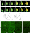
Classification of Stages and Levels of Jasmonates in Developing Carpels of Wild Type (WT) and jai1-1. (A) Developmental stages of opened stamen cones and dissected carpels of wild type and jai1-1 showing the six developmental stages as defined by Dobritzsch et al. (2015). In the picture down left, bar = 5 mm for all photographs. (B) JA and JA-Ile levels in developing carpels. Carpels of the respective stage were extracted, and contents of JA and JA-Ile were determined. Mean values ±sd are shown (n ≥ 3 independent pools of carpels). Data of the same developmental stage were compared between wild type and jai1-1 by Student’s t test (* P ≤ 0.05, ** P ≤ 0.01, *** P ≤ 0.001). (C) and (D) Immunocytochemical detection of JA/JA-Ile in ovaries (C) and ovules (D) of wild type and jai1-1. Carpels of wild type flowers at stages 1, 3, and 6 and of jai1-1 at stage 3 were harvested, fixed with 1-ethyl-3-(3-dimethyl aminopropyl)-carbodiimide hydrochloride (EDC), and processed for immunolabeling as described by Mielke et al. (2011). The occurrence of JA is indicated by the green fluorescence. Note the strongest label in wild-type carpels at stage 3, whereas fluorescence signal is faint in carpels and ovules of jai1-1. Bars = 50 µm for all micrographs. Abbreviations: ov, ovule; pl, placenta; ow, ovary wall.
Figure 2.
Figure 2.
Comparative Analysis of Transcript Accumulation in Dissected Ovules of Wild Type (WT) and jai1-1. Total RNA isolated from three developmental stages of ovules of wild type and jai1-1 was subjected to transcript profiling using the Agilent-Tomato 44K-full genome chip. (A) Venn diagram showing the number of significantly regulated genes (P ≤ 0.01, n = 3 independent pools of dissected ovules). Numbers in parentheses are related to genes with unknown functions. Note that the highest number of differentially regulated genes was found in stage 3. (B) Classification of differentially expressed genes according to functional classes. The bold numbers indicate how many genes in total were differentially regulated in the respective developmental stage, whereas the regular numbers show how many of them exhibited decreased (dark blue) or increased transcript levels in jai1-1 (dark brown). Light blue and light brown colors mark the overall tendency of differential transcript accumulation in each developmental stage/functional class (blue higher in wild type, brown higher in jai1-1). (C) and (D) Relative transcript levels of genes positively (C) and negatively (D) regulated by JA. Note that transcripts of the typical JA-regulated genes shown in (C) are nearly not detectable in ovules of jai1-1. All transcript levels were determined by RT-qPCR and set in relation to SlTIP41 (rel. expression). The inset in each diagram visualizes the absolute signal intensity (abs. signal int.) obtained from microarray analysis. Mean values ±sd are shown (n = 3). Data of the same developmental stage were compared between wild type and jai1-1 by Student’s t test (*P ≤ 0.05, **P ≤ 0.01, ***P ≤ 0.001). ▬○▬ wild type,—▵—jai1-1
Figure 3.
Figure 3.
Ovules of jai1-1 Show Altered Morphology and Exhibit Enhanced Callose Accumulation and PCD in the Nucellus. (A) Semi-thin cross sections of ovules stained with toluidine blue. The developmental stages are indicated by numbers. Note the thickened cell walls in stage 5 (arrow head) and vacuolated cells of the inner layer of nucellus in stage 6 (arrow) of jai1-1. (B) Immunolabeling of callose in ovules of stage 5. Callose visualized by green fluorescence is detectable in small spots between all cells of wild type ovules pointing to plasmodesmata (see inset), but surrounds additionally the innermost cell layer of the nucellus of jai1-1 ovules. (C) Cross sections of ovules at stage 6 were analyzed by the TUNEL assay. The wild type tissues only showed few TUNEL-positive signals. In contrast, a higher number of TUNEL-positive signals appeared in the innermost cell layer of the nucellus of the jai1-1 mutant. Positive and negative controls showed labeling of all and no nuclei, respectively (Supplemental Figure 2B). Bars = 50 µm in all micrographs, except the inset, where it = 10 µm.
Figure 4.
Figure 4.
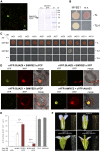
SlMYB21 Encodes an Active Transcription Factor That Interacts With SlJAZ9 and Complements the Arabidopsis Mutant myb21-5. (A) SlMYB21 is located in the nucleus as shown by transient expression of a 35S:gMYB21:GFP construct in N. benthamiana leaves. The green fluorescence of the fusion protein is detectable in nuclei of the epidermal cells. Plastids are visible by their red autofluorescence. Protein gel blot performed with total protein extract from transformed leaves (empty vector and 35S:SlMYB21:GFP) and an anti-GFP antibody shows the correct size of the fusion protein as indicated by the molecular weight marker (M). Bar in the micrograph = 20 µm. (B) Yeast assay used to detect the transcriptional activity of SlMYB21. Yeast cells transformed with SlMYB21 fused to the GAL4 BD and the empty vector encoding the GAL4 AD were grown on synthetic defined (SD) medium without tryptophan and leucine (-TL) or SD medium without tryptophane, leucine and histidine (-TLH). Transformation with the empty vector (e.v.) only served as negative control. (C) Y2H assays testing the interaction of SlMYB21 with SlJAZ proteins. Full-length CDS of all 12 SlJAZ proteins (JAZ1–JAZ11, JAZ13) were fused with the GAL4 BD and CDS of SlMYB21 was fused with the GAL4 AD. Transformed yeast cells were grown on SD/-TLH to determine protein–protein interactions. Positive yeast transformations are visualized by growth on SD/-TL. Omitting SlMYB21 by using the empty vector (e.v.) served as control and did not show yeast growth on SD/-TLH. (D) BiFC assays performed to test the interaction of SlMYB21 with SlJAZ proteins. CDS of all 12 SlJAZ proteins (JAZ1–JAZ11, JAZ13) and of SlMYB21 were introduced in the 2in1 vectors (Grefen and Blatt, 2012), transformed into N. benthamiana protoplasts, and analyzed by confocal laser scanning microscopy. Interaction of SlMYB21 with SlJAZ9 and SlJAZ8 was detectable, whereas all other JAZ proteins did not show interaction as exemplified shown for SlJAZ1. Interaction of AtMYC2 with AtJAZ1 served as positive control. Bars = 5 µm. (E) splitTALE assay showing the interaction of SlMYB21 with SlJAZ9 in planta. CDS of SlMYB21 with or without AD (C-terminal deletion of 25 aa) and SlJAZ9 were fused either to the TALE BD (N-terminal) or to AD (C-terminal), respectively, and expressed together with the reporter construct 4xSTAP1:GUS in leaves of N. benthamiana. Transcriptional activity of full-length SlMYB21 fused to TALE BD is visible by a high GUS activity, which was enhanced by coexpression with SlJAZ9 fused to TALE-AD. Removal of AD of SlMYB21 resulted in lower basal activity, but was significantly increased by coexpression of SlJAZ9 fused to TALE-AD. Expression of complete TALE together with the reporter as well as 35S:GUS served as positive controls. Mean values ±se are shown (n = 3 different plants). Data were compared between expression of SlMYB21 alone and together with SlJAZ9 by Student’s t test (**P ≤ 0.01). (F) Comparison of Arabidopsis flowers from different genotypes as indicated. As shown by one example out of eight and five independent lines, expression of pAtMYB21:AtMYB21 results in partial rescue of the phenotype leading to seed set, which is also visible by expression of pAtMYB21:SlMYB21. Bars = 1 mm for flowers and 1 cm for shoots. WT, wild type.
Figure 5.
Figure 5.
Slmyb21-2 and Slmyb21-3 Mutant Flowers and Fruits Show a Phenotype Similar to jai1-1, but Differ in the JA Content of Carpels. (A) Sequence of genomic DNA encoding MYB21 showing the wild type (WT) sequence and two mutant sequences, which have either a deletion of one T (Slmyb21-2) or an insertion of one T (Slmyb21-3). The resulting peptide sequences are given below the DNA sequencing graph and show that both mutations result in a premature stop. (B) Flowers of homozygous mutant plants exhibited defective opening and a protrusion of the stigma from the stamen cone of mature flowers. Fruits did not develop seeds. (C) Cross-sections of mutant ovules show enhanced callose accumulation around and vacuolated cells in the inner layer of the nucellus as visualized by immuno stain at stage 5 and toluidine blue staining at stage 6, respectively. Bars = 50 µm in all micrographs. (D) JA and JA-Ile levels in carpels of Slmyb21 were diminished in comparison with wild type, but were higher than in jai1-1 carpels. Carpels of stage 3 of wild type, jai1-1, and Slmyb21-1 (left) as well as of wild type, Slmyb21-2, and Slmyb21-3 (right) were extracted and contents of JA and JA-Ile were determined. Mean values ±se are shown (n = 5 independent pools of carpels). Different letters within data for compound indicate significant differences according to one-way-ANOVA with Tukey's HSD test (P < 0.05).
Figure 6.
Figure 6.
Comparative Analysis of Transcript Accumulation in Carpels at Flower Stage 3 of Wild Type (WT), jai1-1, and slmyb21-2. Total RNA isolated from carpels at stage 3 of wild type, jai1-1, and Slmyb21-2 was subjected to transcript profiling using RNA-seq. (A) Venn diagram showing the number of significantly regulated genes (P ≤ 0.01, n = 3 from different plants with two carpels pooled each) in both mutants in comparison with wild type. Note that the highest number of differentially regulated genes was found in jai1-1, and in both mutants 226 and 237 genes were commonly down- and upregulated, respectively. (B) to (D) Classification of differentially expressed genes according to Gene Ontology: B: regulation of jasmonic acid–mediated signaling pathway, C: auxin-activated signaling pathway, D: gibberellic acid–mediated signaling pathway. Selected nodes (biological processes, BP) from the Enrichment Map where the left half corresponds to the comparison of jai1-1 to wild type and the right half of Slmyb21-2 to wild type are shown. Note that light orange symbolizes a high q-value and therefore almost no impact on the enrichment (see Supplemental Figure 7). The corresponding heat maps are based on the row-scaled expression values (log2 FPKM+1) and list the set of genes, which contributed most to the enrichment of the selected biological process. (E) Relative transcript levels of genes commonly upregulated in jai1-1 and Slmyb21-2. (F) Relative transcript levels of genes commonly downregulated in jai1-1 and Slmyb21-2. All transcript levels in (C) and (D) were determined by RT-qPCR and set in relation to SlTIP41. The color code visualizes the ΔCt values from yellow representing high values (= almost no transcripts detectable) up to red representing low values (= high expression). The calculated, relative transcript levels are given in Supplemental Tables 3 and 4.
Figure 7.
Figure 7.
GA Levels in Carpels From Flower Buds at Stage 3 From Wild Type (WT), jai1-1, and myb21-2. Simplified GA biosynthesis pathway and levels of 14 GA isoforms determined in carpels from flower buds at stage 3. Data are means ±se (n = 5 independent pools of carpels). Different letters within data for each compound indicate significant differences according to one-way ANOVA with Tukey's Honestly Significant Difference test (P < 0.05).
Figure 8.
Figure 8.
Jasmonates and SlMYB21 Regulate Ovule Development in Tomato. During flower development in tomato, levels of JA and JA-Ile increase transiently resulting in upregulation of JA-regulated genes including this encoding SlMYB21, which in turn regulates specific genes. In addition, action of SlMYB21 positively feed back into JA/JA-Ile biosynthesis. Both JA- and MYB21-regulated gene expression are necessary to ensure proper ovule development and contribute to coordination of floral organ development. In addition, JA/JA-Ile and SlMYB21 repress biosynthesis and/or function of auxin and GA, thereby preventing a premature switch into fruit development.
None

Similar articles

Cited by

References

    1. Balcke G.U., Handrick V., Bergau N., Fichtner M., Henning A., Stellmach H., Tissier A., Hause B., Frolov A. (2012). An UPLC-MS/MS method for highly sensitive high-throughput analysis of phytohormones in plant tissues. Plant Methods 8: 47. - PMC - PubMed
    1. Bradford K.J., Trewavas A.J. (1994). Sensitivity thresholds and variable time scales in plant hormone action. Plant Physiol. 105: 1029–1036. - PMC - PubMed
    1. Brooks C., Nekrasov V., Lippman Z.B., Van Eck J. (2014). Efficient gene editing in tomato in the first generation using the clustered regularly interspaced short palindromic repeats/CRISPR-associated9 system. Plant Physiol. 166: 1292–1297. - PMC - PubMed
    1. Browse J. (2009a). The power of mutants for investigating jasmonate biosynthesis and signaling. Phytochemistry 70: 1539–1546. - PubMed
    1. Browse J. (2009b). Jasmonate passes muster: A receptor and targets for the defense hormone. Annu. Rev. Plant Biol. 60: 183–205. - PubMed

Publication types

MeSH terms