. 2019 May 15;93(11):e00224-19.
doi: 10.1128/JVI.00224-19. Print 2019 Jun 1.

Two Coselected Distal Mutations in HIV-1 Reverse Transcriptase (RT) Alter Susceptibility to Nonnucleoside RT Inhibitors and Nucleoside Analogs

Affiliations

Two Coselected Distal Mutations in HIV-1 Reverse Transcriptase (RT) Alter Susceptibility to Nonnucleoside RT Inhibitors and Nucleoside Analogs

Paul L Boyer et al. J Virol. .

Abstract

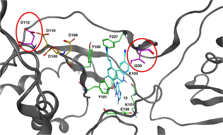
Two mutations, G112D and M230I, were selected in the reverse transcriptase (RT) of human immunodeficiency virus type 1 (HIV-1) by a novel nonnucleoside reverse transcriptase inhibitor (NNRTI). G112D is located near the HIV-1 polymerase active site; M230I is located near the hydrophobic region where NNRTIs bind. Thus, M230I could directly interfere with NNRTI binding but G112D could not. Biochemical and virological assays were performed to analyze the effects of these mutations individually and in combination. M230I alone caused a reduction in susceptibility to NNRTIs, while G112D alone did not. The G112D/M230I double mutant was less susceptible to NNRTIs than was M230I alone. In contrast, both mutations affected the ability of RT to incorporate nucleoside analogs. We suggest that the mutations interact with each other via the bound nucleic acid substrate; the nucleic acid forms part of the polymerase active site, which is near G112D. The positioning of the nucleic acid is influenced by its interactions with the "primer grip" region and could be influenced by the M230I mutation.IMPORTANCE Although antiretroviral therapy (ART) is highly successful, drug-resistant variants can arise that blunt the efficacy of ART. New inhibitors that are broadly effective against known drug-resistant variants are needed, although such compounds might select for novel resistance mutations that affect the sensitivity of the virus to other compounds. Compound 13 selects for resistance mutations that differ from traditional NNRTI resistance mutations. These mutations cause increased sensitivity to NRTIs, such as AZT.

Keywords: drug resistance; nonnucleoside RT inhibitor; nucleic acid repositioning; nucleoside analogs; nucleoside inhibitor.


Figures

FIG 1
FIG 1
Chemical structures of RPV and compound 13.
FIG 2
FIG 2
Selection of RT mutations. HuT-R5 cells were infected with replication-competent WT HIV-1 or HIV-1 with the mutation M230I in the RT. The cells were cultured in the presence of increasing concentrations of RPV or compound (Cpd) 13.
FIG 3
FIG 3
Overview of the HIV-1 RT structure near the polymerase active site. Compound 13 (cyan) is bound at the NNRTI binding pocket. Residues that comprise the NNRTI binding pocket and commonly undergo resistance mutations are K101, K103, E138, Y181, Y188, and F227 and are depicted in green. The three aspartic acid residues (D110, D185, and D186) that bind metal ions at the polymerase active site are shown in gold. The mutated G112D and M230I are modeled into the structure and are shown in magenta and circled red. The RT ribbon backbone is dark gray, while the nitrogen atoms in the residues are blue and the oxygen and hydrogen atoms are red and light gray, respectively.
FIG 4
FIG 4
Effects of various NNRTI concentrations on DNA polymerization by WT RT and the various RT variants. As described in Materials and Methods, the value with no NNRTI present in the reaction is considered 100% activity (for all RT variants) and the other reactions are normalized to this value. Experiments were done in triplicate. (A) Reactions done in the presence of nevirapine. (B) Reactions done in the presence of compound 13.
FIG 5
FIG 5
Competition assays were performed with pairs of HIV-1. HuT-R5 cells were infected with WT HIV-1 and G112D HIV-1 (A), WT HIV-1 and M230I HIV-1 (B), WT HIV-1 and G112D/M230I HIV-1 (double mutant [DM]) (C), G112D HIV-1 and G112D/M230I HIV-1 (DM) (D), or M230I HIV-1 and G112D/M230I HIV-1 (DM) (E). Viral RNA isolated from each culture at days 3, 5, 7, 9, and 11 postinfection was sequenced using Primer ID MiSeq.
FIG 6
FIG 6
DNA-dependent DNA polymerase (DDDP), RNA-dependent DNA polymerase (RDDP), and processivity activities of the RT variants compared to the WT RT. (A) Two different primers (no. 2 and −47) were 5′ end labeled and annealed to single-stranded M13mp18 DNA in the DDDP assays. These primers were then extended by WT RT or by an RT variant in the presence of low (0.5 μM each) and high (10.0 μM each) concentrations of dNTPs. Resulting DNA products were fractionated on a 6% polyacrylamide gel. (B) A 5′-end-labeled PBS primer was annealed to an RNA template (an ∼700-nt RNA generated from a single LTR sequence) for the RDDP assay. The primer were then extended by WT RT or by an RT variant in the presence of low (0.5 μM each) and high (10.0 μM each) concentrations of dNTPs. Resulting DNA products were fractionated on a 6% polyacrylamide gel. (C) The −47 primer was 5′ end labeled and annealed to single-strand M13mp18 DNA. RT was allowed to bind to the labeled T/P. The primer extension reactions by WT RT or by an RT variant was initiated by the addition of dNTPs (final of 10.0 μM each dNTP) and the nucleic acid “cold trap,” which should have limited the extension by RT to one round. Resulting DNA products were fractionated on a 6% polyacrylamide gel. The extension of the labeled primer in the absence of the cold trap is on the far right and labeled “−.”
FIG 7
FIG 7
RT incorporation and processing in HIV-1 particles. The viruses are limited to a single cycle of replication. Equal amounts of WT and mutant virus (as measured by p24) were prepared from cells that were infected at either 37 or 32°C. The viruses were disrupted and were fractionated on an SDS-PAGE gel. The proteins were transferred onto a polyvinylidene difluoride (PVDF) membrane and blotted using a mixture of monoclonal antibodies that detect HIV-1 RT (A) or HIV-1 p24 CA (B). The location of the two subunits of HIV-1 RT (p66 and p51) as well as the p24 CA are marked on the right.
FIG 8
FIG 8
Effects of the various mutations on the ability to incorporate AZTTP into a DNA:DNA substrate. As described in Materials and Methods, the values obtained in the absence of AZTTP are considered 100% activity, and the values obtained with AZTTP in the reaction were normalized to this value.
FIG 9
FIG 9
Effects of the various mutations on the ability to excise the AZTMP blocking group from the end of a primer using ATP as the pyrophosphate donor. As described in Materials and Methods, the primer strand of a DNA:DNA template/primer was blocked with AZTMP. The WT and mutant RTs were incubated with this blocked substrate in the presence of a pyrophosphate donor (ATP) and dNTPs.
FIG 10
FIG 10
Effects of the various mutations on the ability to incorporate FTCTP (A) or 3TCTP (B) into a DNA:DNA substrate. As described in Materials and Methods, the values obtained in the absence of FTCTP or 3TCTP is considered 100% activity, and the other values (with FTCTP or 3TCTP in the reaction) are normalized to this value.
FIG 11
FIG 11
Overview of the RT polymerase active site structure, indicating the position of the nucleic acid and the G112 and M230 positions. The RT ribbon backbone (in cyan) is modeled with the nucleic acid (no ribbon backbone) and an NRTI bound in the RT DNA polymerase active site (aspartic residues labeled). Residues G112 and M230 are shown in relation to the bound nucleic acid and are labeled. The NNRTI binding pocket (signature residues that comprise the binding pocket are labeled) sits behind the RT DNA polymerase active site. Carbon atoms are gray, nitrogen atoms blue, oxygen atoms red, sulfur atoms yellow, and phosphates pink.

Similar articles

Cited by

References

    1. Das K, Martinez SE, Bauman JD, Arnold E. 2012. HIV-1 reverse transcriptase complex with DNA and nevirapine reveals non-nucleoside inhibition mechanism. Nat Struct Mol Biol 19:253–259. doi:10.1038/nsmb.2223. - DOI - PMC - PubMed
    1. Sluis-Cremer N. 2014. The emerging profile of cross-resistance among the nonnucleoside HIV-1 reverse transcriptase inhibitors. Viruses 6:2960–2973. doi:10.3390/v6082960. - DOI - PMC - PubMed
    1. Janssen PA, Lewi PJ, Arnold E, Daeyaert F, de Jonge M, Heeres J, Koymans L, Vinkers M, Guillemont J, Pasquier E, Kukla M, Ludovici D, Andries K, de Bethune MP, Pauwels R, Das K, Clark AD Jr, Frenkel YV, Hughes SH, Medaer B, De Knaep F, Bohets H, De Clerck F, Lampo A, Williams P, Stoffels P. 2005. In search of a novel anti-HIV drug: multidisciplinary coordination in the discovery of 4-[[4-[[4-[(1E)-2-cyanoethenyl]-2,6-dimethylphenyl]amino]-2- pyrimidinyl]amino]benzonitrile (R278474, rilpivirine). J Med Chem 48:1901–1909. doi:10.1021/jm040840e. - DOI - PubMed
    1. Das K, Bauman JD, Clark AD Jr, Frenkel YV, Lewi PJ, Shatkin AJ, Hughes SH, Arnold E. 2008. High-resolution structures of HIV-1 reverse transcriptase/TMC278 complexes: strategic flexibility explains potency against resistance mutations. Proc Natl Acad Sci U S A 105:1466–1471. doi:10.1073/pnas.0711209105. - DOI - PMC - PubMed
    1. Azijn H, Tirry I, Vingerhoets J, de Bethune MP, Kraus G, Boven K, Jochmans D, Van Craenenbroeck E, Picchio G, Rimsky LT. 2010. TMC278, a next-generation nonnucleoside reverse transcriptase inhibitor (NNRTI), active against wild-type and NNRTI-resistant HIV-1. Antimicrob Agents Chemother 54:718–727. doi:10.1128/AAC.00986-09. - DOI - PMC - PubMed

Publication types

Substances

LinkOut - more resources