. 2019 May 15;93(11):e00124-19.
doi: 10.1128/JVI.00124-19. Print 2019 Jun 1.

Foot-and-Mouth Disease Virus Antagonizes NOD2-Mediated Antiviral Effects by Inhibiting NOD2 Protein Expression

Affiliations

Foot-and-Mouth Disease Virus Antagonizes NOD2-Mediated Antiviral Effects by Inhibiting NOD2 Protein Expression

Huisheng Liu et al. J Virol. .

Abstract

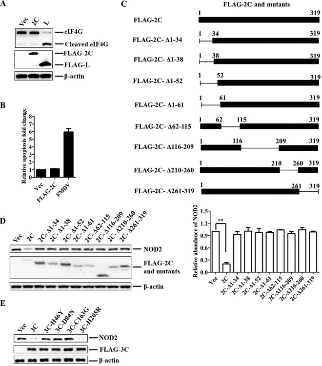
The role of nucleotide-binding oligomerization domain 2 (NOD2) in foot-and-mouth disease virus (FMDV)-infected cells remains unknown. Here, we showed that FMDV infection activated NOD2-mediated beta interferon (IFN-β) and nuclear factor-κB (NF-ĸB) signaling pathways. NOD2 inhibited FMDV replication in the infected cells. FMDV infection triggered NOD2 transcription, while it reduced the abundance of NOD2 protein. Our results revealed that FMDV 2B, 2C, and 3C proteinase (3Cpro) were responsible for the decrease in NOD2 protein levels. 3Cpro is a viral proteinase that can cleave multiple host proteins and limit protein synthesis. Our previous studies determined that FMDV 2B suppressed protein expression of RIG-I and LGP2. Here, we found that 3Cpro and 2B also decreased NOD2 expression. However, this is the first report that 2C induced the reduction of NOD2 protein levels. We determined that both 2B- and 2C-induced decreases in NOD2 were independent of the cleavage of host eukaryotic translation initiation factor 4 gamma (eIF4G), induction of cellular apoptosis, or proteasome, lysosome, and caspase pathways. The interactions between NOD2 and 2B or 2C were observed in the context of viral infection. The carboxyl-terminal amino acids 105 to 114 and 135 to 144 of 2B were essential for the reduction of NOD2, while the residues 105 to 114 were required for the interaction. Amino acids 116 to 260 of the carboxyl terminus of 2C were essential for the interaction, while truncated 2C mutants did not reduce NOD2. These data suggested novel antagonistic mechanisms of FMDV that were mediated by 2B, 2C, and 3Cpro proteins.IMPORTANCE NOD2 was identified as a cytoplasmic viral pattern recognition receptor in 2009. Subsequently, many viruses were reported to activate NOD2-mediated signaling pathways. This study demonstrated that FMDV infection activated NOD2-mediated IFN-β and NF-ĸB signaling pathways. Host cells have developed multiple strategies against viral infection; however, viruses have evolved many strategies to escape host defenses. FMDV has evolved multiple mechanisms to inhibit host type I IFN production. Here, we showed that NOD2 suppressed FMDV replication during viral infection. FMDV 2B, 2C, and 3Cpro decreased NOD2 protein expression by different mechanisms to promote viral replication. This study provided new insight into the immune evasion mechanisms mediated by FMDV and identified 2B, 2C, and 3Cpro as antagonistic factors for FMDV to evade host antiviral responses.

Keywords: 2B; 2C; FMDV; NOD2; antagonistic mechanism.


Figures

FIG 1
FIG 1
FMDV infection activates NOD2-mediated IFN-β and NF-ĸB signaling pathways. (A and B) PK-15 cells were transfected with 0.1 μg/well of IFN-β-Luc or NF-ĸB-Luc, 0.01 μg/well of pRL-TK plasmid, and 75 nM/well of NOD2 or NC siRNA. At 36 hpt, cells were stimulated by MDP (5 μg/ml), mock infected, or infected with FMDV. The promoter activations of IFN-β (A) and NF-ĸB (B) were determined by the specific Dual-Luciferase assay kit. (C) PK-15 cells were transfected with NOD2 or NC siRNA for 36 h, and the cells were then mock infected or infected with equal amounts of FMDV. The cells were collected at 0, 6, and 12 hpi. Expression of viral VP1 protein and host proteins was detected by Western blotting. (D) Transfection and infection experiments performed as described for panels A and B. The cells were collected at 0, 6, or 12 hpi. Expression of NOD2, FMDV, IFN-β, ISG15, IL-1β, and CCL3L1 mRNA was determined by qPCR assay. The viral titers were determined by 50% tissue culture infective dose (TCID50) assay, and the knockdown of NOD2 was confirmed by Western blotting.
FIG 2
FIG 2
FMDV infection triggered NOD2 transcription and reduced NOD2 protein abundance. (A and B) PK-15 cells were infected with FMDV for 0, 4, 8, 12, 16, or 20 h. Expression of NOD2 mRNA and viral RNA was determined by qPCR assay, viral titers were determined by TCID50 assay, and expression of endogenous NOD2 protein was detected by Western blotting. (C) PK-15 cells were mock infected or infected with FMDV type O or A strains (MOI of 0.5) for 12 h. Expression of endogenous NOD2 protein was detected by Western blotting. (D and E) PK-15 cells were infected with FMDV for 0, 4, 8, 12, 16, or 20 h, and the expression of endogenous NOD1, NLRC5, NLRX1, NLRP3, and CIITA protein was detected by Western blotting. (E) Expression of NOD1, NLRC5, NLRX1, NLRP3, and CIITA mRNA was determined by qPCR assay.
FIG 3
FIG 3
NOD2, NLRP3, and CIITA inhibit FMDV replication during viral infection. (A) PK-15 cells were transfected with 0, 1, 2, or 3 μg of HA-NOD2-expressing plasmid. At 24 hpt, the cells were infected with equal amounts of FMDV for 12 h. Expression of the NOD2 and viral VP1 protein was detected by Western blotting, expression of viral RNA was determined by qPCR assay, and the viral titers were determined by TCID50 assay. (B, C, D, and E) PK-15 cells were transfected with 150 nM NOD2 siRNA, NLRP3 siRNA, CIITA siRNA, or NC siRNA. At 36 hpt, the cells were infected with equal amounts of FMDV. The cells were collected at the indicated time points. Expression of NOD2, NLRP3, CIITA, and viral RNA was determined by qPCR assay. Expression of the NOD2, NLRP3, CIITA, and viral VP1 proteins was detected by Western blotting, and viral titers were determined by TCID50 assay.
FIG 4
FIG 4
FMDV 2B, 2C, and 3Cpro proteins were responsible for the reduction of NOD2 expression. (A) PK-15 cells were transfected with 2 μg plasmid expressing various FLAG-tagged viral proteins. At 24 hpt, expression of endogenous NOD2 protein was determined by Western blotting. (B and D) PK-15 cells were seeded in 6-well plates, and the monolayer cells were transfected with 0, 1, 2, and 3 μg FLAG-2B-, FLAG-2C-, or FLAG-3C-expressing plasmid for 24 h. Expression of NOD2 protein was determined by Western blotting (B), and expression of NOD2 mRNA was determined by qPCR assay (D). (C) BHK-21 cells were seeded in 6-well plates, and the monolayer cells were mock infected or infected with FMDV-wt or FMDV-ΔL. Expression of NOD2 protein was determined by Western blotting, and the relative abundance of NOD2 was determined by statistical analyses. (E) PK-15 cells were transfected with 2 μg empty vector, 2 μg FLAG-2BC-expressing plasmid, or 2 μg FLAG-2C-expressing plasmid. At 24 hpt, the cells were infected with equal amounts of FMDV (MOI of 0.5) for 12 h. Viral RNA, VP1 protein, and titers were examined.
FIG 5
FIG 5
2B-induced reduction of NOD2. (A) Schematic representation showing a series of FLAG-tagged truncated 2B mutants. (B and C) PK-15 cells were transfected with 1.5 μg FLAG-2B-expressing plasmid, 1.5 μg FLAG-2B mutant-expressing plasmids, or 1.5 μg empty vector. At 24 hpt, the cells were collected for Western blotting.
FIG 6
FIG 6
2C- and 3C-induced reduction of NOD2. (A) PK-15 cells were seeded in 6-well plates, and the monolayer cells were transfected with 2 μg FLAG-L-expressing plasmid, 2 μg FLAG-2C-expressing plasmid, or 2 μg empty vector. At 24 hpt, the cells were collected for Western blotting. (B) PK-15 cells were transfected with 2 μg empty vector or 2 μg FLAG-2C-expressing plasmid or infected with FMDV (MOI of 0.05). The apoptotic status of the transfected and infected cells was analyzed by annexin V-PI staining and flow cytometry analysis at 24 hpt or hpi. The experiments were repeated three times with similar results. (C) Schematic representation showing a series of FLAG-tagged truncated 2C mutants. (D) PK-15 cells were transfected with 2 μg FLAG-2C-expressing plasmid, 2 μg FLAG-2C mutant-expressing plasmids, or 2 μg empty vector. At 24 hpt, the cells were collected for Western blotting. (E) PK-15 cells were transfected with 1.5 μg FLAG-3C-expressing plasmid, 1.5 μg FLAG-3C mutant-expressing plasmids, or 1.5 μg empty vector. At 24 hpt, the cells were collected for Western blotting.
FIG 7
FIG 7
NOD2 interacts with 2B and 2B truncated mutants. (A) PK-15 cells were transfected with 10 μg FLAG-2B-expressing plasmid or 10 μg empty FLAG vector. The cells were lysed at 30 hpt. The lysates were immunoprecipitated with rabbit anti-NOD2 antibody and rabbit normal IgG antibody and subjected to Western blotting. The whole-cell lysates and IP antibody-antigen complexes were analyzed by IB using anti-NOD2, anti-FLAG, or anti-β-actin antibodies. (B) Transfection in PK-15 cells and IP experiments were carried out as described above. However, the lysates were immunoprecipitated with mouse anti-FLAG or mouse normal IgG antibody and subjected to Western blotting. (C) PK-15 cells were mock infected or infected with FMDV (MOI of 0.5) for 12 h. The cells lysates were immunoprecipitated with anti-NOD2 antibody. The antibody-antigen complexes were detected using anti-NOD2, anti-2B, or anti-2C antibody. (D) A reverse immunoprecipitation assay was performed using anti-2B antibody as described for panel C. (E) PK-15 cells were transfected with 10 μg empty FLAG vector, 10 μg FLAG-2B-expressing plasmid, and 10 μg FLAG-2B mutant-expressing plasmids. At 30 hpt, the cells were lysed and the lysates were immunoprecipitated with rabbit anti-NOD2 antibody and rabbit normal IgG antibody and subjected to Western blotting. The whole-cell lysates and IP antibody-antigen complexes were analyzed by IB using anti-FLAG, anti-NOD2, or anti-β-actin antibodies.
FIG 8
FIG 8
NOD2 interacts with 2C and 2C truncated mutants. (A, B, and C) Similar transfection, infection, and IP assays in PK-15 cells were performed as described for Fig. 7A, B, and D. (D) PK-15 cells were transfected with 10 μg empty FLAG vector, FLAG-2C-expressing plasmid, and FLAG-2C mutant-expressing plasmids. At 30 hpt, the cells were lysed and the lysates were immunoprecipitated with mouse anti-FLAG antibody and mouse normal IgG antibody and subjected to Western blotting. The whole-cell lysates and IP antibody-antigen complexes were analyzed by IB using anti-FLAG, anti-NOD2, or anti-β-actin antibody.

Similar articles

Cited by

References

    1. Grubman MJ, Baxt B. 2004. Foot-and-mouth disease. Clin Microbiol Rev 17:465–493. doi:10.1128/CMR.17.2.465-493.2004. - DOI - PMC - PubMed
    1. Belsham GJ. 1993. Distinctive features of foot-and-mouth disease virus, a member of the picornavirus family; aspects of virus protein synthesis, protein processing and structure. Prog Biophys Mol Biol 60:241–260. doi:10.1016/0079-6107(93)90016-D. - DOI - PMC - PubMed
    1. Moffat K, Howell G, Knox C, Belsham GJ, Monaghan P, Ryan MD, Wileman T. 2005. Effects of foot-and-mouth disease virus nonstructural proteins on the structure and function of the early secretory pathway: 2BC but not 3A blocks endoplasmic reticulum-to-Golgi transport. J Virol 79:4382–4395. doi:10.1128/JVI.79.7.4382-4395.2005. - DOI - PMC - PubMed
    1. Moffat K, Knox C, Howell G, Clark SJ, Yang H, Belsham GJ, Ryan M, Wileman T. 2007. Inhibition of the secretory pathway by foot-and-mouth disease virus 2BC protein is reproduced by coexpression of 2B with 2C, and the site of inhibition is determined by the subcellular location of 2C. J Virol 81:1129–1139. doi:10.1128/JVI.00393-06. - DOI - PMC - PubMed
    1. Moraes MP, Segundo FD, Dias CC, Pena L, Grubman MJ. 2011. Increased efficacy of an adenovirus-vectored foot-and-mouth disease capsid subunit vaccine expressing nonstructural protein 2B is associated with a specific T cell response. Vaccine 29:9431–9440. doi:10.1016/j.vaccine.2011.10.037. - DOI - PubMed

Publication types

MeSH terms

LinkOut - more resources