. 2019 May 15;93(11):e00183-19.
doi: 10.1128/JVI.00183-19. Print 2019 Jun 1.

A Novel Murine Model Expressing a Chimeric mSCARB2/hSCARB2 Receptor Is Highly Susceptible to Oral Infection with Clinical Isolates of Enterovirus 71

Affiliations

A Novel Murine Model Expressing a Chimeric mSCARB2/hSCARB2 Receptor Is Highly Susceptible to Oral Infection with Clinical Isolates of Enterovirus 71

Cheng-Hung Yang et al. J Virol. .

Abstract

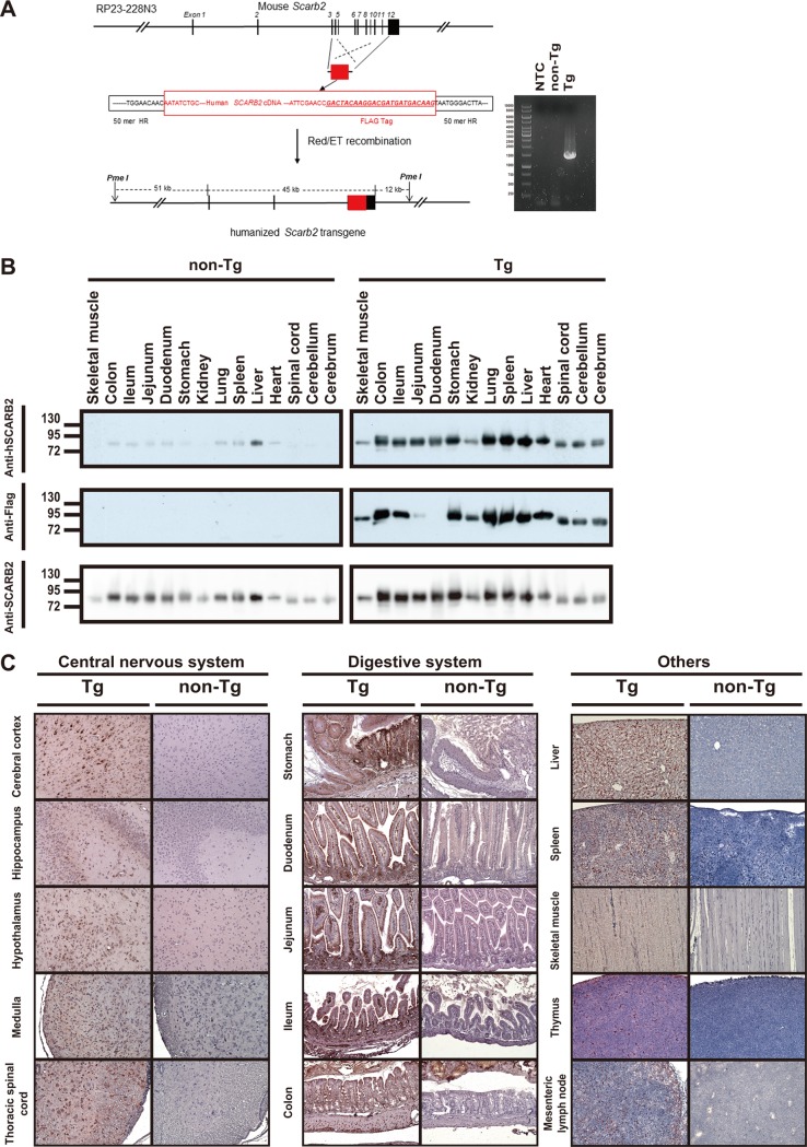
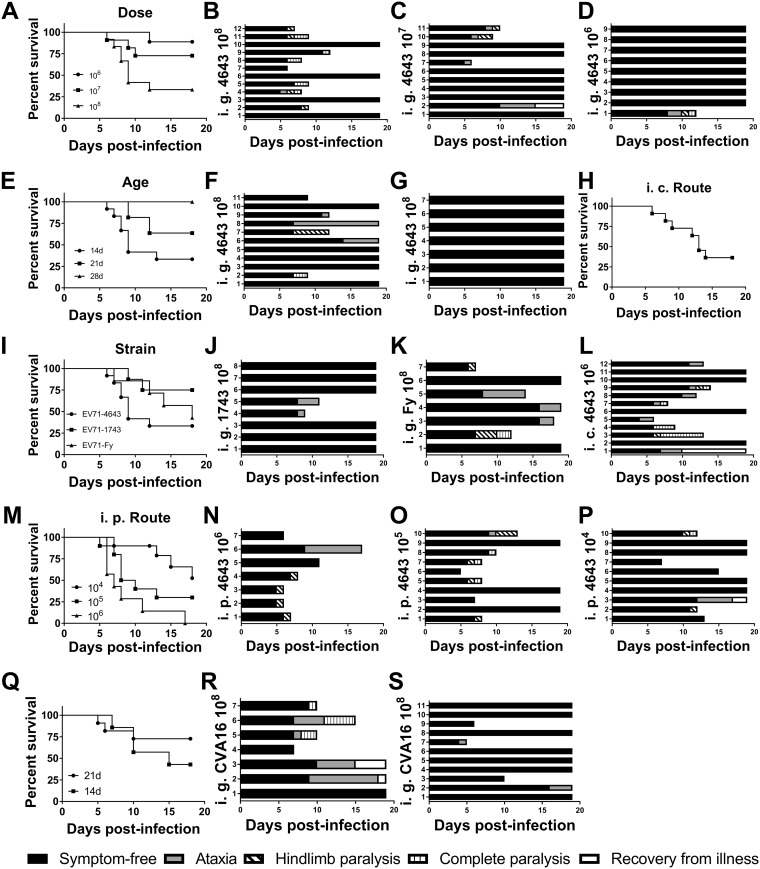
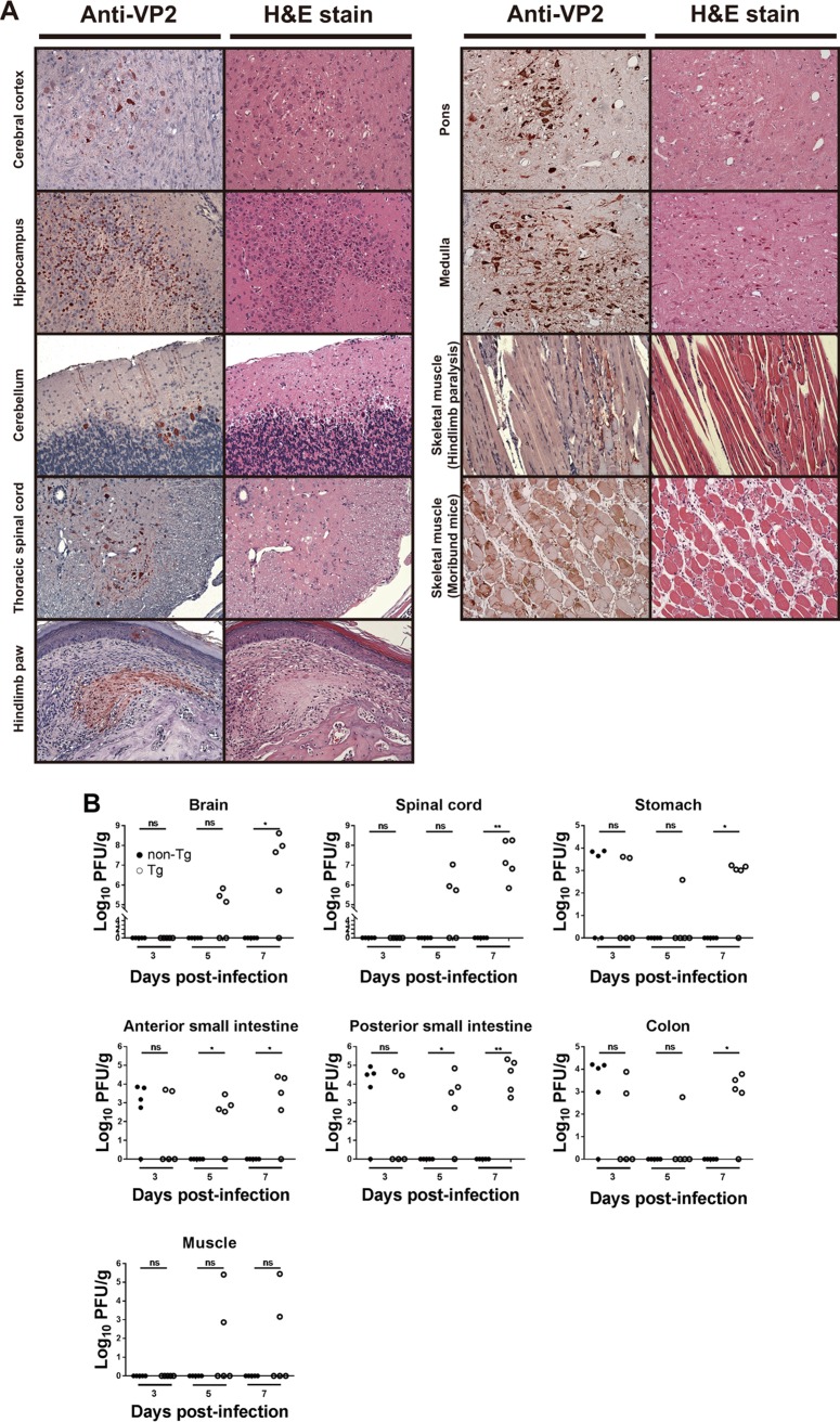
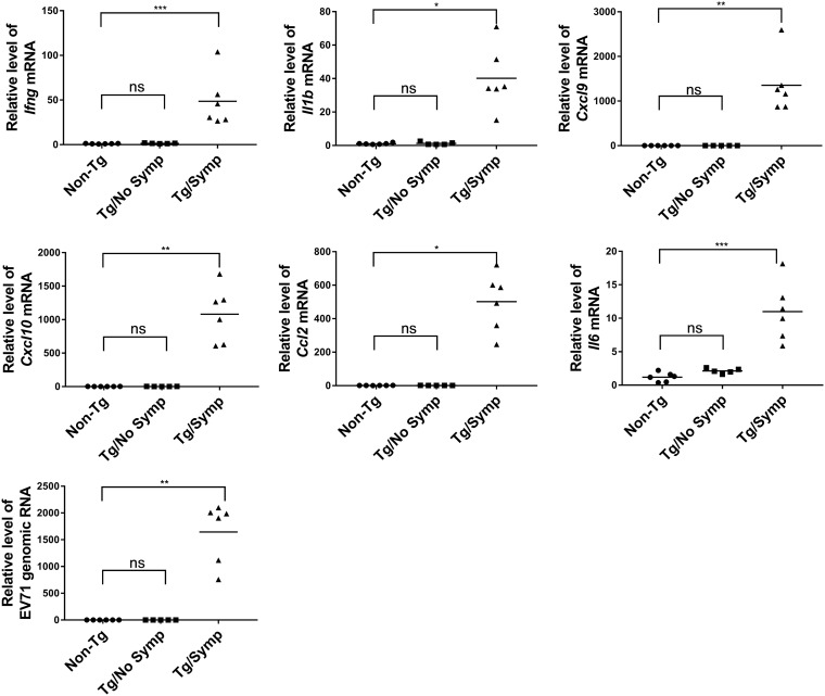
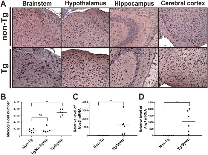
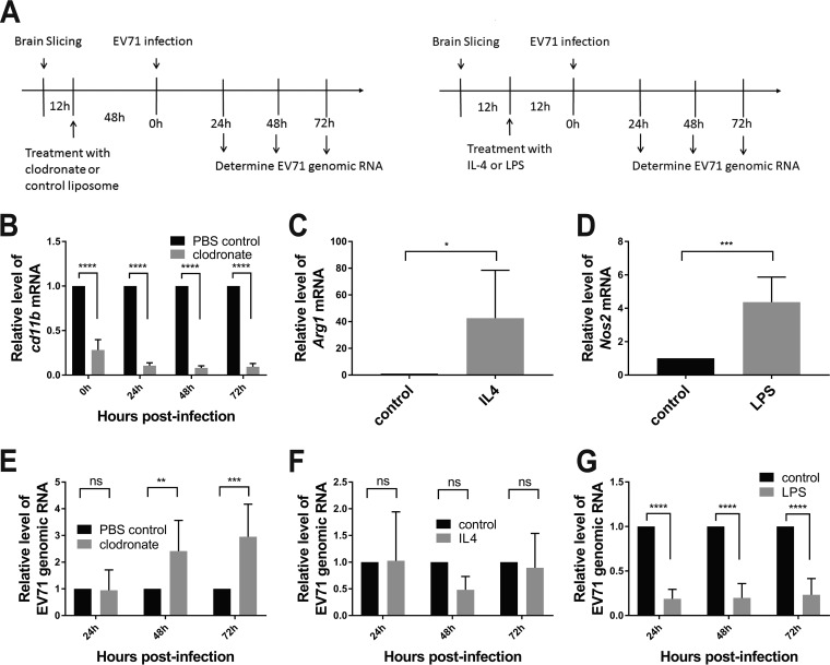
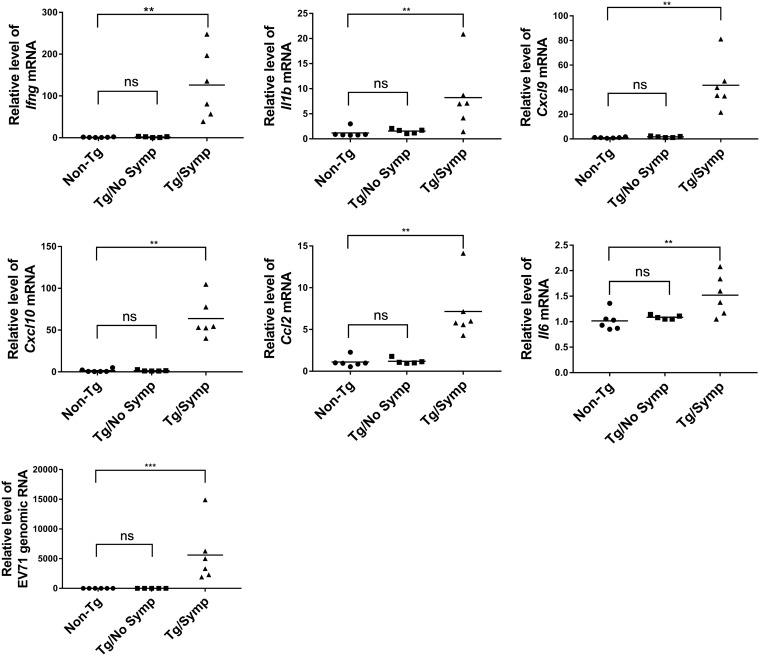
Enterovirus 71 (EV71) infection is generally associated with hand-foot-and-mouth disease (HFMD) and may cause severe neurological disorders and even death. An effective murine oral infection model for studying the pathogenesis of various clinical EV71 isolates is lacking. We developed a transgenic (Tg) mouse that expresses an EV71 receptor, that is, human scavenger receptor class B member 2 (hSCARB2), in a pattern highly similar to that of endogenous murine SCARB2 (mSCARB2) protein. A FLAG-tagged SCARB2 cDNA fragment composed of exons 3 to 12 was inserted into a murine Scarb2 gene-containing bacterial artificial chromosome (BAC) clone, and the resulting transgene was used for establishment of chimeric receptor-expressing Tg mice. Tg mice intragastrically (i.g.) infected with clinical isolates of EV71 showed neurological symptoms, such as ataxia and paralysis, and fatality. There was an age-dependent decrease in susceptibility to viral infection. Pathological characteristics of the infected Tg mice resembled those of encephalomyelitis in human patients. Viral infection was accompanied by microglial activation. Clodronate treatment of the brain slices from Tg mice enhanced viral replication, while lipopolysaccharide treatment significantly inhibited it, suggesting an antiviral role for microglia during EV71 infection. Taken together, this Tg mouse provides a model that closely mimics natural infection for studying EV71 pathogenesis and for evaluating the efficacy of vaccines or other antiviral drugs.IMPORTANCE The availability of a murine model of EV71 infection is beneficial for the understanding of pathogenic mechanisms and the development and assessment of vaccines and antiviral drugs. However, the lack of a murine oral infection model thwarted the study of pathogenesis induced by clinically relevant EV71 strains that are transmitted via the oral-oral or oral-fecal route. Our Tg mice could be intragastrically infected with clinically relevant EV71 strains in an efficient way and developed neurological symptoms and pathological changes strikingly resembling those of human infection. Moreover, these mice showed an age-dependent change in susceptibility that is similar to the human case. This Tg mouse, when combined with the use of other genetically modified mice, potentially contributes to studying the relationship between developmental changes in immunity and susceptibility to virus.

Keywords: SCARB2; enterovirus 71; oral infection.


Figures

FIG 1
FIG 1
Expression of mSCARB2/hSCARB2 in Scarb2-SCARB2 BAC Tg mice. (A) Schematic diagram showing the construction of the recombinant BAC. The RP23-228N3 clone is shown at the top. The genomic sequence spanning from exon 3 to exon 12 was replaced with the corresponding part of human SCARB2 cDNA that carries a FLAG tag-coding sequence. The resulting clone carrying the humanized Scarb2 transgene is displayed at the bottom. PCR genotyping of the Tg mice is also shown. Genomic DNAs isolated from Tg3 and non-Tg mice were subjected to PCR genotyping as described in Materials and Methods. The amplified product for the Tg sample is 1,511 bp long. NTC, no-template control. (B) Tissue distribution of the mSCARB2/hSCARB2 chimeric protein in Tg mice. (Top and middle) Tg and non-Tg mice were sacrificed, and cerebrum, cerebellum, spinal cord, heart, liver, spleen, lung, stomach, duodenum, jejunum, ileum, colon, kidney, and skeletal muscle were harvested for Western blotting with anti-hSCARB2 (top) and anti-FLAG (middle) antibodies. It should be noted that the anti-hSCARB2 antibody is known to cross-react with the murine endogenous protein. (Bottom) The immunoblot membrane shown in the top panel was stripped and reprobed with anti-SCARB2 antibody, which can react with both murine and human proteins. A representative result out of three experiments is shown. (C) IHC analysis of expression of the chimeric protein in Tg mice. Cerebral cortex, hippocampus, hypothalamus, medulla, thoracic spinal cord, stomach, duodenum, jejunum, ileum, colon, liver, spleen, skeletal muscle, thymus, and mesenteric lymph nodes were collected from the Tg mice; paraffinized; sectioned for immunohistochemical staining with the anti-hSCARB2 antibody; and counterstained with hematoxylin. Original magnification, ×200. A representative result out of three experiments is shown.
FIG 2
FIG 2
Tg mice are susceptible to oral EV71 and CVA16 infection. (A to P) Tg mice were infected with EV71 under different conditions, such as viral doses (A to D), mouse ages (E, B, F, and G), clinical isolates (I, B, J, and K), and inoculation routes (intraperitoneal [i.p.] route [M to P] and intracerebral [i.c.] route [H and L]). Survival curves (A, E, H, I, and M) and neurological signs (B to D, F, G, J to L, and N to P) are shown. For the charts showing neurological signs, the numbers alongside the ordinate axis refer to the identification numbers of mice within the particular experimental group. (A to D) For testing the effect of viral dosage (A), Tg mice were intragastrically (i.g.) infected with 1 × 106 PFU (D), 1 × 107 PFU (C), or 1 × 108 PFU (B) of the EV71-4643 strain at the age of 14 days. (E to G) Tg mice were i.g. infected with 1 × 108 PFU of the EV71-4643 strain (E) at the age of 14 days (B), 21 days (F), or 28 days (G). (H and L) Tg mice were i.c. infected with 1 × 106 PFU of the EV71-4643 strain at the age of 1 year. (I to K) Tg mice were i.g. infected (I) with 1 × 108 PFU of the EV71-4643 (B), EV71-1743 (J), or EV71-Fy (K) strain at the age of 14 days. (M to P) Tg mice were intraperitoneally (i.p.) infected (M) with 1 × 104 PFU (P), 1 × 105 PFU (O), or 1 × 106 PFU (N) of the EV71-4643 strain at the age of 14 days. Survival rates and clinical symptoms were recorded daily for 18 consecutive days. (Q to S) Tg mice are also susceptible to oral CVA16 infection. Tg mice were i.g. infected with 1 × 108 PFU of CVA16 at the ages of 14 days (Q and R) and 21 days (Q and S). Survival rates (Q) and clinical symptoms (R and S) were recorded daily for 18 consecutive days.
FIG 3
FIG 3
Replication of EV71 in orally infected Tg mice. (A) Histopathological changes and expression of EV71 VP2 protein in infected mice. Two-week-old Tg mice were inoculated i.g. with the EV71-4643 strain and euthanized at 6 days postinfection. (Left) Cerebral cortex, hippocampus, cerebellum, thoracic spinal cord, pons, medulla, skeletal muscle (mice with hind-limb paralysis and moribund mice with complete paralysis), and hind-limb paw of Tg mice were retrieved for sample processing, IHC staining with the anti-EV71 VP2 antibody, and hematoxylin counterstaining. (Right) In parallel, sections were stained with H&E. Original magnification, ×200. A representative result out of six experiments is shown. (B) Viral titers in various parts of the CNS and GI tract and in skeletal muscle of infected mice. Two-week-old Tg and non-Tg mice were infected i.g. with 1 × 108 PFU of the EV71-4643 strain and euthanized at the indicated times. Brains and GI tracts were collected and dissected into parts, as indicated in the figure. The anterior small intestine includes the duodenum and parts of the jejunum, while the posterior small intestine includes parts of the jejunum and ileum. Viral titers were determined using a plaque-forming assay and are expressed as log PFU per gram of tissue. ns, not significant; *, P < 0.05; **, P < 0.01 (versus non-Tg mice [by a Mann-Whitney test]).
FIG 4
FIG 4
Expression of proinflammatory cytokines in the brains of EV71-infected mice. Tg (n = 11) and non-Tg (n = 5) (circles) mice were infected i.g. with 1 × 108 PFU of the EV71-4643 strain at the age of 14 days and sacrificed at 7 days p.i. Tg mice were divided into mice without neurological symptoms (Tg/No Symp) (squares) (n = 5) and those with symptoms (Tg/Symp) (triangles) (n = 6). Brains were isolated from these mice for total RNA extraction. Levels of Ifng, Il1b, Cxcl9, Cxcl10, Ccl2, and Il6 mRNAs and of EV71 genomic RNA were determined by RT-qPCR. Data are expressed as fold changes relative to the levels in non-Tg mice. ns, not significant; *, P < 0.05; **, P < 0.01; ***, P < 0.001 (versus non-Tg mice [by a Kruskal-Wallis test with Dunn’s multiple-comparison test]).
FIG 5
FIG 5
Microglial activation in infected Tg mice. (A) IHC analysis of activated microglia in various parts of the brains of infected Tg mice. Two-week-old Tg and non-Tg mice were inoculated i.g. with 1 × 108 PFU of the EV71-4643 strain and sacrificed at 6 days p.i. Brains were retrieved for sample processing, IHC staining with the anti-Iba1 antibody, and hematoxylin counterstaining. Original magnification, ×200. (B) Increases in microglia numbers in infected Tg mice with symptoms. Tg and non-Tg (n = 6) (circles) mice were infected i.g. with 1 × 108 PFU of the EV71-4643 strain at the age of 14 days and sacrificed at 7 days p.i. Tg mice were divided into mice without neurological symptoms (Tg/No Symp) (squares) (n = 5) and those with symptoms (Tg/Symp) (triangles) (n = 6). Brains were isolated from these mice, and microglia were purified by Percoll gradient centrifugation. Microglia numbers were determined by hemocytometer counting. ns, not significant; **, P < 0.01 (versus non-Tg mice [by a Kruskal-Wallis test with Dunn’s multiple-comparison test]). (C and D) Microglia in the Tg/Symp and non-Tg groups were isolated as described above for panel B. Total RNA was purified and subjected to RT-qPCR analysis of the expression levels of the M1 activation marker Nos2 (C) and the M2 activation marker Arg1 (D). Data are expressed as fold changes relative to the value for non-Tg mice. **, P < 0.01 (versus non-Tg mice [by a Mann-Whitney test]).
FIG 6
FIG 6
Antiviral response of activated microglia. (A) Brain slices were prepared from Tg mice. The workflow of clodronate treatment of brain slices (left) and induction of microglial polarization in these slices (right) is shown. (B and E) Twelve hours after brain slicing, the slices were treated with clodronate or control liposome for 48 h prior to infection with 104 PFU of the EV71-4643 strain per slice (A, left). Cd11b mRNA (B) and EV71 genomic RNA (E) were quantified at the indicated times p.i. by RT-qPCR. Data are expressed as fold changes relative to the value for control treatment. Data are means ± SD (n = 6). ns, not significant; **, P < 0.01; ***, P < 0.001; ****, P < 0.0001 (versus control treatment). (C, D, F, and G) Twelve hours after brain slicing, the slices were treated with IL-4 or LPS for 12 h (A, right). (C and D) The change in the level of Arg1 (C) or Nos2 (D) mRNA in response to IL-4 (C) or LPS (D) treatment was determined using RT-qPCR. (E and F) IL-4 (F)- and LPS (G)-treated brain slices were infected with 104 PFU of the EV71-4643 strain per slice, and the levels of EV71 genomic RNA were quantified at the indicated times p.i. by RT-qPCR. Data are expressed as fold changes relative to the value for control treatment. Data are means ± SD (n = 6). ns, not significant; *, P < 0.05; ***, P < 0.001; ****, P < 0.0001 (versus control treatment [by two-way ANOVA with Sidak’s multiple-comparison test and by Student’s t test]).
FIG 7
FIG 7
Expression of proinflammatory cytokines in microglia from EV71-infected Tg mice. Two-week-old Tg and non-Tg (n = 6) (circles) mice were inoculated i.g. with 1 × 108 PFU of the EV71-4643 strain and sacrificed at 7 days p.i. Tg mice were divided into mice without neurological symptoms (Tg/No Symp) (squares) (n = 5) and those with symptoms (Tg/Symp) (triangles) (n = 6). Brains were isolated from these mice, and microglia were purified by Percoll gradient centrifugation. Total RNA from microglia was purified for RT-qPCR analysis of the levels of Ifng, Il1b, Cxcl9, Cxcl10, Ccl2, and Il6 mRNAs and of EV71 genomic RNA. Data are expressed as fold changes relative to the values for microglia from non-Tg mice. ns, not significant; **, P < 0.01; ***, P < 0.001 (versus non-Tg mice [by a Kruskal-Wallis test with Dunn’s multiple-comparison test]).

Similar articles

Cited by

References

    1. Solomon T, Lewthwaite P, Perera D, Cardosa MJ, McMinn P, Ooi MH. 2010. Virology, epidemiology, pathogenesis, and control of enterovirus 71. Lancet Infect Dis 10:778–790. doi:10.1016/S1473-3099(10)70194-8. - DOI - PubMed
    1. Weng KF, Chen LL, Huang PN, Shih SR. 2010. Neural pathogenesis of enterovirus 71 infection. Microbes Infect 12:505–510. doi:10.1016/j.micinf.2010.03.006. - DOI - PubMed
    1. Yang F, Ren L, Xiong Z, Li J, Xiao Y, Zhao R, He Y, Bu G, Zhou S, Wang J, Qi J. 2009. Enterovirus 71 outbreak in the People’s Republic of China in 2008. J Clin Microbiol 47:2351–2352. doi:10.1128/JCM.00563-09. - DOI - PMC - PubMed
    1. Yang F, Zhang T, Hu Y, Wang X, Du J, Li Y, Sun S, Sun X, Li Z, Jin Q. 2011. Survey of enterovirus infections from hand, foot and mouth disease outbreak in China, 2009. Virol J 8:508. doi:10.1186/1743-422X-8-508. - DOI - PMC - PubMed
    1. Mao LX, Wu B, Bao WX, Han FA, Xu L, Ge QJ, Yang J, Yuan ZH, Miao CH, Huang XX, Zhang C, Xu H. 2010. Epidemiology of hand, foot, and mouth disease and genotype characterization of enterovirus 71 in Jiangsu, China. J Clin Virol 49:100–104. doi:10.1016/j.jcv.2010.07.009. - DOI - PubMed

Publication types

MeSH terms

LinkOut - more resources