. 2019 May 15;93(11):e00068-19.
doi: 10.1128/JVI.00068-19. Print 2019 Jun 1.

An Immunodominant and Conserved B-Cell Epitope in the Envelope of Simian Foamy Virus Recognized by Humans Infected with Zoonotic Strains from Apes

Affiliations

An Immunodominant and Conserved B-Cell Epitope in the Envelope of Simian Foamy Virus Recognized by Humans Infected with Zoonotic Strains from Apes

Caroline Lambert et al. J Virol. .

Abstract

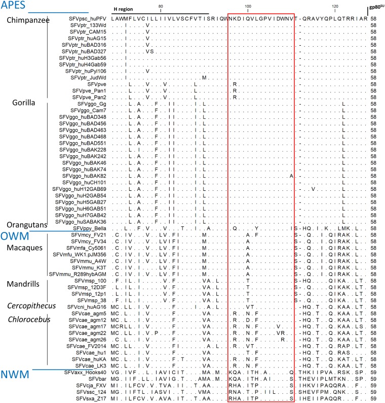
Cross-species transmission of simian foamy viruses (SFVs) from nonhuman primates (NHPs) to humans is currently ongoing. These zoonotic retroviruses establish lifelong persistent infection in their human hosts. SFV are apparently nonpathogenic in vivo, with ubiquitous in vitro tropism. Here, we aimed to identify envelope B-cell epitopes that are recognized following a zoonotic SFV infection. We screened a library of 169 peptides covering the external portion of the envelope from the prototype foamy virus (SFVpsc_huHSRV.13) for recognition by samples from 52 Central African hunters (16 uninfected and 36 infected with chimpanzee, gorilla, or Cercopithecus SFV). We demonstrate the specific recognition of peptide N96-V110 located in the leader peptide, gp18LP Forty-three variant peptides with truncations, alanine substitutions, or amino acid changes found in other SFV species were tested. We mapped the epitope between positions 98 and 108 and defined six amino acids essential for recognition. Most plasma samples from SFV-infected humans cross-reacted with sequences from apes and Old World monkey SFV species. The magnitude of binding to peptide N96-V110 was significantly higher for samples of individuals infected with a chimpanzee or gorilla SFV than those infected with a Cercopithecus SFV. In conclusion, we have been the first to define an immunodominant B-cell epitope recognized by humans following zoonotic SFV infection.IMPORTANCE Foamy viruses are the oldest known retroviruses and have been mostly described to be nonpathogenic in their natural animal hosts. SFVs can be transmitted to humans, in whom they establish persistent infection, like the simian lenti- and deltaviruses that led to the emergence of two major human pathogens, human immunodeficiency virus type 1 and human T-lymphotropic virus type 1. This is the first identification of an SFV-specific B-cell epitope recognized by human plasma samples. The immunodominant epitope lies in gp18LP, probably at the base of the envelope trimers. The NHP species the most genetically related to humans transmitted SFV strains that induced the strongest antibody responses. Importantly, this epitope is well conserved across SFV species that infect African and Asian NHPs.

Keywords: antibody; emergence; retrovirus; zoonotic infections.


Figures

FIG 1
FIG 1
Reactivity of plasma samples from SFV-uninfected and SFV-infected individuals to peptide pools covering the external portion of the chimpanzee SFV envelope. (A) Scheme of the organization of the SFV envelope protein. The amino acid positions are those of the SFVpsc_huPFV strain. The 988-residue envelope precursor is cleaved by furins (arrows) into the leader peptide (gp18LP, aa 1 to 126), surface protein (gp80SU, aa 127 to 571), and transmembrane protein (gp48™, aa 572 to 988). The hydrophobic region (h region, aa 68 to 84), receptor binding domain (RBD, aa 225 to 555), genotype-specific variable region (SUvar, aa 241 to 491), fusion peptide (FP, aa 572 to 598), and membrane-spanning domain (MSD, aa 939 to 975) are drawn in orange. The black line indicates the region covered by the overlapping peptides. (B) Binding of 1:200-diluted plasma samples of six SFV-uninfected (AKO255, BAD19, BAD449, BAD459, BAD552, and BAK165) and seven SFV-infected individuals (SFVggo: BAD448, BAD468, BAK74, BAK133, and LOBAK2; SFVptr: BD327 and PYL106) to 28 peptide pools was quantified by ELISA. Peptides from CMV and HTLV-1 were used as controls. ΔOD, defined as the difference between the OD of peptide-coated wells and diluent-treated wells, is presented. Bars at ΔOD of 0.2 and 0.5 indicate values used to identify samples with high or moderate seroreactivity, as described in the text.
FIG 2
FIG 2
Plasma sample and peptide N96-V110 titration shows the specificity of recognition. Six dilutions (1:50 to 1:51,200) of the plasma sample from individual BAD468 were tested for binding to peptide N96-V110 coated at five concentrations (5 to 0.008 μg/ml). The ΔOD (A) and ratioOD (B) are shown on the graph. Red lines indicate positivity thresholds (ΔOD ≥ 0.13; ratioOD ≥ 2).
FIG 3
FIG 3
Mapping of amino acids involved in plasma sample binding to peptide N96-V110. Plasma samples from five SFV-infected individuals were tested for binding to peptide N96-V110 and its variants (Table 2). Wells coated with peptide N96-V110 provided the reference value. Relative binding was calculated for wells coated with variant peptides and is expressed as the percentage of the reference value. Bars indicate 33, 66, 100, and 133% levels. For each peptide, the position of the first and last amino acids or the position of the amino acid replaced by an alanine are indicated on the graph. (A) The first peptide set comprised the two 15mers adjacent to peptide N96-V110, ten N- and C-terminal truncated variants, and three peptides in which three consecutive amino acids were replaced by an alanine. (B) The second peptide set comprised peptides with combined N- and C-terminal deletions and single alanine mutations. (C to E) Plasma samples from individuals BAD348 (gray symbols), BAD448 (blue), and BAD468 (green) were titrated for the recognition of peptides N96-V110 (C), V101-V110 (D), and N96-A100to102-V110 (E), coated at 1 μg/ml. The ΔOD values are presented as a function of sample dilution, and the red lines indicate the positivity threshold (ΔOD ≥ 0.13).
FIG 4
FIG 4
Alignment of the C-terminal gp18LP protein sequences from SFV. The gp18LP protein sequences extending from the hydrophobic region to the gp18LP/gp80SU cleavage site were aligned. We included all sequences from our previous envelope analysis (20) and those published and/or deposited in GenBank since SFVsxa_Z17 (KP143760.1), SFVmfa_Cy5061 (LC094267.1), SFVmmu_K3T (MF280817.1), SFVcae_FV214 (MF582544.1), SFVbar (MH368762.1), SFVmsp_100 (MK014759), SFVmsp_12D3F (MK014758), SFVmsp_12p1 (MK014761), and SFVmsp_38 (MK014760). Identical residues are indicated by dots. The red square highlights the N96-V110 sequence. The sequences are ordered according to the phylogeny of host species and within each species in alphabetical order.
FIG 5
FIG 5
Recognition of variant peptides by plasma samples from SFV-infected individuals. Plasma samples from SFV-infected individuals were tested for binding to peptide N96-V110 and its naturally occurring variants. The ΔOD is presented for peptide N96-V110 and 13 peptides with sequences found in SFV from apes, OWM, and NWM. The peptide sequences are presented in Table 2. (A to C) Plasma samples were obtained from Cameroonian hunters infected with gorilla (A, n = 9), chimpanzee (B, n = 8), or Cercopithecus (C, n = 6) SFV. (D) The ΔOD are presented for peptide N96-V110, of which the sequence is found in most chimpanzee and gorilla SFV strains circulating in Cameroon and the Cercopithecus SFV cni_huAG16 strain obtained from a Cameroonian hunter (9). Plasma samples were obtained from Cameroonian hunters infected with gorilla (green symbols), chimpanzee (blue), or Cercopithecus (brown) SFV. Bars represent the median values and red lines the positivity threshold (ΔOD ≥ 0.13). P values from the Wilcoxon signed-rank test are shown.
FIG 6
FIG 6
Avidity of plasma antibodies for peptide N96-V110 and its variants. Plasma samples from the individuals BAD468 (A), BAD448 (B), BAD348 (C), and BAK232 (D) were tested at a 1:200 dilution for the recognition of serial dilutions of peptide N96-V110 (red symbols), apes and OWM SFV variants (pve, ggo_huBAK82 and msp., cae_LK3; orange symbols), NWM variants (ssc and cja/sxa; green symbols), frequently recognized mutated peptides (K97-N109, N96-W108, and N96-A99-V110; blue symbols), and rarely recognized mutated peptides (N96-V105, N96-A103-V110, N96-A105-V110, and N96-A106-V110; purple symbols). The peptide sequences are presented in Table 2. For each sample, the titration was performed for reactive peptides only (Fig. 3 and 5). The nonlinear three-parameter regression lines are shown. Red lines indicate the positivity threshold (ΔOD ≥ 0.13).
FIG 7
FIG 7
The binding magnitude of peptide N96-V110 is strongly correlated with titer, avidity, and breadth. (A) Fourfold dilutions (1:200 to 1:12,800) of plasma samples from 19 gorilla SFV-infected hunters were tested for binding to peptide N96-V110 (1 nM). The ΔOD is presented as a function of plasma dilution (log10 transformed). Plasma sample titers were defined as the inverse of the plasma dilution required to achieve the positivity threshold, as described in Materials and Methods. Titers (B), avidity (C), and breadth (D) are presented as a function of binding magnitude for plasma samples from the 19 SFV-infected individuals presented in panel A. (E) Binding magnitude of the samples from 19 gorilla SFV-infected individuals to six peptides (N96-V110, msp., cae_LK3, cja/sxa, ssx, and cae_agm22). The peptide concentration was 1 nM, and the plasma dilution was 1:200. In panels A and E, the ΔOD is presented, and the red line indicates the positivity threshold (ΔOD ≥ 0.13).
FIG 8
FIG 8
Plasma sample binding to peptide N96-V110 is correlated with neutralization magnitude and breadth, and the binding magnitude of plasma samples to peptide N96-V110 is presented according to ethnicity (A), to HTLV-1 infection (B), and to the number of viral genotypes detected with a genotype-specific PCR (15) (C). Mann-Whitney P values are shown above the graphs. The binding magnitude to peptide N96-V110 is presented as a function of age (D), duration of infection (E), SFV DNA levels in blood cells for the two ethnic groups (F), neutralization titer (G), neutralization breadth (H), hemoglobin level (I), and urea level (J). The lines represent the linear regression curves. Results from Spearman tests are indicated on the graphs, and statistically significant results are shown in boldface.

Similar articles

Cited by

References

    1. Gessain A, Rua R, Betsem E, Turpin J, Mahieux R. 2013. HTLV-3/4 and simian foamy retroviruses in humans: discovery, epidemiology, cross-species transmission, and molecular virology. Virology 435:187–199. doi:10.1016/j.virol.2012.09.035. - DOI - PMC - PubMed
    1. Pinto-Santini DM, Stenbak CR, Linial ML. 2017. Foamy virus zoonotic infections. Retrovirology 14:55. doi:10.1186/s12977-017-0379-9. - DOI - PMC - PubMed
    1. Stenbak CR, Craig KL, Ivanov SB, Wang X, Soliven KC, Jackson DL, Gutierrez GA, Engel G, Jones-Engel L, Linial ML. 2014. New World simian foamy virus infections in vivo and in vitro. J Virol 88:982–991. doi:10.1128/JVI.03154-13. - DOI - PMC - PubMed
    1. Muniz CP, Cavalcante LTF, Jia H, Zheng H, Tang S, Augusto AM, Pissinatti A, Fedullo LP, Santos AF, Soares MA, Switzer WM. 2017. Zoonotic infection of Brazilian primate workers with New World simian foamy virus. PLoS One 12:e0184502. doi:10.1371/journal.pone.0184502. - DOI - PMC - PubMed
    1. Betsem E, Rua R, Tortevoye P, Froment A, Gessain A. 2011. Frequent and recent human acquisition of simian foamy viruses through apes’ bites in Central Africa. PLoS Pathog 7:e1002306. doi:10.1371/journal.ppat.1002306. - DOI - PMC - PubMed

Publication types

MeSH terms