. 2019 Apr 2;116(14):7033-7042.
doi: 10.1073/pnas.1819534116. Epub 2019 Mar 20.

PTPN14 degradation by high-risk human papillomavirus E7 limits keratinocyte differentiation and contributes to HPV-mediated oncogenesis

Affiliations

PTPN14 degradation by high-risk human papillomavirus E7 limits keratinocyte differentiation and contributes to HPV-mediated oncogenesis

Joshua Hatterschide et al. Proc Natl Acad Sci U S A. .

Abstract

High-risk human papillomavirus (HPV) E7 proteins enable oncogenic transformation of HPV-infected cells by inactivating host cellular proteins. High-risk but not low-risk HPV E7 target PTPN14 for proteolytic degradation, suggesting that PTPN14 degradation may be related to their oncogenic activity. HPV infects human keratinocytes but the role of PTPN14 in keratinocytes and the consequences of PTPN14 degradation are unknown. Using an HPV16 E7 variant that can inactivate retinoblastoma tumor suppressor (RB1) but cannot degrade PTPN14, we found that high-risk HPV E7-mediated PTPN14 degradation impairs keratinocyte differentiation. Deletion of PTPN14 from primary human keratinocytes decreased keratinocyte differentiation gene expression. Related to oncogenic transformation, both HPV16 E7-mediated PTPN14 degradation and PTPN14 deletion promoted keratinocyte survival following detachment from a substrate. PTPN14 degradation contributed to high-risk HPV E6/E7-mediated immortalization of primary keratinocytes and HPV+ but not HPV- cancers exhibit a gene-expression signature consistent with PTPN14 inactivation. We find that PTPN14 degradation impairs keratinocyte differentiation and propose that this contributes to high-risk HPV E7-mediated oncogenic activity independent of RB1 inactivation.

Keywords: HPV; PTPN14; carcinogenesis; differentiation; papillomavirus.


Conflict of interest statement

The authors declare no conflict of interest.

Figures

Fig. 1.
Fig. 1.
The HPV16 E10K variant is impaired in PTPN14 degradation but binds RB1 and promotes the expression of E2F-regulated genes. (A) Schematic of protein complexes including HPV E7/RB1 and HPV E7/PTPN14/UBR4. (B) N/Tert-1 keratinocytes were transduced with control and HPV16 E7 retroviruses. Total cell lysates were analyzed by SDS/PAGE/Western blotting and probed with antibodies to PTPN14, RB1, HA, and actin (Upper). HPV16 E7-FlagHA was immunoprecipitated with anti-HA from N/Tert lysates and coimmunoprecipitation of UBR4 and RB1 was assessed by SDS/PAGE/Western blotting (Lower). (C) qRT-PCR for E2F-regulated genes in primary HFK transduced with control and HPV16 E7 retroviruses. Bar graphs display the mean ± SD of two (16E7 Δ21–24) or three (empty vector, 16E7 WT, and 16E7 E10K) independent experiments. Statistical significance was determined by ANOVA followed by multiple t tests with the Holm-Šídák family-wise error rate correction (**P < 0.01; ****P < 0.0001). (D) NTert-1 keratinocytes expressing HPV16 E7 WT were treated with control siRNAs or siRNAs targeting UBR4. Protein levels were assessed by Western blot.
Fig. 2.
Fig. 2.
HPV16 E7-mediated degradation of PTPN14 inhibits keratinocyte differentiation. Primary HFK were transduced with retroviruses encoding HPV16 E7, HPV16 E7 E10K, HPV16 E7 Δ21–24, or an empty vector control. PolyA selected RNA was analyzed by RNA-seq. (A) GO enrichment analysis of genes with ≥1.5-fold lower expression in HPV16 E7 WT cells relative to HPV16 E7 E10K cells and P ≤ 0.05. (B) Scatter plot of log2(fold-change) in gene expression compares the gene expression changes of HPV16 E7 E10K relative to HPV16 E7 WT to those of PTPN14 KO relative to control. Colors denote whether genes are altered by PTPN14 KO only (blue), by HPV16 E7 WT more than HPV16 E7 E10K only (light red), or both (dark red). (C) Same analysis as A of (Upper) genes with ≥1.5-fold higher expression in HPV16 E7 WT than HPV16 E7 Δ21–24 cells, and (Lower) genes ≥1.5-fold lower expression in HPV16 E7 Δ21–24 cells relative to empty vector control cells and P ≤ 0.05. (D) Impacts of HPV16 E7 WT, HPV16 E7 E10K, and HPV16 E7 Δ21–24 on gene expression in primary HFK cells were validated by qRT-PCR targeting markers of differentiation. Bar graphs display the mean ± SD of two (16E7 Δ21–24) or three (empty vector, 16E7 WT, and 16E7 E10K) independent experiments. Statistical significance was determined by ANOVA followed by multiple t tests with the Holm-Šídák family-wise error rate correction (*P < 0.05; **P < 0.01; ***P < 0.001; ns, not significant).
Fig. 3.
Fig. 3.
The ability of HPV E7 to degrade PTPN14 correlates with its ability to inhibit differentiation and promote survival upon detachment. N/Tert-1 stably transduced with retroviruses encoding HPV16 E7, HPV16 E7 E10K, HPV6 E7, or an empty vector control were subjected to growth in suspension for 12 h and assayed for markers of differentiation, YAP/TEAD targets, and survival after detachment. (A and B) Gene-expression changes induced by suspension were assayed by qRT-PCR targeting markers of differentiation: KRT16 and IVL (A), and YAP/TEAD targets: CTGF and CYR61 (B). mRNA expression was calculated relative to GAPDH. Bar graphs display the mean ± SD of three independent experiments. (C) Survival after detachment was assayed by replating 1,000 cells from suspension and measuring the surface area covered after 5 d of growth by Crystal violet staining. Three independent experiments are displayed along with mean ± SD. Statistical significance was determined by ANOVA followed by multiple t tests with the Holm-Šídák family-wise error rate correction (*P < 0.05; **P < 0.01).
Fig. 4.
Fig. 4.
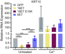
The ability of HPV E7 to degrade PTPN14 correlates with its ability to inhibit differentiation induced by Ca2+. N/Tert-1 stably transduced with retroviruses encoding HPV16 E7, HPV16 E7 E10K, HPV6 E7, or an empty vector control were treated with 1.5 mM Ca2+ and assayed for KRT10 as a marker of differentiation. Bar graph displays the mean ± SD of four independent experiments. Statistical significance was determined by ANOVA followed by multiple t tests with the Holm-Šídák family-wise error rate correction (***P < 0.001; ****P < 0.0001).
Fig. 5.
Fig. 5.
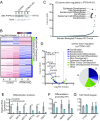
PTPN14 depletion impairs differentiation-related gene expression in primary human keratinocytes. Primary HFK were transduced with LentiCRISPRv2 lentiviral vectors encoding SpCas9 and nontargeting or PTPN14-directed sgRNAs and analyzed for changes in gene expression. (A) Cell lysates were subjected to SDS/PAGE/Western analysis and probed with anti-PTPN14 and antiactin antibodies. (B) PolyA selected RNA was analyzed by RNA-seq. Genes differentially expressed by ≥1.5-fold with P value ≤ 0.05 are displayed in the heat map. Color coding on the right side denotes whether genes are related to epidermis development (blue), other developmental processes (green), or neither (gray). (C) GO enrichment analysis of genes down-regulated in HFK-PTPN14 KO compared with HFK-control. (D) Volcano plot of gene-expression changes in HFK-control vs. HFK-PTPN14 KO. Dots colored by GO terms. Pie chart displays the fraction of genes down-regulated in the absence of PTPN14 that fall into enriched GO Terms. (EG) Transcript abundance for selected genes in HFK-control and HFK-PTPN14 KO was measured by qRT-PCR detecting differentiation markers (E), differentiation promoting TFs (F), and YAP/TEAD targets (G). Bar graphs display the mean ± SD of two or three independent experiments. Statistical significance was determined by Welch’s t tests (*P < 0.05; **P < 0.01).
Fig. 6.
Fig. 6.
PTPN14 loss reduces the expression of differentiation markers after detachment. Adherent N/Tert-mock and -PTPN14 KO cells were detached by trypsinization and replated in ultralow adherence plates before harvesting 12-h postdetachment, then assayed for markers of differentiation, YAP/TEAD targets, and survival after detachment. (A and B) Gene-expression changes induced by suspension were assayed by qRT-PCR targeting markers of differentiation: KRT16 and IVL (A), and YAP/TEAD targets: CTGF and CYR61 (B). mRNA expression was calculated relative to GAPDH. Bar graphs display the mean ± SD of three independent experiments. (C) Survival after detachment was assessed by replating 1,000 cells from suspension, allowing cells to grow for 5 d, and measuring total viable cell area by Crystal violet staining. Three independent experiments are displayed along with mean ± SD. Statistical significance was determined by ANOVA followed by multiple t tests with the Holm-Šídák family-wise error rate correction (*P < 0.05; **P < 0.01; ***P < 0.001).
Fig. 7.
Fig. 7.
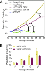
PTPN14 degradation contributes to high-risk HPV E6/E7 immortalization of primary human keratinocytes. Primary HFK cells were transduced with pairs of retroviruses encoding various HPV E6 and E7 and passaged for up to 75 d. (A) Growth curves from representative immortalization experiment. Population doublings were calculated based upon the number of cells harvested at each passage. Statistical significance was determined from three independent experiments by repeated measures two-way ANOVA. Displayed P values represent the column (cell line) factor (*P < 0.05; **P < 0.01). (B) Bar chart shows mean ± SD of selected passages from growth curves in A.
Fig. 8.
Fig. 8.
Keratinocyte differentiation gene signature describes the major differences between HPV+ and HPV HNSCC. Data from HNSCC samples on the TCGA database were determined to be HPV+ or HPV and analyzed for differences in gene expression. Bar chart portrays the ranked −log10(P values) of enriched GO terms among genes down-regulated in HPV+ HNSCC. Pie chart displays fraction of total down-regulated genes that fall into selected GO categories.

Similar articles

Cited by

References

    1. Doorbar J, Egawa N, Griffin H, Kranjec C, Murakami I. Human papillomavirus molecular biology and disease association. Rev Med Virol. 2015;25:2–23. - PMC - PubMed
    1. Graham SV. The human papillomavirus replication cycle, and its links to cancer progression: A comprehensive review. Clin Sci (Lond) 2017;131:2201–2221. - PubMed
    1. McBride AA, Münger K. Expert views on HPV infection. Viruses. 2018;10:E94. - PMC - PubMed
    1. Gillison ML, Chaturvedi AK, Anderson WF, Fakhry C. Epidemiology of human papillomavirus-positive head and neck squamous cell carcinoma. J Clin Oncol. 2015;33:3235–3242. - PMC - PubMed
    1. Muñoz N, et al. International Agency for Research on Cancer Multicenter Cervical Cancer Study Group Epidemiologic classification of human papillomavirus types associated with cervical cancer. N Engl J Med. 2003;348:518–527. - PubMed

Publication types

MeSH terms

Substances