. 2019 Apr 2;116(14):6812-6817.
doi: 10.1073/pnas.1817246116. Epub 2019 Mar 20.

Structure-guided design fine-tunes pharmacokinetics, tolerability, and antitumor profile of multispecific frizzled antibodies

Affiliations

Structure-guided design fine-tunes pharmacokinetics, tolerability, and antitumor profile of multispecific frizzled antibodies

Swetha Raman et al. Proc Natl Acad Sci U S A. .

Abstract

Aberrant activation of Wnt/β-catenin signaling occurs frequently in cancer. However, therapeutic targeting of this pathway is complicated by the role of Wnt in stem cell maintenance and tissue homeostasis. Here, we evaluated antibodies blocking 6 of the 10 human Wnt/Frizzled (FZD) receptors as potential therapeutics. Crystal structures revealed a common binding site for these monoclonal antibodies (mAbs) on FZD, blocking the interaction with the Wnt palmitoleic acid moiety. However, these mAbs displayed gastrointestinal toxicity or poor plasma exposure in vivo. Structure-guided engineering was used to refine the binding of each mAb for FZD receptors, resulting in antibody variants with improved in vivo tolerability and developability. Importantly, the lead variant mAb significantly inhibited tumor growth in the HPAF-II pancreatic tumor xenograft model. Taken together, our data demonstrate that anti-FZD cancer therapeutic antibodies with broad specificity can be fine-tuned to navigate in vivo exposure and tolerability while driving therapeutic efficacy.

Keywords: Frizzled receptors; Wnt signaling; X-ray crystallography; antibody therapeutic; protein engineering.


Conflict of interest statement

Conflict of interest statement: The authors have filed a patent application for the antibodies described in this work.

Figures
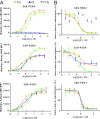
Fig. 1.
Fig. 1.
Binding and functional characteristics of lead FZD mAbs. (A) Binding curves for mAbs F2.I, F7.B, and F6 to 32D cells overexpressing FZD4, -5, or -7. MFI indicates median fluorescence intensity. (B) Dose–response curves for mAbs F2.I, F7.B, and F6 inhibition of Wnt3a-induced β-catenin stabilization in 32D cells overexpressing FZD4, -5, or -7. As F6 was not found to inhibit FZD4, no trendline could be drawn.
Fig. 2.
Fig. 2.
Structural insights into antibody recognition of FZD–CRD and mechanism of Wnt-signaling inhibition. Cocrystal structures of (A) F2.I Fab in complex with FZD5, (B) F7.B Fab in complex with FZD7, and (C) F6 Fab in complex with FZD7. (D) Angle of approach of three antibodies against the same subsite on FZDs. (E) mAbs F2.I (green), F7.B (wheat), and F6 (cyan) block the Wnt–FZD interaction at site 1. The modeled Wnt (pink) is represented as cartoon and the FZD–CRDs (gray) are shown as ribbons. The palmitoyl group is shown in yellow as ball and stick. (F) Outline of the epitope for mAbs F2.I (green), F7.B (wheat), and F6 (cyan) traced onto the surface of FZD7 show overlapping sites of interaction and the lipid-binding pocket with the Wnt palmitoyl group modeled in yellow. Molecular basis of lipid blocking by mAbs (G) F2.I (green), (H) F7.B (wheat), and (I) F6 (cyan). FZD5 is shown in purple and FZD7 in gray.
Fig. 3.
Fig. 3.
Lead FZD mAbs display poor exposure or gut toxicity in vivo. (A) Body weights of mice treated with 30 mg/kg of mAbs F2.I (green), F7.B (orange), or F6 (blue). Arrows indicate when mice were dosed. n = 3 for mAbs F6 and F7.B; n = 10 for mAb F2.I. (B) mAb plasma exposures corresponding to C, presented as mean ± SD. *Mice treated with mAb F2.I were killed on day 5 due to dramatic body weight loss. (C) Histological cross-sections stained with H&E of the duodenum of mice treated with 30 mg/kg of either control IgG, mAbs F2.I, F7.B, or F6. Shown are 10× and 20× magnifications. (Scale bars, 500 μm.)
Fig. 4.
Fig. 4.
Structure-based design of mAb F2.I variants reduces in vivo toxicity. (A) Mutants designed to reduce the affinity toward FZD5. (B) The corresponding binding curves for F2.I Fab and Fab variants, F2.Iv1 (V92A, light chain), F2.Iv2 (Y100AS, heavy chain/V92A, light chain) and F2.Iv3 (H97S, heavy chain/V92A, light chain) are shown. The data (red) were fit using a 1:1 model (black). (C) β-Catenin stabilization assays in 32D–FZD5 cells for mAb F2.I and its variants, mAbs F2.Iv1 (red), F2.Iv2 (brown), and F2.Iv3 (purple). (D) HPAF-II cell proliferation assay for mAb F2.I and its variants. (E) Body weights of mice treated with 30 mg/kg of mAbs F2.I, F2.Iv1, F2.Iv2, and F2.Iv3. Arrows indicate when mice were dosed; n = 5 per group. (F) IgG plasma exposure corresponding to E, presented as mean ± SD. Note: one of five mice from mAb F2.Iv1, two of five mice from mAb F2.Iv2, and one of five mice from mAb F2.Iv3 had undetectable plasma IgG exposure on day 4, indicating that these mice likely did not receive their first dose (technical error).
Fig. 5.
Fig. 5.
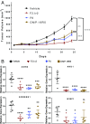
In vivo efficacy of engineered FZD antibody variant shows significant tumor growth inhibition. (A) HPAF-II xenograft study showing tumor volume for mice treated with 30 mg/kg of mAbs OMP-18R5, F6, F2.Iv2, or vehicle i.p. twice per week (n = 12 per group). (B) Tumor gene expression for mice treated with mAbs OMP-18R5, F6, F2.Iv2, or vehicle showing representative genes for Wnt-pathway gene modulation at study endpoint (29 d after HPAF-II cell injection) (*P < 0.05, **P < 0.01, ***P < 0.001, ****P < 0.0001). ns, not significant.

Similar articles

Cited by

References

    1. Dravid G, et al. Defining the role of Wnt/beta-catenin signaling in the survival, proliferation, and self-renewal of human embryonic stem cells. Stem Cells. 2005;23:1489–1501. - PubMed
    1. Logan CY, Nusse R. The Wnt signaling pathway in development and disease. Annu Rev Cell Dev Biol. 2004;20:781–810. - PubMed
    1. Clevers H, Loh KM, Nusse R. Stem cell signaling. An integral program for tissue renewal and regeneration: Wnt signaling and stem cell control. Science. 2014;346:1248012. - PubMed
    1. MacDonald BT, Tamai K, He X. Wnt/beta-catenin signaling: Components, mechanisms, and diseases. Dev Cell. 2009;17:9–26. - PMC - PubMed
    1. Komiya Y, Habas R. Wnt signal transduction pathways. Organogenesis. 2008;4:68–75. - PMC - PubMed

Publication types

MeSH terms