. 2019 Apr 2;116(14):7043-7052.
doi: 10.1073/pnas.1814850116. Epub 2019 Mar 20.

OR14I1 is a receptor for the human cytomegalovirus pentameric complex and defines viral epithelial cell tropism

Affiliations

OR14I1 is a receptor for the human cytomegalovirus pentameric complex and defines viral epithelial cell tropism

Xiaofei E et al. Proc Natl Acad Sci U S A. .

Abstract

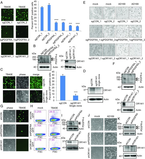
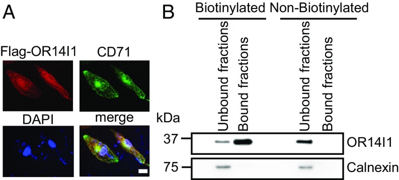
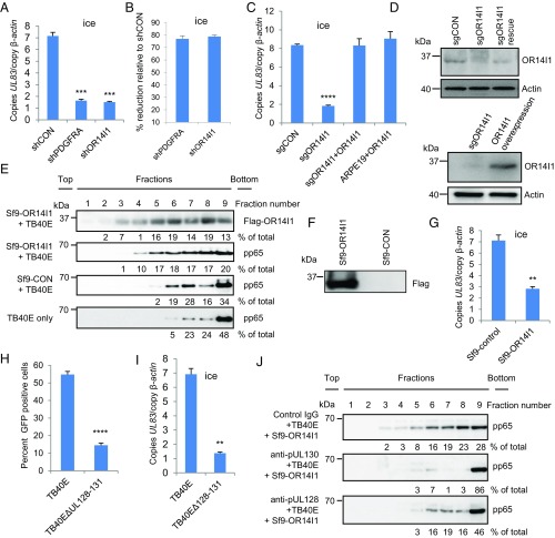
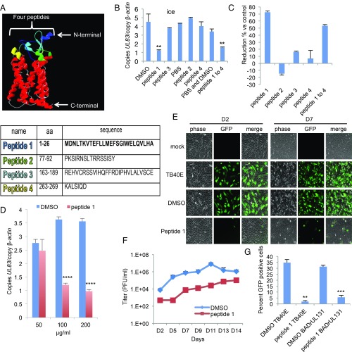
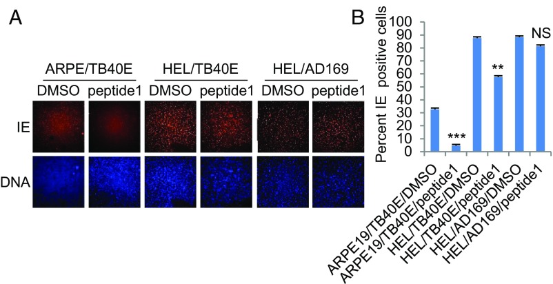
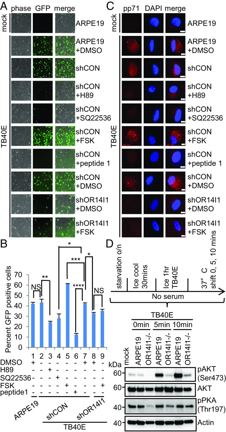
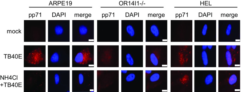
A human cytomegalovirus (HCMV) pentameric glycoprotein complex (PC), gH-gL-UL128-UL130-UL131A, is necessary for viral infection of clinically relevant cell types, including epithelial cells, which are important for interhost transmission and disease. We performed genome-wide CRISPR/Cas9 screens of different cell types in parallel to identify host genes specifically required for HCMV infection of epithelial cells. This effort identified a multipass membrane protein, OR14I1, as a receptor for HCMV infection. This olfactory receptor family member is required for HCMV attachment, entry, and infection of epithelial cells and is dependent on the presence of viral PC. OR14I1 is required for AKT activation and mediates endocytosis entry of HCMV. We further found that HCMV infection of epithelial cells is blocked by a synthetic OR14I1 peptide and inhibitors of adenylate cyclase and protein kinase A (PKA) signaling. Identification of OR14I1 as a PC-dependent HCMV host receptor associated with epithelial tropism and the role of the adenylate cyclase/PKA/AKT-mediated signaling pathway in HCMV infection reveal previously unappreciated targets for the development of vaccines and antiviral therapies.

Keywords: CRISPR screen; OR14I1; human cytomegalovirus; pentameric glycoprotein complex; virus receptor.


Conflict of interest statement

Conflict of interest statement: X.E., A.L.B., and T.F.K. are listed as coinventors in a patent application based in part on data from this study.

Figures

Fig. 1.
Fig. 1.
OR14I1 and PDGRFA are required for HCMV infection of epithelial cells. (A) ARPE-19 cells stably expressing the indicated sgRNAs were infected with TB40E-GFP virus (MOI 3.0). Cells were imaged (10×) for GFP expression (green) as an indicator of viral infection at 2 dpi, and the percent GFP-positive cells was quantified. CON, negative control. (B) Immunoblots of lysates from cells in A. (C) Clonally derived sgOR14I1-expressing ARPE-19 cells were infected with TB40E-GFP virus (MOI 3.0). Cells were imaged (10×) for GFP expression (green) as an indicator of viral infection at 2 dpi, and the GFP-positive cells were quantified. (D) Immunoblots of lysates from cells in C. (E) HEL fibroblasts stably expressing the indicated sgRNAs were infected with AD169 virus (MOI 3.0). Infectivity was determined by cytopathic effect at 2 dpi. (F) Immunoblots of lysates from cells in E. (G) ARPE-19 cells stably expressing the indicated shRNAs were infected with TB40E-GFP virus (MOI 2.0) and then imaged (10×) as in A. (H) ARPE-19 cells stably expressing the indicated shRNAs were infected with TB40E-GFP virus (MOI 2.0). GFP expression was determined at 2 dpi using flow cytometry. The plot depicts GFP versus forward scatter (FSC). (I) Immunoblots of lysates from cells in G. (J) HEL fibroblasts stably expressing the indicated shRNAs were infected with AD169 virus (MOI 2.0). Infectivity was determined by cytopathic effect at 2 dpi. (K) Immunoblots of lysates from cells in J. Actin serves as a loading control throughout. Representative images of three independent experiments are shown. Data represent the mean of n = 3 experiments ±SD. ***P < 0.001, ****P < 0.0001.
Fig. 2.
Fig. 2.
OR14I1 localizes to the plasma and additional cellular membranes. (A) Flag-tagged OR14I1 plasmid was transiently transfected into ARPE-19 cells. Thirty-six hours after transfection, cells were fixed, permeabilized, and immunostained with anti-Flag (red) and anti-transferrin receptor, CD71 (green) antibodies, and then imaged by confocal microscopy. CD71 served as a surrogate for membrane proteins. DAPI staining was used to define nuclei. (Scale bar, 20 µm.) (B) Immunoblot analysis for OR14I1 in the cell surface-bound fractions of biotinylated proteins and the flow-through (unbound fraction representing proteins that did not bind to the EZ-Link Sulfo-NHS-SS-Biotin column). Calnexin immunodetection was included to define the intracellular protein pool.
Fig. 3.
Fig. 3.
OR14I1 is necessary for PC+ HCMV binding to and infection of epithelial cells. (A) Virus binding assay. ARPE-19 cells expressing the indicated shRNAs were incubated with TB40E-GFP virus on ice (MOI 3.0). After washing, cell surface-bound viral DNA (UL83) was quantified using qPCR and normalized to cellular DNA (β-actin). (B) The results in A are presented as the relative reduction of viral DNA in the knockdown cell lines relative to shCON. (C) Binding assays as in A using ARPE-19 cells expressing the indicated sgRNAs and/or cDNAs: sgCON, clonal sgOR14I1 cells, sgOR14I1 cells expressing sgRNA-resistant OR14I1, or WT cells overexpressing OR14I1 (MOI 3.0). (D) Immunoblots of cells in C. (E) Membrane flotation assay. TB40E-GFP virus was incubated with membrane vesicles from control Sf9 cells (Sf9-CON) or Sf9 cells expressing human Flag-OR14I1 (Sf9-OR14I1). After centrifugation, fractions underwent immunoblotting to determine the levels of TB40E-GFP virus (virion protein pp65) and location of membrane vesicles (Flag-OR14I1). (F) Immunoblot of cells in E. (G) TB40E-GFP virus was preincubated with Sf9-control or Sf9-Flag-OR14I1 membrane vesicles before being used in a virus binding assay with ARPE-19 cells (MOI 3.0). Viral (UL83) and cellular (β-actin) DNA levels were quantified by qPCR. (H) ARPE-19 cells infected with TB40E-GFP or a PC-deleted TB40E-GFP (TB40E∆UL128–131; MOI 3.0). Cells were fixed at 2 dpi and assessed for GFP (green)-positive cells. (I) ARPE-19 cells were incubated with either TB40E-GFP or TB40E∆UL128–131 virus (MOI 3.0) on ice. After washing, cell surface-bound viral DNA (UL83) was quantified using qPCR and normalized to cellular DNA (β-actin). (J) Control IgG antibody, anti-pUL128, or anti-pUL130 was preincubated with purified TB40E-GFP virus in a membrane flotation assay. Data represent the mean of n = 3 experiments ±SD. **P < 0.01, ***P < 0.001, ****P < 0.0001.
Fig. 4.
Fig. 4.
OR14I1 peptide inhibits HCMV binding and infection of epithelial cells. (A) OR14I1 model depicting four extracellular domains (blue, green, aqua, yellow) with their amino acid (aa) sequences listed below. (B) Binding assay. TB40E-GFP virus was preincubated with the indicated OR14I1 peptides (100 μg/mL) or relevant solvent followed by incubation of the virus with ARPE-19 cells on ice (MOI 2.0). After washing, cell surface-bound viral DNA (UL83) was quantified using qPCR and normalized to cellular DNA (β-actin). (C) The results in B are presented as the relative reduction in cell-bound viral DNA by peptide treatment relative to the relevant control. (D) Cell binding assay using the indicated concentrations of peptide 1 (MOI 2.0). (E) TB40E-GFP virus was preincubated with peptide 1 (100 μg/mL) or DMSO, followed by infection of ARPE-19 cells (MOI 2.0). Cells were imaged (10×) for GFP expression at 2 dpi (D2) and 7 dpi (D7). (F) Culture media supernatants from E were harvested on the indicated dpi and assayed for infectious virus by plaque assay. (G) TB40E-GFP or BADrUL131-GFP virus, both expressing the PC, was preincubated with peptide 1 (100 μg/mL) or DMSO before infection of ARPE-19 cells (MOI 2.0). Cells were fixed and assessed for GFP (green)-positive cells at 2 dpi. Data represent the mean of n = 3 experiments ±SD. **P < 0.01, ***P < 0.001, ****P < 0.0001.
Fig. 5.
Fig. 5.
Synthetic N-terminal peptide of OR14I1 blocks HCMV infection of ARPE-19 epithelial cells and is dependent on the presence of viral PC. (A) TB40E-GFP encoding the viral PC or AD169 lacking the PC was preincubated with peptide 1 (100 μg/mL) and then infected on ARPE-19 epithelial cells or HEL fibroblasts (MOI 2.0). Cells were fixed, permeabilized, stained for DNA (blue), immunostained for viral immediate-early protein (IE), and imaged (4×). (B) Quantitation of data in A indicating the percent IE-positive cells. Data represent the mean of n = 3 experiments ±SD. **P < 0.01, ***P < 0.001; NS, not significant.
Fig. 6.
Fig. 6.
AC/PKA/AKT signaling is required for HCMV entry and infection of epithelial cells. (A) ARPE-19 cells expressing the indicated shRNAs were pretreated with PKA inhibitor H-89 (20 μM), adenylate cyclase antagonist SQ22536 (150 μM), AC agonist forskolin (20 μM), or DMSO solvent for 2 h before TB40E-GFP infection (MOI 2.0). Peptide 1 was preincubated with TB40E for 2 h at 37 °C before addition to cells. Cells were imaged (10×) for GFP at 2 dpi. (B) Quantification of data in A after cell fixation and DNA staining. Results are presented as the percent GFP-positive cells. Data represent the mean of n = 3 experiments ±SD. *P < 0.05, **P < 0.01, ***P < 0.001, ****P < 0.0001. (C) ARPE-19 cells expressing the indicated shRNAs were pretreated with the indicated drug for 2 h before TB40E-GFP infection (MOI 2.0); peptide 1 was preincubated with TB40E for 2 h at 37 °C. Cells were cooled on ice for 30 min and then infected with cold TB40E (MOI 2.0) containing the noted small molecules for 1 h on ice. Cells were then transferred to 37 °C. At 2 hpi, cells were washed and treated briefly with trypsin to remove surface-bound virus. They were then fixed, permeabilized, stained with anti-pp71, and imaged (63×). (Scale bars, 10 µm.) Representative images of three independent experiments are shown. (D) ARPE-19 and OR14I1−/− cells were infected with cold TB40E (MOI 2.0) for 1 h on ice. Cells were transferred to 37 °C for 0, 5, and 10 min. Levels of p-AKT (S473), total AKT, p-PKA (T197), and actin were detected by immunoblotting from whole-cell lysates. o/n, overnight.
Fig. 7.
Fig. 7.
OR14I1 mediates endocytosis for entry of HCMV. ARPE-19, OR14I1−/− ARPE-19, and HEL cells were pretreated with ammonium chloride (30 mM) for 2 h. Cells and virus were cooled on ice for 30 min and infected with cold TB40E (MOI 2.0) in the presence or absence of ammonium chloride for 1 h on ice. Cells were transferred to 37 °C. At 2 hpi, cells were washed and treated briefly with trypsin to remove surface-bound virus. The cells were then fixed, permeabilized, stained with anti-pp71, and imaged (63×). (Scale bar, 10 µm.)

Similar articles

Cited by

References

    1. Britt W. Manifestations of human cytomegalovirus infection: Proposed mechanisms of acute and chronic disease. Curr Top Microbiol Immunol. 2008;325:417–470. - PubMed
    1. Dollard SC, Grosse SD, Ross DS. New estimates of the prevalence of neurological and sensory sequelae and mortality associated with congenital cytomegalovirus infection. Rev Med Virol. 2007;17:355–363. - PubMed
    1. Kenneson A, Cannon MJ. Review and meta-analysis of the epidemiology of congenital cytomegalovirus (CMV) infection. Rev Med Virol. 2007;17:253–276. - PubMed
    1. Sinzger C, Digel M, Jahn G. Cytomegalovirus cell tropism. Curr Top Microbiol Immunol. 2008;325:63–83. - PubMed
    1. Chan G, Nogalski MT, Yurochko AD. Activation of EGFR on monocytes is required for human cytomegalovirus entry and mediates cellular motility. Proc Natl Acad Sci USA. 2009;106:22369–22374. - PMC - PubMed

Publication types

MeSH terms

Substances