Clinical Trial
. 2019 Mar 20;11(484):eaav0891.
doi: 10.1126/scitranslmed.aav0891.

Delta-like protein 3 expression and therapeutic targeting in neuroendocrine prostate cancer

Affiliations
Clinical Trial

Delta-like protein 3 expression and therapeutic targeting in neuroendocrine prostate cancer

Loredana Puca et al. Sci Transl Med. .

Abstract

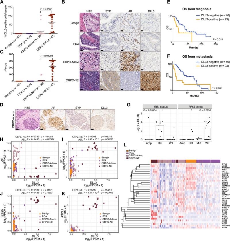
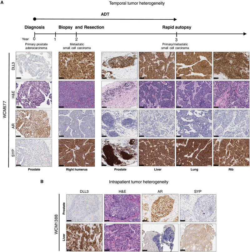
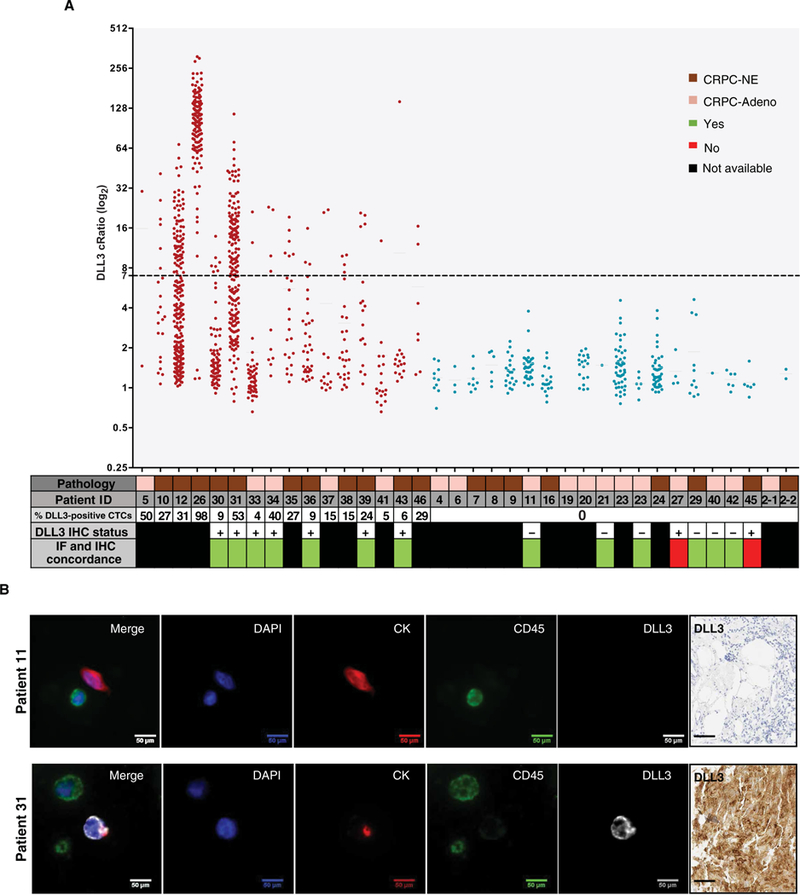
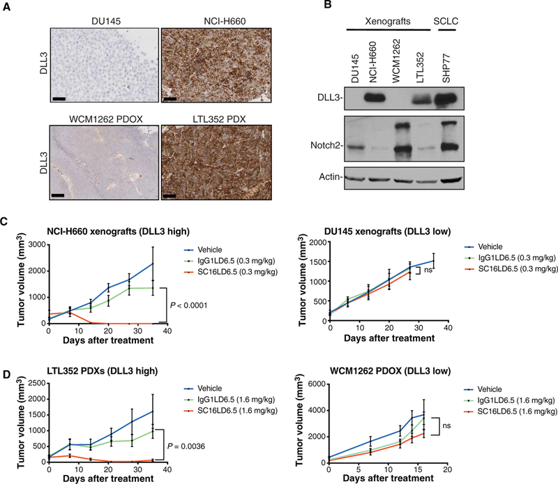
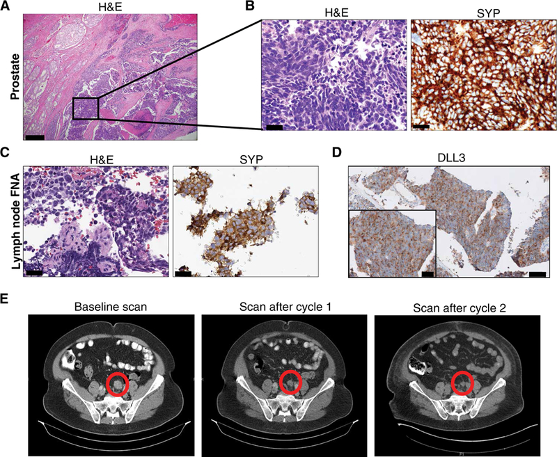
Histologic transformation to small cell neuroendocrine prostate cancer occurs in a subset of patients with advanced prostate cancer as a mechanism of treatment resistance. Rovalpituzumab tesirine (SC16LD6.5) is an antibody-drug conjugate that targets delta-like protein 3 (DLL3) and was initially developed for small cell lung cancer. We found that DLL3 is expressed in most of the castration-resistant neuroendocrine prostate cancer (CRPC-NE) (36 of 47, 76.6%) and in a subset of castration-resistant prostate adenocarcinomas (7 of 56, 12.5%). It shows minimal to no expression in localized prostate cancer (1 of 194) and benign prostate (0 of 103). DLL3 expression correlates with neuroendocrine marker expression, RB1 loss, and aggressive clinical features. DLL3 in circulating tumor cells was concordant with matched metastatic biopsy (87%). Treatment of DLL3-expressing prostate cancer xenografts with a single dose of SC16LD6.5 resulted in complete and durable responses, whereas DLL3-negative models were insensitive. We highlight a patient with neuroendocrine prostate cancer with a meaningful clinical and radiologic response to SC16LD6.5 when treated on a phase 1 trial. Overall, our findings indicate that DLL3 is preferentially expressed in CRPC-NE and provide rationale for targeting DLL3 in patients with DLL3-positive metastatic prostate cancer.


Conflict of interest statement

Competing interests: AbbVie Stemcentrx LLC provided research funding for this study. K.I. and L.S. are employees of AbbVie Stemcentrx LLC. L.S. is listed on patent WO 2017/201442 and related filings. L.F. R.D., and M.K. are employed by Epic Sciences Inc. D.M.N. has declared financial relationship with Roche-Genentech (served on DSMB). S.T.T. and H.B. have received advisory/consulting fees from AbbVie Stemcentrx LLC. All other authors declare that they have no competing interests.

Figures

Fig. 1.
Fig. 1.. DLL3 expression and clinical outcomes in neuroendocrine prostate cancer.
(A) Benign prostate samples (n = 103), PCA (n = 194), CRPC-Adeno (n = 56), and CRPC-NE (n = 47) were evaluated for the percentage of DLL3-positive cells per sample. Median value for CRPC-Adeno was 10.37% of DLL3-positive cells per tumor sample. Median value for CRPC-NE was 63.95% of DLL3-positive cells per tumor sample. Two-tailed t test was performed (P < 0.0001). The error bars indicate the SEM. (B) Representative IHC images of benign, PCA, CRPC-Adeno, and CRPC-NE stained by hematoxylin and eosin (H&E) and with AR, synaptophysin (SYP), and DLL3 antibodies. Scale bars, 50 μm. (C) H-score, combining the percentage of DLL3-positive cells and intensity of the signal for samples stained with DLL3 antibody (H-score: 0 to 300), two-tailed t test was used to compare CRPC-Adeno versus CRPC-NE samples (P < 0.0001). (D) Representative IHC image of a CRPC-Adeno sample stained by H&E and with AR, SYP, and DLL3 antibodies. Scale bars, 100 μm. (E) OS of patients with prostate cancer (n = 63) evaluated from the time of prostate cancer diagnosis. Patients are divided into DLL3-positive (n = 23) (orange) and DLL3-negative (n = 40) (blue) based on IHC, RNA-seq, and NanoString data. Log-rank test was used (P = 0.015). (F) OS of patients with prostate cancer (n = 63) evaluated from the time of metastasis. Patients are divided into DLL3-positive (n = 23) (orange) and DLL3-negative (n = 40) (blue). Log-rank test was used (P = 0.032). (G) Correlation of DLL3 expression and RB1 and TP53 genomic status (amplification, Amp; deletion, Del; mutation, Mut; wild-type, WT) (RB1 deletion versus no deletion: Mann-Whitney, P = 0.03404; TP53 WT versus no WT: Mann-Whitney, P = 0.2256). (H) Correlation of DLL3 and AR expression [log2 (FPKM + 1)] in benign, PCA, CRPC-Adeno, and CRPC-NE patient samples evaluated by RNA-seq (Spearman correlation within CRPC-NE samples, ρ = –0.4211, P = 0.0740; in all samples, ρ = −0.07334, P = 0.3433). (I) Correlation of DLL3 and SYP expression [log2 (FPKM + 1)] in benign, PCA, CRPC-Adeno, and CRPC-NE patient samples evaluated by RNA-seq (Spearman correlation within CRPC-NE samples, ρ = 0.5316, P = 0.0208; in all samples, ρ = 0.38760, P = 1.93E-07). FPKM, fragments per kilobase of transcript per million mapped reads. (J) Correlation of DLL3 and CHGA expression [log2 (FPKM + 1)] in benign, PCA, CRPC-Adeno, and CRPC-NE patient samples evaluated by RNA-seq (Spearman correlation within CRPC-NE samples, ρ = 0.5667, P = 0.0128; in all samples ρ = 0.15592, P = 0.0429). (K) Correlation of DLL3 and ASCL1 expression [log2 (FPKM + 1)] in benign, PCA, CRPC-Adeno, and CRPC-NE patient samples evaluated by RNA-seq (Spearman correlation within CRPC-NE samples, ρ = 0.7211, P = 0.0007; in all samples, ρ = 0.33012, P = 1.17E-05). (L) Supervised cluster analysis of Notch signaling genes in benign, PCA, CRPC-Adeno, and CRPC-NE patient samples.
Fig. 2.
Fig. 2.. Temporal and intrapatient tumor heterogeneity in two patients.
(A) Representative H&E histology images with DLL3, AR, and SYP IHC of a patient with prostate cancer, followed from initial diagnosis of prostate cancer (year 0) to autopsy (year 3). Prostate (year 0), right humerus (year 2), and autopsy (year 3). Scale bars, 100 μm. Samples were obtained from the prostate, liver, lung, and rib. Androgen deprivation therapy (ADT) treatment is indicated by the arrow. (B) Representative H&E and representative DLL3, AR, and SYP IHC images of prostate and concurrent liver biopsies derived from the same patient at the same time point. Scale bars, 100 μm.
Fig. 3.
Fig. 3.. DLL3 detection in CTCs.
(A) Graph showing DLL3 positivity in CTCs of 36 patients with CRPC-Adeno and CRPC-NE tested with the DLL3 Epic four-color immunofluorescence (IF) assay. DLL3 cRatio (signal-to-noise ratio) is plotted along the y axis, and patient ID is plotted along the x axis. The x axis also includes a data table with additional patient-specific information including pathology [CRPC-NE (brown) or CRPC-Adeno (light brown)], percentage of DLL3-positive CTCs, DLL3 IHC status, and IF and IHC concordance. Samples expressing DLL3 are indicated in red, and samples negative for DLL3 are indicated in blue. Each dot represents a detected cell, and the dashed line at 7 along the y axis indicates the analytical threshold of positivity for DLL3. (B) Representative images of patient 11 (DLL3-negative) and patient 31 (DLL3-positive) CTCs. In the four-color composite image, blue represents 4′,6-diamidino-2-phenylindole (DAPI), and red is used for CK, green for CD45, and white for DLL3. Other images show the independent images for each channel used to create the four-color composite (DAPI, CK, CD45, and DLL3). Scale bars, 50 μm. Corresponding DLL3 IHC image is also included for both patients. Scale bars, 50 μm.
Fig. 4.
Fig. 4.. DLL3 antibody drug-conjugate activity in DLL3-expressing neuroendocrine prostate tumors in vivo.
(A) Representative DLL3 IHC images of DU145 cells and WCM1262 PDOX (DLL3-negative) and NCI-H660 cells and LTL352 PDX (DLL3-positive). Scale bars, 100 μm. (B) Western blot analysis of tissue derived from DU145, NCI-H660, WCM1262, and LTL352 xenografts using DLL3 and Notch2 antibodies. Actin is used as a loading control. The SHP77 cell line is used as a control for DLL3 expression. (C) DU145 and NCI-H660 tumor volume (mm3) measurements after single-dose treatment of vehicle, IgG1LD6.5 (0.3 mg/kg), and SC16LD6.5 (0.3 mg/kg) in NU/J mice evaluated for 35 days after treatment. Two-way analysis of variance (ANOVA) test was performed (P < 0.0001). ns, not significant. (D) LTL352 PDX and WCM1262 PDOX tumor volume (mm3) measurements after single-dose treatment of vehicle, IgG1LD6.5 (1.6 mg/kg), and SC16LD6.5 (1.6 mg/kg) in NOD scid gamma mice evaluated for 35 days after treatment. Two-way ANOVA test was performed (P = 0.0036).
Fig. 5.
Fig. 5.. Clinical response in a patient enrolled on a phase 1 SC16LD6.5 basket trial.
(A) Representative H&E image of radical prostatectomy specimen that shows a PCA (left) with an adjacent component of small cell neuroendocrine carcinoma (inset). Scale bar, 200 μm. (B) H&E and strong diffuse synaptophysin (SYP) IHC of the small cell carcinoma component. Scale bars, 50 μm. (C) Lymph node biopsy performed 2 years after prostatectomy. Representative images of H&E and SYP IHC are shown. Scale bars, 50 μm. Fine-needle aspiration, FNA. (D) Representative images of DLL3 IHC are shown (scale bar, 200 μm; inset scale bar, 50 μm). (E) CT scans before therapy and after cycles 1 and 2 of SC16LD6.5 (nodal metastasis highlighted by a circle).

Comment in

References

    1. Beltran H, Rickman DS, Park K, Chae SS, Sboner A, MacDonald TY, Wang Y, Sheikh KL, Terry S, Tagawa ST, Dhir R, Nelson JB, de la Taille A, Allory Y, Gerstein MB, Perner S, Pienta KJ, Chinnaiyan AM, Wang Y, Collins CC, Gleave ME, Demichelis F, Nanus DM, Rubin MA, Molecular characterization of neuroendocrine prostate cancer and identification of new drug targets. Cancer Discov. 1, 487–495 (2011). - PMC - PubMed
    1. Beltran H, Prandi D, Mosquera JM, Benelli M, Puca L, Cyrta J, Marotz C, Giannopoulou E, Chakravarthi BVSK, Varambally S, Tomlins SA, Nanus DM, Tagawa ST, van Allen EM, Elemento O, Sboner A, Garraway LA, Rubin MA, Demichelis E, Divergent clonal evolution of castration-resistant neuroendocrine prostate cancer. Nat. Med. 22, 298–305 (2016). - PMC - PubMed
    1. Bluemn EG, Coleman IM, Lucas JM, Coleman RT, Hernandez-Lopez S, Tharakan R, Bianchi-Frias D, Dumpit RF, Kaipainen A, Corella AN, Yang YC, Nyquist MD, Mostaghel D, Hsieh AC, Zhang X, Corey E, Brown LG, Nguyen HM, Pienta K, Ittmann M, Schweizer M, True LD, Wise D, Rennie PS, Vessella RL, Morrissey C, Nelson PS, Androgen receptor pathway-independent prostate cancer is sustained through FGF signaling. Cancer Cell 32, 474–489.e6 (2017). - PMC - PubMed
    1. Aggarwal R, Huang J, Alumkal JJ, Zhang L, Feng FY, Thomas GV, Weinstein AS, Friedl V, Zhang C, Witte ON, Lloyd P, Gleave M, Evans CP, Youngren J, Beer TM, Rettig M, Wong CK, True L, Foye A, Playdle D, Ryan CJ, Lara P, Chi KN, Uzunangelov V, Sokolov A, Newton Y, Beltran H, Demichelis F, Rubin MA, Stuart JM, Small EJ, Clinical and genomic characterization of treatment-emergent small-cell neuroendocrine prostate cancer: A multi-institutional prospective study. J. Clin. Oncol. 36, 2492–2503 (2018). - PMC - PubMed
    1. Ku SY, Rosario S, Wang Y, Mu P, Seshadri M, Goodrich ZW, Goodrich MM, Labbé DP, Gomez EC, Wang J, Long HW, Xu B, Brown M, Loda M, Sawyers CL, Ellis L, Goodrich DW, Rb1 and Trp53 cooperate to suppress prostate cancer lineage plasticity, metastasis, and antiandrogen resistance. Science 355, 78–83 (2017). - PMC - PubMed

Publication types

MeSH terms