

Increasing Size of Cholesterol Granuloma of the Breast in the Vicinity of a Previous Breast Biopsy: Imaging Features and Review of the Literature
- PMID: 30894505
- PMCID: PMC6439936
- DOI: 10.12659/AJCR.914282
Increasing Size of Cholesterol Granuloma of the Breast in the Vicinity of a Previous Breast Biopsy: Imaging Features and Review of the Literature
Abstract
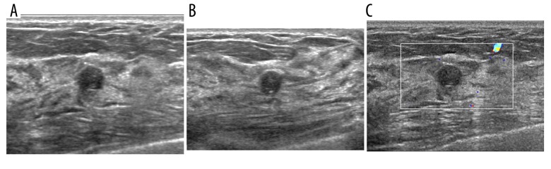
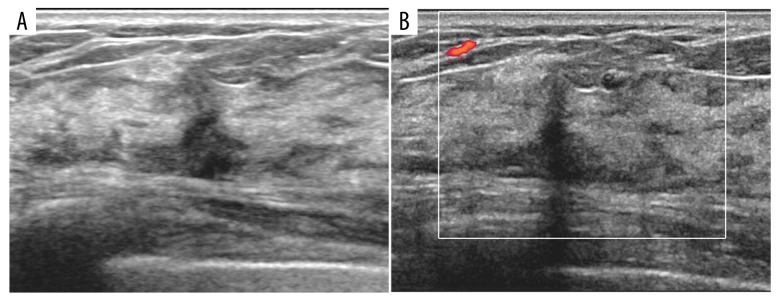
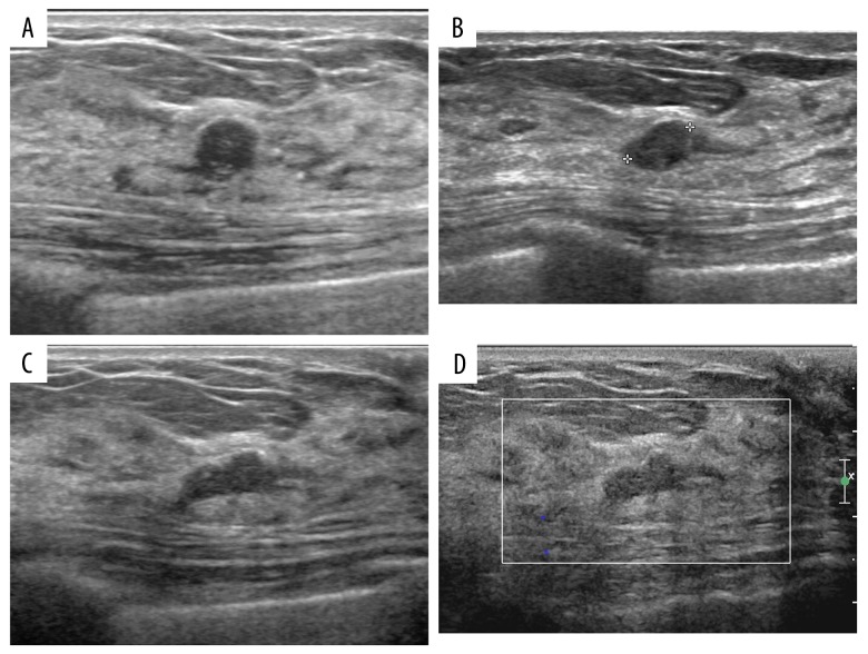
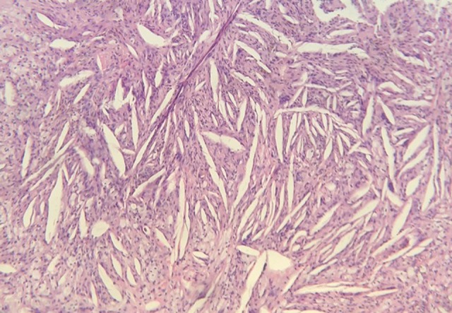
BACKGROUND Cholesterol granuloma is a benign condition that can occur in the breast following trauma, breast biopsy, or surgical procedures. Cholesterol granuloma can mimic breast cancer on imaging studies. This report is of a case of an enlarging breast mass due to a repeat needle biopsy in the vicinity of a previous biopsy that resulted in a cholesterol granuloma that appeared to increase in size on ultrasound imaging. The mammographic and high-resolution sonographic imaging features of cholesterol granuloma of the breast are described, with a review of the literature of published cases of cholesterol granuloma of the breast diagnosed on imaging. CASE REPORT A 52-year-old woman had undergone multiple rounds of breast ultrasound scans for multiple breast nodules. At six-month follow-up with a needle biopsy was performed that showed benign cystic change. At 18-months follow-up high-resolution sonographic imaging showed a circumscribed, oval, hypoechoic mass with a benign morphology in the vicinity of the biopsy site. The size of the hypoechoic nodule increased progressively, and histopathology confirmed the diagnosis of a benign cholesterol granuloma. CONCLUSIONS This case demonstrated that cholesterol granuloma of the breast can increase in size after repeats adjacent breast biopsy, and may retain benign morphological features on ultrasound imaging, despite increasing in size.
Conflict of interest statement
None.
Figures
Similar articles
-
Cholesterol Granuloma of the Breast Mimicking Cancer: A Case Report and Review of the Literature.J Investig Med High Impact Case Rep. 2023 Jan-Dec;11:23247096231193275. doi: 10.1177/23247096231193275. J Investig Med High Impact Case Rep. 2023. PMID: 37565689 Free PMC article. Review.
-
Sonographic appearance of a cholesterol granuloma mimicking breast cancer.J Clin Ultrasound. 2017 Nov 12;45(9):608-611. doi: 10.1002/jcu.22453. Epub 2017 Feb 21. J Clin Ultrasound. 2017. PMID: 28220943
-
The unusual ultrasound features of a breast cholesterol granuloma manifesting as an intracystic mass: case report and literature review.Korean J Radiol. 2013 Mar-Apr;14(2):179-82. doi: 10.3348/kjr.2013.14.2.179. Epub 2013 Feb 22. Korean J Radiol. 2013. PMID: 23482884 Free PMC article. Review.
-
A case of breast cholesterol granuloma accompanied by cancer.Breast Cancer. 2004;11(2):210-3. doi: 10.1007/BF02968304. Breast Cancer. 2004. PMID: 15550870
-
T1-Weighted Dynamic Contrast-Enhanced Magnetic Resonance Imaging (DCE-MRI) to Distinguish Between Concurrent Cholesterol Granuloma and Invasive Ductal Carcinoma of the Breast: A Case Report.Am J Case Rep. 2018 May 23;19:593-598. doi: 10.12659/AJCR.909161. Am J Case Rep. 2018. PMID: 29789520 Free PMC article.
Cited by
-
Multiple cholesterol granulomas of the breast: A case report and review of the literature.Medicine (Baltimore). 2023 Feb 22;102(8):e33084. doi: 10.1097/MD.0000000000033084. Medicine (Baltimore). 2023. PMID: 36827045 Free PMC article. Review.
-
Cholesterol granuloma mimicking ovarian cancer.Int J Clin Exp Pathol. 2021 Jun 15;14(6):741-745. eCollection 2021. Int J Clin Exp Pathol. 2021. PMID: 34239676 Free PMC article.
-
Cholesterol Granuloma of the Breast Mimicking Cancer: A Case Report and Review of the Literature.J Investig Med High Impact Case Rep. 2023 Jan-Dec;11:23247096231193275. doi: 10.1177/23247096231193275. J Investig Med High Impact Case Rep. 2023. PMID: 37565689 Free PMC article. Review.
References
-
- Kim YS, Chang JM. Sonographic appearance of a cholesterol granuloma mimicking breast cancer. J Clin Ultrasound. 2017;45(9):608–11. - PubMed
-
- Jeong SH, Lee EH, Hong HS, et al. Cholesterol granuloma of the breast incidentally detected on dynamic abdominal CT: A case report. J Korean Soc Radiol. 2016;74(1):22–25.
-
- Martineau P, Pelletier-Galarneau M, Leung E. Cholesterol granuloma: An unusual and benign cause of focal FDG uptake in the breast detected on PET/CT. Clin Nucl Med. 2015;40(11):e511–13. - PubMed
Publication types
MeSH terms
Substances
LinkOut - more resources
Full Text Sources
Medical
