. 2019 Mar 20;10(4):278.
doi: 10.1038/s41419-019-1514-4.

The histone demethylase LSD1 promotes renal inflammation by mediating TLR4 signaling in hepatitis B virus-associated glomerulonephritis

Affiliations

The histone demethylase LSD1 promotes renal inflammation by mediating TLR4 signaling in hepatitis B virus-associated glomerulonephritis

Yi-Tong Yang et al. Cell Death Dis. .

Abstract

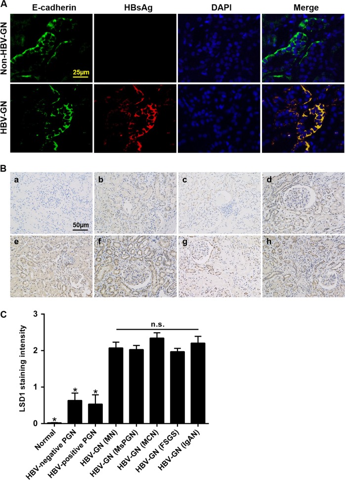
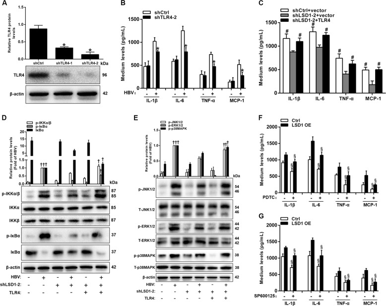
Renal inflammation significantly contributes to the progression of hepatitis B virus (HBV)-associated glomerulonephritis (HBV-GN), but the mechanisms that control its precise regulation remain largely unknown. In this study, we showed that the lysine-specific demethylase 1 (LSD1) was significantly upregulated in renal tissue of HBV-GN patients, and its expression was positively correlated with inflammation. Functionally, LSD1 could promote HBV-induced release of proinflammatory mediators in HK-2 cells, a human renal tubular epithelial (RTE) cell line. Mechanistic investigations suggested that LSD1 directly promoted the transcription of the inflammatory-related gene Tlr4 by eliminating the mono- or di-methylation of H3K9 near its promoter. Knockdown of Lsd1 further inhibited TLR4-NF-κB/JNK signaling cascades, and subsequently decreased HBV-induced production of proinflammatory mediators in HK-2 cells. Co-transfection with Tlr4-expressing plasmids counteracted these effects. Meanwhile, downregulation of abovementioned TLR4-related pathways using small-molecule inhibitors attenuated inflammation. Importantly, LSD1 inhibitor tranylcypromine (TCP) could inhibit TLR4-NF-κB/JNK signaling axis and alleviate renal inflammation in HBV transgenic mice. Taken together, our data identify LSD1 as a novel regulator of renal inflammation and as a potential therapeutic target in HBV-GN.


Conflict of interest statement

The authors declare that they have no conflict of interest.

Figures

Fig. 1
Fig. 1. Elevated lysine-specific demethylase 1 (LSD1) expression levels in patients with hepatitis B virus-associated glomerulonephritis (HBV-GN).
a Immunofluorescence for HBsAg and E-cadherin in HBV-GN and non-HBV-GN groups. E-cadherin was used as a tubule epithelial marker. b Immunohistochemistry for LSD1 in HBV-GN group and other pathological conditions. a Normal control group, b HBV-negative primary glomerulonephritis (PGN) group, c HBV-positive PGN group, d membranous nephropathy (MN), e mesangial-proliferative glomerulonephritis (MsPGN), f minimal change nephropathy (MCN), g focal segmental glomerulosclerosis (FSGS), and h IgA nephropathy (IgAN). d–h The different pathology types for HBV-GN group. c Mean LSD1 staining intensity in each group. Staining intensity was graded on a scale of 0 (no staining) to 3+ (intense staining). Data are expressed as the mean ± SD. *P < 0.05 versus HBV-GN. n.s. not significant
Fig. 2
Fig. 2. Increased inflammatory responses in renal tissues of hepatitis B virus-associated glomerulonephritis (HBV-GN).
a Immunohistochemistry for interleukin (IL)-1β, IL-6, monocyte chemoattractant protein-1 (MCP-1), and CD68 in HBV-GN and non-HBV-GN groups. b Mean IL-1β, IL-6, MCP-1, and CD68 staining intensity in each group. Staining intensity was graded as indicated in Fig. 1c. Data are expressed as the mean ± SD. *P < 0.05 versus HBV-GN
Fig. 3
Fig. 3. Lysine-specific demethylase 1 (LSD1) increases the production of proinflammatory mediators in hepatitis B virus (HBV)-infected HK-2 cells.
a, b Quantitative real-time PCR (qRT-PCR) and western blot analysis of LSD1 mRNA (a) and protein (b) in HK-2 cells transfected with pCMV-HBV1.3 at different times. c, d Enzyme-linked immunosorbent assay analysis of proinflammatory mediators including interleukin (IL)-1β, IL-6, tumor necrosis factor-alpha (TNF-α), and monocyte chemoattractant protein-1 (MCP-1) levels in culture medium of HK-2 cells co-transfected with pCMV-HBV1.3 and LSD1 overexpressed (OE) plasmid (c) or shLSD1-2 (d) for 48 h. e, f qRT-PCR analysis of proinflammatory mediators mRNA expression in HK-2 cells co-transfected as indicated in c and d, respectively. Data are presented as the mean ± SD (N = 3). *P < 0.05 versus Ctrl; #P < 0.05 versus shCtrl; P < 0.05 versus no HBV infection
Fig. 4
Fig. 4. RNA-sequencing after Lsd1 knockdown in hepatitis B virus (HBV)-infected HK-2 cells.
a Heat map shows top 30 differentially expressed genes in shCtrl- or shLSD1-treated cells with three repeats. Differential gene expression is displayed as Z-score. b Gene ontology analysis for all genes with altered expressions. c The altered mRNA levels of inflammatory-related genes were selectively confirmed by quantitative real-time PCR in knockdown Lsd1. Data are presented as the mean ± SD (N = 3). *P < 0.05 versus shCtrl
Fig. 5
Fig. 5. Lsd1 directly regulates Tlr4 expression in hepatitis B virus (HBV)-infected HK-2 cells.
a Western blot analysis of H3K4 and H3K9 methylation in HK-2 cells co-transfected with pCMV-HBV1.3 and shCtrl or shLSD1-2 for 48 h. b, d, e Chromatin immunoprecipitation-quantitative PCR analysis of LSD1 (b), H3K9me1/2 (d), and H3K4me1/2 (e) enrichment in the Tlr4 promoter regions. Signals are shown as a percentage of the input. IgG immunoglobulin G. c Luciferase activity in lysates of HK-2 cells transfected with luciferase reporter plasmids of pGL3-basic vector (vector), toll-like receptor 4 (TLR4) promoter (pTLR4-wt), or TLR4 promoter with mutation on the predicted LSD1-binding site (pTLR4-mut). Relative luciferase activity for each group was standardized using the value from the control cells transfected with pGL3-basic vector. f Western blot analysis of TLR4 in HBV-infected HK-2 cells transfected with shCtrl or shLSD1-2 for 48 h. g Western blot analysis of TLR4 in HBV-infected HK-2 cells treated with saline (Ctrl) or tranylcypromine (TCP, 10 µM) for 12 h. Data are presented as the mean ± SD (N = 3). *P < 0.05 versus shCtrl; #P < 0.05 versus HBV + shCtrl group; P< 0.05 versus Ctrl. n.s. not significant
Fig. 6
Fig. 6. Lysine-specific demethylase 1 (LSD1) promotes the release of proinflammatory mediators in hepatitis B virus (HBV)-infected HK-2 cells via toll-like receptor 4 (TLR4).
a Western blot analysis of TLR4 in HK-2 cells co-transfected with pCMV-HBV1.3 and shCtrl or shTLR4 for 48 h. b Enzyme-linked immunosorbent assay (ELISA) analysis of proinflammatory mediator levels in culture medium of HK-2 cells co-transfected with pCMV-HBV1.3 and shTLR4-2 for 48 h. c ELISA analysis of proinflammatory mediators levels in culture medium of HK-2 cells infected by HBV treated with shLSD1-2 along with pcDNA3.1/myc or pcDNA3.1/myc-TLR4 for 48 h. d, e Western blot analysis of p-IKKα/β, IKKα, IKKβ, p-IκBα, IκBα (d), p-JNK1/2, T-JNK1/2, p-ERK1/2, T-ERK1/2, p-p38MAPK, and T-p38MAPK (e) in HK-2 cells co-transfected with pCMV-HBV1.3 and shLSD1-2 and pcDNA3.1/myc-TLR4 for 12 h. f, g ELISA analysis of proinflammatory mediators levels in cell culture medium. HK-2 cells were pre-incubated with or without 30 µm PDTC (f) or 20 µm SP600125 (g) for 2 h and then treated with pCMV-HBV1.3 along with pcDNA3.1/myc or pcDNA3.1/myc-LSD1 for 48 h. Data are presented as the mean ± SD (N = 4). *P < 0.05 versus shCtrl; #P < 0.05 versus shLSD1-2 + vector group; P < 0.05 versus HBV + shLSD1-2 group; §P < 0.05 versus no PDTC or SP600125 treatment
Fig. 7
Fig. 7. Inhibition of lysine-specific demethylase 1 (LSD1) attenuates renal inflammation in vivo.
a, b Quantitative real-time PCR (qRT-PCR) and western blot analysis of LSD1 mRNA (a) and protein (b) in renal cortex of wild-type (WT) and hepatitis B virus-transgenic (HBV-Tg) mice. c Immunohistochemistry for LSD1 and H3K9me1/2 in the renal cortex. dh WT or HBV-Tg mice were treated with saline or tranylcypromine (TCP). d qRT-PCR analysis of proinflammatory mediator mRNA levels. e Blood urea nitrogen (BUN) and Scr were examined by Roche Modular P800. f Western blot analysis of Kim-1 and cystatin C. g Western blot analysis of p-IKKα/β, IKKα, IKKβ, p-IκBα, IκBα, p-JNK1/2, and T-JNK1/2. h Immunohistochemistry for toll-like receptor 4 (TLR4) in the renal cortex. Data are presented as the mean ± SD (N = 3). *P < 0.05 versus WT; #P < 0.05 versus Ctrl; P< 0.05 versus HBV-Tg alone group
Fig. 8
Fig. 8. The proposed mechanism of lysine-specific demethylase 1 (LSD1) involved in promoting hepatitis B virus (HBV)-induced production of proinflammatory mediators via toll-like receptor 4 (TLR4).
LSD1 promotes HBV-induced production of proinflammatory mediators in vitro by epigenetically upregulating TLR4, thereby contributing to the activation of nuclear factor-κB (NF-κB) and JNK pathways

Similar articles

Cited by

References

    1. Yi Z, Jie YW, Nan Z. The efficacy of anti-viral therapy on hepatitis B virus-associated glomerulonephritis: a systematic review and meta-analysis. Ann. Hepatol. 2011;10:165–173. - PubMed
    1. Takekoshi Y, Tochimaru H, Nagata Y, Itami N. Immunopathogenetic mechanisms of hepatitis B virus-related glomerulopathy. Kidney Int. Suppl. 1991;35:S34–S39. - PubMed
    1. Lai KN, Ho RT, Tam JS, Lai FM. Detection of hepatitis B virus DNA and RNA in kidneys of HBV related glomerulonephritis. Kidney Int. 1996;50:1965–1977. doi: 10.1038/ki.1996.519. - DOI - PubMed
    1. Chen L, et al. Replication and infectivity of hepatitis B virus in HBV-related glomerulonephritis. Int. J. Infect. Dis. 2009;13:394–398. doi: 10.1016/j.ijid.2008.08.014. - DOI - PubMed
    1. Zhou Y, et al. The role of the toll-like receptor TLR4 in hepatitis B virus-associated glomerulonephritis. Arch. Virol. 2013;158:425–433. doi: 10.1007/s00705-012-1508-3. - DOI - PubMed

Publication types

MeSH terms