. 2019 Mar 18;9(1):4745.
doi: 10.1038/s41598-019-39993-7.

Hemodynamic responses to emotional speech in two-month-old infants imaged using diffuse optical tomography

Affiliations

Hemodynamic responses to emotional speech in two-month-old infants imaged using diffuse optical tomography

Shashank Shekhar et al. Sci Rep. .

Abstract

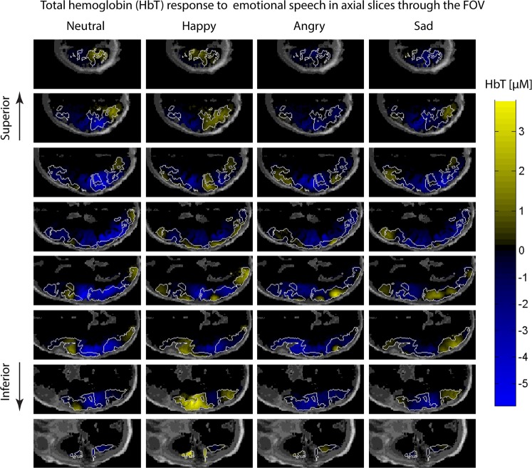
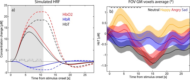
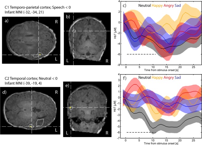
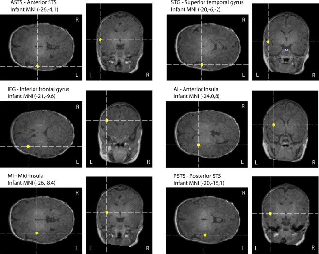
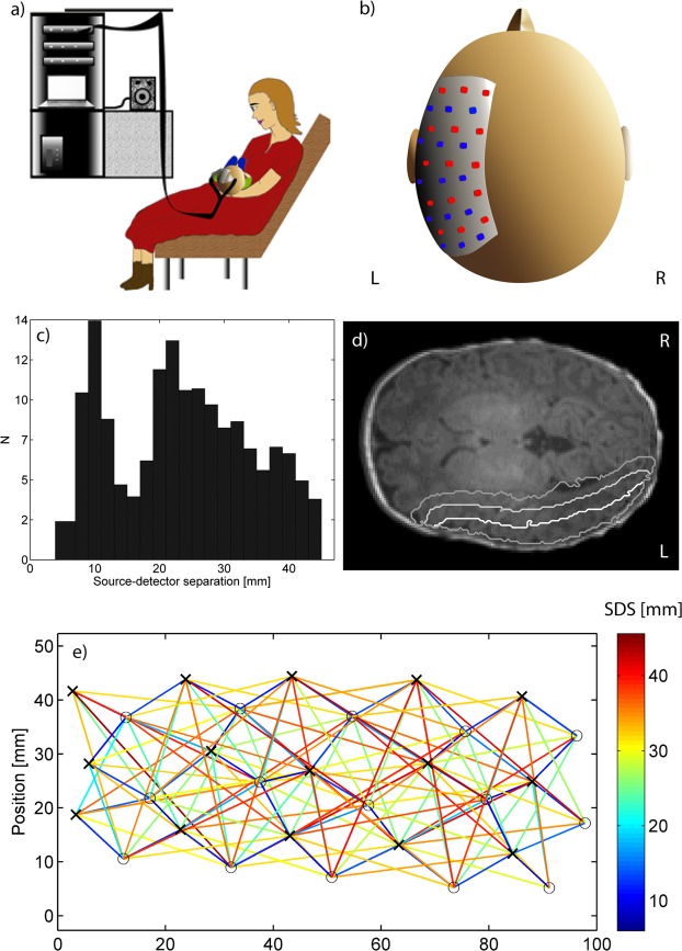
Emotional speech is one of the principal forms of social communication in humans. In this study, we investigated neural processing of emotional speech (happy, angry, sad and neutral) in the left hemisphere of 21 two-month-old infants using diffuse optical tomography. Reconstructed total hemoglobin (HbT) images were analysed using adaptive voxel-based clustering and region-of-interest (ROI) analysis. We found a distributed happy > neutral response within the temporo-parietal cortex, peaking in the anterior temporal cortex; a negative HbT response to emotional speech (the average of the emotional speech conditions < baseline) in the temporo-parietal cortex, neutral > angry in the anterior superior temporal sulcus (STS), happy > angry in the superior temporal gyrus and posterior superior temporal sulcus, angry < baseline in the insula, superior temporal sulcus and superior temporal gyrus and happy < baseline in the anterior insula. These results suggest that left STS is more sensitive to happy speech as compared to angry speech, indicating that it might play an important role in processing positive emotions in two-month-old infants. Furthermore, happy speech (relative to neutral) seems to elicit more activation in the temporo-parietal cortex, thereby suggesting enhanced sensitivity of temporo-parietal cortex to positive emotional stimuli at this stage of infant development.


Conflict of interest statement

The authors declare no competing interests.

Figures

Figure 1
Figure 1
Axial slices of the left hemisphere showing the grand average responses over the 21 subjects within the time window [2 s, 18 s] from stimulus onset from top of the head to bottom in 10 mm intervals. Warm colors (yellow) indicate an increase in total hemoglobin (HbT) and cool colors (blue) indicate a decrease in HbT in response to the stimulus averaged in the 2 s to 18 s post stimulus onset time window. The scalp and skull are shown in dark gray. White line surrounds gray matter voxels within the field of view which are positive at least for one stimulus condition.
Figure 2
Figure 2
(a) The simulated shape of the hemodynamic response (HbR in blue, HbO2 in red, and HbT in black) obtained by convolving a square wave depicting the stimulus train consisting of four sentences and the adult canonical hemodynamic response function (HRF). Default parameters for the HRF were used for HbR, and the delay of onset of the response and undershoot for HbO2 were set to −1 s relative to corresponding parameters for the HbR. The ratio of HbR and HbO2 amplitudes was set to −1:6. HbT was obtained as a sum of HbO2 and HbR. The stimulus epoch is shaded in gray and the individual stimuli indicated with a dark gray line, with habituation after the first stimulus of a train factored into the amplitude (solid lines) and no habituation (dashed ‘--’ lines). (b) Time course of HbT responses to each of the four stimulus conditions (black = neutral, orange = happy, red = angry, and blue = sad speech) in the GM voxels of the region showing activation for at least one stimulus condition. Shading indicates standard error mean (SEM). Black dashed line (‘--’) indicates the time of stimulus presentation.
Figure 3
Figure 3
Axial and coronal slices for clusters C1-C2, with voxels that satisfy p < 0.001 marked in yellow and p = 0.01 marked with white contour line, and the corresponding HbT response time courses for each stimulus condition. C1 shows a significant negative response to speech (ac); C2 a significant negative response to neutral speech (df). Neutral speech is marked in black, happy speech in orange, angry speech in red and sad speech in blue. The shaded area shows the standard error mean (SEM).
Figure 4
Figure 4
Locations of the regions of interest (ROIs) used in this study.
Figure 5
Figure 5
(a) Illustration of the measurement session (drawn by author SS). (b) Position of the probe on the subject’s head. (c) Histogram of source-detector separations used in the imaging. (d) The relative sensitivity map thresholded at 0.1 (white line), 0.01 (light gray line) and 0.001 (dark gray line) superimposed on an axial slice. (e) Relative positions of the sources (black crosses) and detectors (gray circles) with color-coding of the interconnecting lines indicating source-detector distances (SDS) up to 45 mm.

References

    1. Grossmann T, Oberecker R, Koch SP, Friederici AD. The developmental origins of voice processing in the human brain. Neuron. 2010;65:852–858. doi: 10.1016/j.neuron.2010.03.001. - DOI - PMC - PubMed
    1. Köchel A, Schöngaßner F, Feierl-Gsodam S, Schienle A. Processing of affective prosody in boys suffering from attention deficit hyperactivity disorder: A near-infrared spectroscopy study. Soc Neurosci. 2015;10:583–591. doi: 10.1080/17470919.2015.1017111. - DOI - PubMed
    1. Grossmann T, Striano T, Friederici AD. Developmental changes in infants’ processing of happy and angry facial expressions: a neurobehavioral study. Brain Cogn. 2007;64:30–41. doi: 10.1016/j.bandc.2006.10.002. - DOI - PubMed
    1. Nakato E, Otsuka Y, Kanazawa S, Yamaguchi MK, Kakigi R. Distinct differences in the pattern of hemodynamic response to happy and angry facial expressions in infants–a near-infrared spectroscopic study. NeuroImage. 2011;54:1600–1606. doi: 10.1016/j.neuroimage.2010.09.021. - DOI - PubMed
    1. Jönsson EH, et al. Affective and non-affective touch evoke differential brain responses in 2-month-old infants. Neuroimage. 2018;169:162–171. doi: 10.1016/j.neuroimage.2017.12.024. - DOI - PubMed

Publication types