. 2019 Mar 18;9(1):4750.
doi: 10.1038/s41598-019-41202-4.

Moderate Nucleoporin 133 deficiency leads to glomerular damage in zebrafish

Affiliations

Moderate Nucleoporin 133 deficiency leads to glomerular damage in zebrafish

Chiara Cianciolo Cosentino et al. Sci Rep. .

Erratum in

Abstract

Although structural nuclear pore proteins (nucleoporins) are seemingly required in every cell type to assemble a functional nuclear transport machinery, mutations or deregulation of a subset of them have been associated with specific human hereditary diseases. In particular, previous genetic studies of patients with nephrotic syndrome identified mutations in Nup107 that impaired the expression or the localization of its direct partner at nuclear pores, Nup133. In the present study, we characterized the zebrafish nup133 orthologous gene and its expression pattern during larval development. Using a morpholino-mediated gene knockdown, we show that partial depletion of Nup133 in zebrafish larvae leads to the formation of kidney cysts, a phenotype that can be rescued by co-injection of wild type mRNA. Analysis of different markers for tubular and glomerular development shows that the overall kidney development is not affected by nup133 knockdown. Likewise, no gross defect in nuclear pore complex assembly was observed in these nup133 morphants. On the other hand, nup133 downregulation results in proteinuria and moderate foot process effacement, mimicking some of the abnormalities typically featured by patients with nephrotic syndrome. These data indicate that nup133 is a new gene required for proper glomerular structure and function in zebrafish.


Conflict of interest statement

The authors declare no competing interests.

Figures

Figure 1
Figure 1
Expression of nup133 in the developing zebrafish detected by in situ hybridization (ISH). Whole mount ISH with nup133 antisense probe of embryos at: (a) sphere stage (4 hpf; embryo shown with animal pole to the top); (b) 24 hfp (lateral view); (cf) 3 and 5 dpf (left panels: dorsal view; right panels: lateral view). Arrows point to tissues with enriched expression of nup133. Abbreviations: E: eyes; T: tectum; C: cerebellum; L: liver; I: intestine; N: neuromasts; SB: swim bladder. Scale bars, 200 µm. (g) Transverse section of a 5 dpf embryo at the level of the pectoral fins (as shown in the dotted line in e) confirms nup133 expression in the liver, and show in addition a diffuse staining in the proximal tubules (PT) and a faint signal in the glomerulus (G). Scale bar, 50 µm.
Figure 2
Figure 2
Splice Morpholinos (MO) targeting nup133 lead to a partial degradation of nup133 mRNAs. (a) Exon structure of Danio rerio (Dr) nup133 around the binding sites of the E3I3 and I3E4 splice morpholinos. Blue arrowheads above the scheme indicate the position of the RT-PCR primers used in (b) and blue/red/orange arrows below indicate the position of primers and RT-qPCR products used in (c). The size of intron 3 is indicated. (b) RT-PCR from total RNAs of 48 hpf uninjected embryos (control) and of embryos coinjected with the two splicing morpholinos (nup133 MO) reveal an additional band caused by retention of intron 3. (c) nup133 mRNA levels relative to actb2 expression was determined by RT-qPCR on 24 and 48 hpf embryos, uninjected, injected with control MO or nup133 MO, or sequentially injected with 3xHA-mCherry-Dr nup133 mRNA (wt mRNA) and nup133 MO. The E3-E4/5 (red) and E3/4-E4/5′ (orange) primer pairs only amplify nup133 mRNA with properly spliced intron 3 (i.e. wt nup133 mRNA not targeted by the morpholinos) while the E1/2-E3 and E7/8-E9 primer pairs (blue bars) recognize both the spliced and unspliced mRNAs.
Figure 3
Figure 3
Partial knockdown of nup133 causes glomerular expansion in zebrafish. (a) Gross morphology of 3 dpf control and nup133 MO embryos (left panels: lateral view; middle panel: dorso-lateral view from two distinct embryos). Scale bar, 500 µm. Two-fold magnification of the indicated area is shown in the rightmost panels. Arrows indicate the pronephric cysts detected in the nup133 MO embryos. (b) Dorsal view of 3dpf Tg(wt1b:EGFP) embryos uninjected (control, top panels), injected with nup133 MO (middle panels), or sequentially injected with 3xHA-mCherry-Dr nup133 mRNA (wt mRNA) and nup133 MO (bottom panels). Overlays of transmission (gray) and GFP-signal (green) images reveal the glomerulus, proximal tubules, and exocrine pancreas. Scale bar, 500 µm. Two-fold magnification of the indicated area and of the same area from a distinct larvae are shown in the right and rightmost panels, respectively. The glomerular structure is indicated in brackets. Asterisks point to cystic dilations of the pronephros in nup133 MO (middle panels). Note that the nup133 MO + wt mRNA embryo displays a left-sided exocrine pancreas (arrowhead) (see also Supplemental Fig. S5). (c) Relative proportion of embryos showing or not kidney cysts at 3 dpf. For each condition, the total number of embryos analyzed is indicated (n=, quantified in 2 distinct experiments for control MO injections and 5 experiments for nup133 MO injections. See also Supplementary Fig. S3). Unlike the embryos injected with control MO, those injected with nup133 MO frequently feature kidney cysts. On the other hand nup133 MO + wt mRNA showed significantly fewer cysts than nup133 MO alone. ***P < 0.0001 using a Fisher exact probability test.
Figure 4
Figure 4
Nup133 depletion does not result in major nuclear pore complex assembly defects. (a) Representative transverse sections of the glomerulus of 3 dpf Tg(wt1b:EGFP) embryos treated with either control or nup133 morpholinos (MO) and stained with mAb414 (not shown in these panels) and DAPI. In these sections, GFP-positive cells mark the glomerulus and the neck region of the proximal tubules. Scale bars, 100 μm. (b) Fivefold magnification of the areas indicated in (a) encompassing the glomerulus (G, indicated by the dashed lines) and the liver (L). mAb414 antibody, that recognizes a subset of FG-nucleoporins, shows specific staining around the nucleus of all cells (visualized with DAPI), with however cell-type-dependent variations in intensity. Note for instance the more prominent staining of liver as compared to glomerular cells. In contrast, no major difference can be seen between control and nup133 MO-treated embryos. Insets show a fourfold magnification of representative liver nuclei, revealing the punctate staining typical of NPC staining (mAb414). Scale bars, 20 μm.
Figure 5
Figure 5
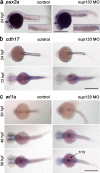
Normal development of the pronephric tubules and glomerulus in nup133 morphants. (a) Lateral view of pax2a mRNA expression in pronephric tubules at 24 hpf in uninjected (control) and nup133 MO-injected embryos reveal that the developmental expression of pax2a is not altered in nup133 morphants. Two-fold magnification of the pronephric tubules is also shown. (b) Pronephros marker cdh17 mRNA expression in nup133 MO is comparable to that of uninjected controls at 24 and 72 hpf (dorsal view). (c) Glomerular development in control and nup133 MO embryos visualized using the podocyte differentiation marker wt1a (dorsal view). At 30 hpf (upper panels), wt1a marks future podocytes with two distinct domains in both control and nup133 MO. At 48 hpf (middle panels), the glomerular primordia merge to the midline to form a single glomerulus. At these stages, mRNA expression does not differ between control and nup133 MO (middle panels). At 58 hpf (lower panels), after the onset of glomerular filtration, the increased area labeled by the wt1a probe (arrow) reflects the glomerular expansion observed in 7 out of 13 nup133 morphants analyzed. Scale bars, 500 µm.
Figure 6
Figure 6
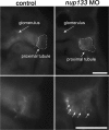
Glomerular filtration is impaired in nup133 morphants. Representative images of cross sections of 4 dpf control (left panels) and nup133 morphants larvae (right panels) fixed 20 minutes after injection of Alexa Fluor™ 647 conjugated-BSA into the common cardinal vein. Lower panels represent a higher magnification view of the proximal tubule region (dotted lines). Note the uptake of fluorescent BSA in the apical endosomes of the proximal tubules of the Nup133-depleted larva (arrows). Scale bars 50 µm.
Figure 7
Figure 7
nup133 knockdown induces moderate foot processes effacement. (a,d) Transmission electron micrographs showing the glomerular capillary wall of 5 dpf uninjected control (a) and nup133 MO injected larvae (d). (b,e) Two-fold magnification of the areas indicated in (a,d), and (c,f,h) representative images from distinct larvae. Images were pseudocolored to better highlight the glomerular filtration barrier components (podocyte foot processes in green, fenestrated endothelium in blue). Arrows point to irregular-shaped foot processes that are more frequently found in nup133 MO larvae. (g) Quantification of foot processes (FP) density along the basement membrane, presented as average number of FP/µm, was performed as described under materials and methods. The distinct labels reflect values measured on the 3 wt or morphant embryos. Statistical analyses were performed using Wilcoxon-Mann-Whitney Rank Sum Test. **P ≤ 0.01 (p = 0.002486). Image (h) corresponds to a basement membrane surrounding a large cyst (presented in Supplemental Fig. S6e). Note that statistical analyses performed without the latter nup133 morphant larva (values indicated by black squares on the graph) still revealed a significant difference between the control and the nup133 morphants (p = 0.01855). Abbreviations: BS: bowman space; C: capillary lumen; EC: endothelial cells; FP: foot processes. Scale bars 1 µm.

Similar articles

  • Biallelic Mutations in Nuclear Pore Complex Subunit NUP107 Cause Early-Childhood-Onset Steroid-Resistant Nephrotic Syndrome.
    Miyake N, Tsukaguchi H, Koshimizu E, Shono A, Matsunaga S, Shiina M, Mimura Y, Imamura S, Hirose T, Okudela K, Nozu K, Akioka Y, Hattori M, Yoshikawa N, Kitamura A, Cheong HI, Kagami S, Yamashita M, Fujita A, Miyatake S, Tsurusaki Y, Nakashima M, Saitsu H, Ohashi K, Imamoto N, Ryo A, Ogata K, Iijima K, Matsumoto N. Miyake N, et al. Am J Hum Genet. 2015 Oct 1;97(4):555-66. doi: 10.1016/j.ajhg.2015.08.013. Epub 2015 Sep 24. Am J Hum Genet. 2015. PMID: 26411495 Free PMC article.
  • Homozygous splicing mutation in NUP133 causes Galloway-Mowat syndrome.
    Fujita A, Tsukaguchi H, Koshimizu E, Nakazato H, Itoh K, Kuraoka S, Komohara Y, Shiina M, Nakamura S, Kitajima M, Tsurusaki Y, Miyatake S, Ogata K, Iijima K, Matsumoto N, Miyake N. Fujita A, et al. Ann Neurol. 2018 Dec;84(6):814-828. doi: 10.1002/ana.25370. Ann Neurol. 2018. PMID: 30427554
  • Mutations in multiple components of the nuclear pore complex cause nephrotic syndrome.
    Braun DA, Lovric S, Schapiro D, Schneider R, Marquez J, Asif M, Hussain MS, Daga A, Widmeier E, Rao J, Ashraf S, Tan W, Lusk CP, Kolb A, Jobst-Schwan T, Schmidt JM, Hoogstraten CA, Eddy K, Kitzler TM, Shril S, Moawia A, Schrage K, Khayyat AIA, Lawson JA, Gee HY, Warejko JK, Hermle T, Majmundar AJ, Hugo H, Budde B, Motameny S, Altmüller J, Noegel AA, Fathy HM, Gale DP, Waseem SS, Khan A, Kerecuk L, Hashmi S, Mohebbi N, Ettenger R, Serdaroğlu E, Alhasan KA, Hashem M, Goncalves S, Ariceta G, Ubetagoyena M, Antonin W, Baig SM, Alkuraya FS, Shen Q, Xu H, Antignac C, Lifton RP, Mane S, Nürnberg P, Khokha MK, Hildebrandt F. Braun DA, et al. J Clin Invest. 2018 Oct 1;128(10):4313-4328. doi: 10.1172/JCI98688. Epub 2018 Sep 4. J Clin Invest. 2018. PMID: 30179222 Free PMC article.
  • Moonlighting nuclear pore proteins: tissue-specific nucleoporin function in health and disease.
    Jühlen R, Fahrenkrog B. Jühlen R, et al. Histochem Cell Biol. 2018 Dec;150(6):593-605. doi: 10.1007/s00418-018-1748-8. Epub 2018 Oct 25. Histochem Cell Biol. 2018. PMID: 30361777 Review.
  • Glomerular changes in hereditary single-gene diseases.
    Sessa A, Meroni M, Battini G, Maglio A, Righetti M, Fabris E, Nebuloni M, Pallotti F, Giordano F, Tosoni A. Sessa A, et al. J Nephrol. 2002 Nov-Dec;15 Suppl 6:S47-56. J Nephrol. 2002. PMID: 12515374 Review.

Cited by

References

    1. Beck M, Hurt E. The nuclear pore complex: understanding its function through structural insight. Nat Rev Mol Cell Biol. 2017;18:73–89. - PubMed
    1. Nofrini V, Di Giacomo D, Mecucci C. Nucleoporin genes in human diseases. Eur J Hum Genet. 2016;24:1388–1395. - PMC - PubMed
    1. Hezwani M, Fahrenkrog B. The functional versatility of the nuclear pore complex proteins. Semin Cell Dev Biol. 2017;68:2–9. - PubMed
    1. Sakuma S, D’Angelo MA. The roles of the nuclear pore complex in cellular dysfunction, aging and disease. Semin Cell Dev Biol. 2017;68:72–84. - PMC - PubMed
    1. Patrakka J, Tryggvason K. Molecular make-up of the glomerular filtration barrier. Biochem Biophys Res Commun. 2010;396:164–169. - PubMed

Publication types

Substances