. 2019 Mar 20;9(1):4902.
doi: 10.1038/s41598-019-41277-z.

A reference collection of patient-derived cell line and xenograft models of proneural, classical and mesenchymal glioblastoma

Affiliations

A reference collection of patient-derived cell line and xenograft models of proneural, classical and mesenchymal glioblastoma

Brett W Stringer et al. Sci Rep. .

Erratum in

Abstract

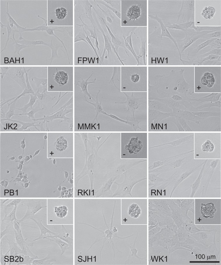
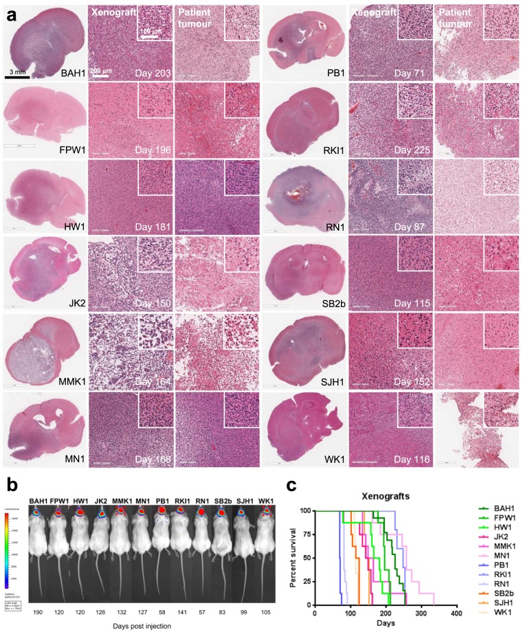
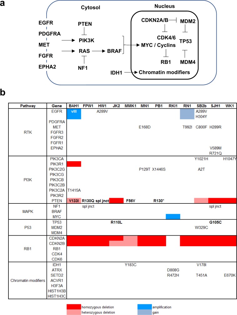
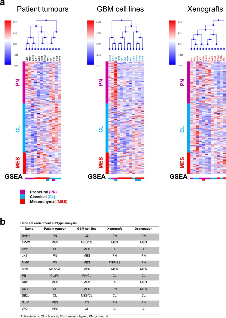
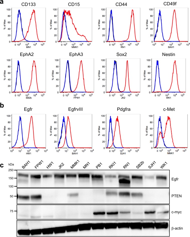
Low-passage, serum-free cell lines cultured from patient tumour tissue are the gold-standard for preclinical studies and cellular investigations of glioblastoma (GBM) biology, yet entrenched, poorly-representative cell line models are still widely used, compromising the significance of much GBM research. We submit that greater adoption of these critical resources will be promoted by the provision of a suitably-sized, meaningfully-described reference collection along with appropriate tools for working with them. Consequently, we present a curated panel of 12 readily-usable, genetically-diverse, tumourigenic, patient-derived, low-passage, serum-free cell lines representing the spectrum of molecular subtypes of IDH-wildtype GBM along with their detailed phenotypic characterisation plus a bespoke set of lentiviral plasmids for bioluminescent/fluorescent labelling, gene expression and CRISPR/Cas9-mediated gene inactivation. The cell lines and all accompanying data are readily-accessible via a single website, Q-Cell (qimrberghofer.edu.au/q-cell/) and all plasmids are available from Addgene. These resources should prove valuable to investigators seeking readily-usable, well-characterised, clinically-relevant, gold-standard models of GBM.


Conflict of interest statement

The authors declare no competing interests.

Figures

Figure 1
Figure 1
A diverse set of low-passage, serum-free, patient-derived GBM cell lines exhibiting a range of cell morphologies. Bright field images of GBM cell lines growing as adherent cultures on matrigel (main images) or as non-adherent tumourspheres (inset). Plus signs (+) indicate cell lines that readily form tumourspheres; minus signs (−) indicate cell lines that form tumourspheres poorly or not at all. Scale bar, 100 μm.
Figure 2
Figure 2
Bioluminescent xenograft tumour formation in NOD/SCID mice. (a) Comparative histology of xenograft tumours formed by the GBM cell lines and the patient tumours they were established from. First column, haematoxylin and eosin (H&E)-stained, coronal sections of mouse brains showing invasive xenograft tumours. Scale bar, 3 mm. Comparative H&E-stained sections of matched xenograft (middle column) and patient tumours (right column). Scale bar 200 μm, inset 100 μm. (b) Bioluminescent intracranial xenograft tumours produced by the GBM cell lines expressing firefly luciferase. Stably-transduced lines were created by transduction with lentivirus containing pF CAG luc puro and selection with puromycin. Time to develop the bioluminescent xenograft tumours detected is shown below each mouse. (c) Kaplan-Meier survival curves for NOD/SCID mice harbouring xenografts initiated by intracranial injection of each of the GBM cell lines.
Figure 3
Figure 3
Genomic profiling of the GBM cell lines. (a) Pathways most frequently affected by genomic changes in GBM showing the relationships of the genes involved. (b) Single nucleotide variants (SNVs), intragenic deletions and gene copy number changes in genes in these pathways detected in the GBM cell lines. SNVs shown in bold are homozygous, otherwise heterozygous. Blanks indicate wild type genes.
Figure 4
Figure 4
Molecular subtyping of matched patient tumours, GBM cell lines and xenografts. (a) Hierarchical cluster analysis (top), corresponding heatmaps (middle) and gene set enrichment analysis (GSEA) (bottom) of patient tumours, derived GBM cell lines and matched murine xenografts initiated by the cell lines identifying gene expression patterns characteristic of proneural, classical and mesenchymal GBM. (b) Summary of GSEA results.
Figure 5
Figure 5
GBM-associated protein expression in the GBM cell lines. Representative flow cytometry analysis of (a) stem cell markers and (b) selected receptor tyrosine kinases in the GBM cell lines cultured as adherent monolayers on matrigel. (c) Western blot analysis of Egfr, Pten and c-myc expression in the GBM cell lines. kDa, kilodalton.

Similar articles

Cited by

References

    1. Omuro A, DeAngelis LM. Glioblastoma and other malignant gliomas: a clinical review. JAMA. 2013;310:1842–1850. - PubMed
    1. Stupp R, et al. Radiotherapy plus concomitant and adjuvant temozolomide for glioblastoma. N Engl J Med. 2005;352:987–996. doi: 10.1056/NEJMoa043330. - DOI - PubMed
    1. Phillips HS, et al. Molecular subclasses of high-grade glioma predict prognosis, delineate a pattern of disease progression, and resemble stages in neurogenesis. Cancer Cell. 2006;9:157–173. - PubMed
    1. Verhaak RG, et al. Integrated genomic analysis identifies clinically relevant subtypes of glioblastoma characterized by abnormalities in PDGFRA, IDH1, EGFR, and NF1. Cancer Cell. 2010;17:98–110. - PMC - PubMed
    1. Bao S, et al. Glioma stem cells promote radioresistance by preferential activation of the DNA damage response. Nature. 2006;444:756–760. - PubMed

Publication types